Journals > > Topics > Biophotonics
Biophotonics|21 Article(s)
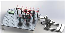
Noncontact ultrasound sensing based on Mach–Zehnder homodyne interferometer for photoacoustic imaging
Xing Long, Yicheng Hu, Yibing Wang, and Changhui Li
We present a novel noncontact ultrasound (US) and photoacoustic imaging (PAI) system, overcoming the limitations of traditional coupling media. Using a long coherent length laser, we employ a homodyne free-space Mach–Zehnder setup with zero-crossing triggering, achieving a noise equivalent pressure of 703 Pa at 5 MHz and a -6 dB bandwidth of 1 to 8.54 MHz. We address the phase uncertainty inherent in the homodyne method. Scanning the noncontact US probe enables photoacoustic computed tomography (PACT). Phantom studies demonstrate imaging performance and system stability, underscoring the potential of our system for noncontact US sensing and PAI. We present a novel noncontact ultrasound (US) and photoacoustic imaging (PAI) system, overcoming the limitations of traditional coupling media. Using a long coherent length laser, we employ a homodyne free-space Mach–Zehnder setup with zero-crossing triggering, achieving a noise equivalent pressure of 703 Pa at 5 MHz and a -6 dB bandwidth of 1 to 8.54 MHz. We address the phase uncertainty inherent in the homodyne method. Scanning the noncontact US probe enables photoacoustic computed tomography (PACT). Phantom studies demonstrate imaging performance and system stability, underscoring the potential of our system for noncontact US sensing and PAI.
Chinese Optics Letters
- Publication Date: Mar. 28, 2024
- Vol. 22, Issue 3, 031702 (2024)


Stimulated emission–depletion-based point-scanning structured illumination microscopy
Lei Wang, Meiting Wang, Luwei Wang, Xiaomin Zheng, Jiajie Chen, Wenshuai Wu, Wei Yan, Bin Yu, Junle Qu, Bruce Zhi Gao, and Yonghong Shao
Wide-field linear structured illumination microscopy (LSIM) extends resolution beyond the diffraction limit by moving unresolvable high-frequency information into the passband of the microscopy in the form of moiré fringes. However, due to the diffraction limit, the spatial frequency of the structured illumination pattern cannot be larger than the microscopy cutoff frequency, which results in a twofold resolution improvement over wide-field microscopes. This Letter presents a novel approach in point-scanning LSIM, aimed at achieving higher-resolution improvement by combining stimulated emission depletion (STED) with point-scanning structured illumination microscopy (psSIM) (STED-psSIM). The according structured illumination pattern whose frequency exceeds the microscopy cutoff frequency is produced by scanning the focus of the sinusoidally modulated excitation beam of STED microscopy. The experimental results showed a 1.58-fold resolution improvement over conventional STED microscopy with the same depletion laser power. Wide-field linear structured illumination microscopy (LSIM) extends resolution beyond the diffraction limit by moving unresolvable high-frequency information into the passband of the microscopy in the form of moiré fringes. However, due to the diffraction limit, the spatial frequency of the structured illumination pattern cannot be larger than the microscopy cutoff frequency, which results in a twofold resolution improvement over wide-field microscopes. This Letter presents a novel approach in point-scanning LSIM, aimed at achieving higher-resolution improvement by combining stimulated emission depletion (STED) with point-scanning structured illumination microscopy (psSIM) (STED-psSIM). The according structured illumination pattern whose frequency exceeds the microscopy cutoff frequency is produced by scanning the focus of the sinusoidally modulated excitation beam of STED microscopy. The experimental results showed a 1.58-fold resolution improvement over conventional STED microscopy with the same depletion laser power.
Chinese Optics Letters
- Publication Date: Mar. 13, 2024
- Vol. 22, Issue 3, 031701 (2024)
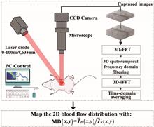
Laser speckle contrast imaging based on uniting spatiotemporal Fourier transform
Linjun Zhai, Yongzhao Du, Xunxun Wu, Yong Diao, and Yuqing Fu
We propose a laser speckle contrast imaging method based on uniting spatiotemporal Fourier transform. First, the raw speckle images are entirely transformed to the spatiotemporal frequency domain with a three-dimensional (3D) fast Fourier transform. Second, the dynamic and static speckle components are extracted by applying 3D low-pass and high-pass filtering in the spatiotemporal frequency domain and inverse 3D Fourier transform. Third, we calculate the time-averaged modulation depth with the average of both components to map the two-dimensional blood flow distribution. The experiments demonstrate that the proposed method could effectively improve computational efficiency and imaging quality. We propose a laser speckle contrast imaging method based on uniting spatiotemporal Fourier transform. First, the raw speckle images are entirely transformed to the spatiotemporal frequency domain with a three-dimensional (3D) fast Fourier transform. Second, the dynamic and static speckle components are extracted by applying 3D low-pass and high-pass filtering in the spatiotemporal frequency domain and inverse 3D Fourier transform. Third, we calculate the time-averaged modulation depth with the average of both components to map the two-dimensional blood flow distribution. The experiments demonstrate that the proposed method could effectively improve computational efficiency and imaging quality.
Chinese Optics Letters
- Publication Date: Jan. 08, 2024
- Vol. 22, Issue 1, 011701 (2024)
Aberration correction for multiphoton microscopy using covariance matrix adaptation evolution strategy
Ke Wang, Lei Zheng, Mengyuan Qin, Wanjian Zhang, Xiangquan Deng, Shen Tong, Hui Cheng, Jie Huang, Jincheng Zhong, Yingxian Zhang, and Ping Qiu
Multiphoton microscopy is the enabling tool for biomedical research, but the aberrations of biological tissues have limited its imaging performance. Adaptive optics (AO) has been developed to partially overcome aberration to restore imaging performance. For indirect AO, algorithm is the key to its successful implementation. Here, based on the fact that indirect AO has an analogy to the black-box optimization problem, we successfully apply the covariance matrix adaptation evolution strategy (CMA-ES) used in the latter, to indirect AO in multiphoton microscopy (MPM). Compared with the traditional genetic algorithm (GA), our algorithm has a greater improvement in convergence speed and convergence accuracy, which provides the possibility of realizing real-time dynamic aberration correction for deep in vivo biological tissues. Multiphoton microscopy is the enabling tool for biomedical research, but the aberrations of biological tissues have limited its imaging performance. Adaptive optics (AO) has been developed to partially overcome aberration to restore imaging performance. For indirect AO, algorithm is the key to its successful implementation. Here, based on the fact that indirect AO has an analogy to the black-box optimization problem, we successfully apply the covariance matrix adaptation evolution strategy (CMA-ES) used in the latter, to indirect AO in multiphoton microscopy (MPM). Compared with the traditional genetic algorithm (GA), our algorithm has a greater improvement in convergence speed and convergence accuracy, which provides the possibility of realizing real-time dynamic aberration correction for deep in vivo biological tissues.
Chinese Optics Letters
- Publication Date: Apr. 12, 2023
- Vol. 21, Issue 5, 051701 (2023)
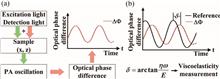
A novel needle probe for deeper photoacoustic viscoelasticity measurement
Daoqian Yang, Zhongjiang Chen, and Da Xing
We present for the first time, to the best of our knowledge, a needle probe for photoacoustic viscoelasticity (PAVE) measurements at a depth of 1 cm below the sample surface. The probe uses a gradient index rod lens, encased within a side-facing needle (0.7 mm outer diameter), to direct excitation light (532 nm) and detection light (1325 nm) focused on the sample, collecting and directing the returned detection light in a spectral domain low coherence interferometry system, which allows for obtaining optical phase differences due to photoacoustic oscillations. The feasibility of needle probe for PAVE depth characterization was investigated on gelatin phantoms and in vitro biological tissues. The experimental results in an in vivo animal model predict the great potential of this technique for in vivo tumor boundary detection. We present for the first time, to the best of our knowledge, a needle probe for photoacoustic viscoelasticity (PAVE) measurements at a depth of 1 cm below the sample surface. The probe uses a gradient index rod lens, encased within a side-facing needle (0.7 mm outer diameter), to direct excitation light (532 nm) and detection light (1325 nm) focused on the sample, collecting and directing the returned detection light in a spectral domain low coherence interferometry system, which allows for obtaining optical phase differences due to photoacoustic oscillations. The feasibility of needle probe for PAVE depth characterization was investigated on gelatin phantoms and in vitro biological tissues. The experimental results in an in vivo animal model predict the great potential of this technique for in vivo tumor boundary detection.
Chinese Optics Letters
- Publication Date: May. 26, 2022
- Vol. 20, Issue 8, 081701 (2022)

Secondary structure changes of ox-LDL by photoirradiation in an optofluidic resonator
Yuxi Shang, Hailang Dai, Daopeng Dai, Jinmao Gu, Meng Zhang, Qiheng Wei, and Xianfeng Chen
Atherosclerotic cardio-cerebral vascular disease is the most common disease that threatens human health. Many researches indicated that oxidatively modified low-density lipoprotein (ox-LDL) is a key pathogenic factor of atherosclerosis. Here, we report the change of the secondary structure of ox-LDL caused by photoirradiation in an optofluidic resonator. The content ratios of amphipathic α-helices and β-sheets of ox-LDL are changed under laser beam illumination, resulting in an increasing binding rate of ox-LDL and ox-LDL antibodies. Our findings may provide a potential way for clinical atherosclerosis treatment and prompt recovery rate of atherosclerotic cardio-cerebral vascular disease by optical technology and immunotherapy. Atherosclerotic cardio-cerebral vascular disease is the most common disease that threatens human health. Many researches indicated that oxidatively modified low-density lipoprotein (ox-LDL) is a key pathogenic factor of atherosclerosis. Here, we report the change of the secondary structure of ox-LDL caused by photoirradiation in an optofluidic resonator. The content ratios of amphipathic α-helices and β-sheets of ox-LDL are changed under laser beam illumination, resulting in an increasing binding rate of ox-LDL and ox-LDL antibodies. Our findings may provide a potential way for clinical atherosclerosis treatment and prompt recovery rate of atherosclerotic cardio-cerebral vascular disease by optical technology and immunotherapy.
Chinese Optics Letters
- Publication Date: Jan. 24, 2022
- Vol. 20, Issue 3, 031702 (2022)
Fluo-Fluo translation based on deep learning
Zhengfen Jiang, Boyi Li, Tho N. H. T. Tran, Jiehui Jiang, Xin Liu, and Dean Ta
Fluorescence microscopy technology uses fluorescent dyes to provide highly specific visualization of cell components, which plays an important role in understanding the subcellular structure. However, fluorescence microscopy has some limitations such as the risk of non-specific cross labeling in multi-labeled fluorescent staining and limited number of fluorescence labels due to spectral overlap. This paper proposes a deep learning-based fluorescence to fluorescence (Fluo-Fluo) translation method, which uses a conditional generative adversarial network to predict a fluorescence image from another fluorescence image and further realizes the multi-label fluorescent staining. The cell types used include human motor neurons, human breast cancer cells, rat cortical neurons, and rat cardiomyocytes. The effectiveness of the method is verified by successfully generating virtual fluorescence images highly similar to the true fluorescence images. This study shows that a deep neural network can implement Fluo-Fluo translation and describe the localization relationship between subcellular structures labeled with different fluorescent markers. The proposed Fluo-Fluo method can avoid non-specific cross labeling in multi-label fluorescence staining and is free from spectral overlaps. In theory, an unlimited number of fluorescence images can be predicted from a single fluorescence image to characterize cells. Fluorescence microscopy technology uses fluorescent dyes to provide highly specific visualization of cell components, which plays an important role in understanding the subcellular structure. However, fluorescence microscopy has some limitations such as the risk of non-specific cross labeling in multi-labeled fluorescent staining and limited number of fluorescence labels due to spectral overlap. This paper proposes a deep learning-based fluorescence to fluorescence (Fluo-Fluo) translation method, which uses a conditional generative adversarial network to predict a fluorescence image from another fluorescence image and further realizes the multi-label fluorescent staining. The cell types used include human motor neurons, human breast cancer cells, rat cortical neurons, and rat cardiomyocytes. The effectiveness of the method is verified by successfully generating virtual fluorescence images highly similar to the true fluorescence images. This study shows that a deep neural network can implement Fluo-Fluo translation and describe the localization relationship between subcellular structures labeled with different fluorescent markers. The proposed Fluo-Fluo method can avoid non-specific cross labeling in multi-label fluorescence staining and is free from spectral overlaps. In theory, an unlimited number of fluorescence images can be predicted from a single fluorescence image to characterize cells.
Chinese Optics Letters
- Publication Date: Jan. 04, 2022
- Vol. 20, Issue 3, 031701 (2022)
Sample entropy analysis of laser speckle fluctuations to suppress motion artifact on blood flow monitoring|Editors' Pick
Sungchul Kim, Evgenii Kim, Eloise Anguluan, and Jae Gwan Kim
Laser speckle imaging is a common technique to monitor blood flow. The fluctuations in speckle intensity can be related to the blood flow by calculating the speckle contrast, the ratio between the standard deviation of speckle fluctuations and the average intensity. However, this simple statistic calculation is easily affected by motion artifacts. In this study, we applied sample entropy analysis instead of calculating standard deviations of the speckle fluctuations. Similar to the traditional method, sample entropy-based speckle contrast increases linearly with flow rate but was shown to be more immune to sudden movements during an upper arm occlusion test. Laser speckle imaging is a common technique to monitor blood flow. The fluctuations in speckle intensity can be related to the blood flow by calculating the speckle contrast, the ratio between the standard deviation of speckle fluctuations and the average intensity. However, this simple statistic calculation is easily affected by motion artifacts. In this study, we applied sample entropy analysis instead of calculating standard deviations of the speckle fluctuations. Similar to the traditional method, sample entropy-based speckle contrast increases linearly with flow rate but was shown to be more immune to sudden movements during an upper arm occlusion test.
Chinese Optics Letters
- Publication Date: Nov. 17, 2021
- Vol. 20, Issue 1, 011702 (2022)
Dual-beam delay-encoded all fiber Doppler optical coherence tomography for in vivo measurement of retinal blood flow
Mingming Wan, Shanshan Liang, Xinyu Li, Zhengyu Duan, Jiebin Zou, Jun Chen, Jin Yuan, and Jun Zhang
In this paper, we propose and demonstrate a dual-beam delay-encoded Doppler spectral domain optical coherence tomography (OCT) system for in vivo measurement of absolute retinal blood velocity and flow with arbitrary orientation. The incident beam is split by a beam displacer into two probe beams of the single-spectrometer spectral domain OCT system with orthogonal polarization states and an optical path length delay. We validate our approach with a phantom and in vivo experiments of human retinal blood flow, respectively. In this paper, we propose and demonstrate a dual-beam delay-encoded Doppler spectral domain optical coherence tomography (OCT) system for in vivo measurement of absolute retinal blood velocity and flow with arbitrary orientation. The incident beam is split by a beam displacer into two probe beams of the single-spectrometer spectral domain OCT system with orthogonal polarization states and an optical path length delay. We validate our approach with a phantom and in vivo experiments of human retinal blood flow, respectively.
Chinese Optics Letters
- Publication Date: Nov. 08, 2021
- Vol. 20, Issue 1, 011701 (2022)
Nonlinear photoacoustic imaging dedicated to thermal-nonlinearity characterization
Yujiao Shi, and Zhenhui Zhang
We proposed a nonlinear photoacoustic (PA) technique as a new imaging contrast mechanism for tissue thermal-nonlinearity characterization. When a sine-modulated Gaussian temperature field is introduced by a laser beam, in view of the temperature dependence of the thermal diffusivity, the nonlinear PA effect occurs, which leads to the production of second-harmonic PA (SHPA) signals. By extracting the fundamental frequency PA and SHPA signal amplitudes of samples through the lock-in technique, a parameter that only reflects nonlinear thermal-diffusivity characteristics of the sample then can be obtained. The feasibility of the technique for thermal-nonlinearity characterization has been studied on phantom samples. In vitro biological tissues have been studied by this method to demonstrate its medical imaging capability, prefiguring great potential of this new method in medical imaging applications. We proposed a nonlinear photoacoustic (PA) technique as a new imaging contrast mechanism for tissue thermal-nonlinearity characterization. When a sine-modulated Gaussian temperature field is introduced by a laser beam, in view of the temperature dependence of the thermal diffusivity, the nonlinear PA effect occurs, which leads to the production of second-harmonic PA (SHPA) signals. By extracting the fundamental frequency PA and SHPA signal amplitudes of samples through the lock-in technique, a parameter that only reflects nonlinear thermal-diffusivity characteristics of the sample then can be obtained. The feasibility of the technique for thermal-nonlinearity characterization has been studied on phantom samples. In vitro biological tissues have been studied by this method to demonstrate its medical imaging capability, prefiguring great potential of this new method in medical imaging applications.
Chinese Optics Letters
- Publication Date: Jul. 10, 2021
- Vol. 19, Issue 7, 071702 (2021)
Topics
© Copyright 2018-2021 | Chinese Laser Press. All Rights Reserved 沪ICP备15018463号-20