
Mingming Wan, Shanshan Liang, Xinyu Li, Zhengyu Duan, Jiebin Zou, Jun Chen, Jin Yuan, Jun Zhang, "Dual-beam delay-encoded all fiber Doppler optical coherence tomography for in vivo measurement of retinal blood flow," Chin. Opt. Lett. 20, 011701 (2022)
- Chinese Optics Letters
- Vol. 20, Issue 1, 011701 (2022)
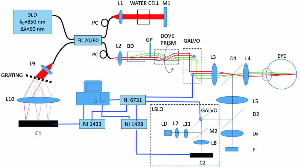
Fig. 1. Schematic of the dual-beam delay-encoded fiber-based DOCT system. L1–L10, lens; L11, cylindrical lens; M1, mirror; M2, strip mirror; D1, D2, dichroic mirror; C1, C2, line scan camera; FC, fiber coupler; PC, polarization controller; F, fixation screen; GP, glass plate; BD, beam displacer; LD, 780 nm laser diode; LSLO, line scanning laser ophthalmoscopy system; SLD, superluminescent diode.
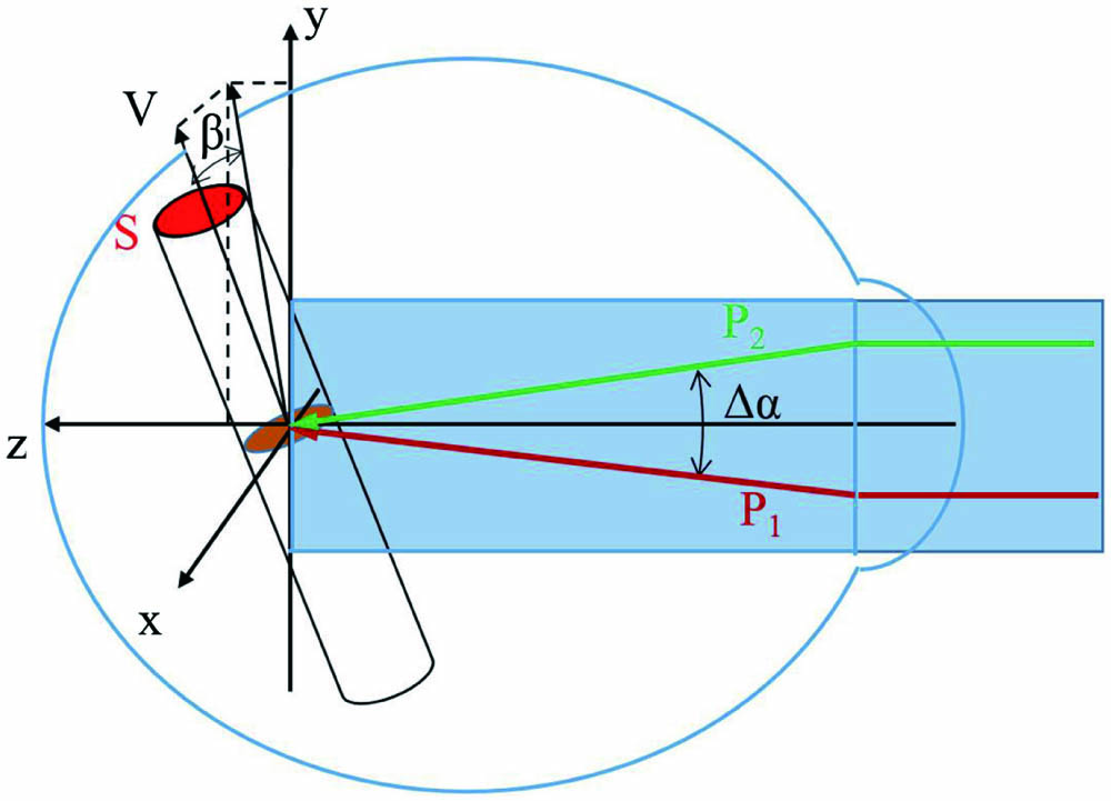
Fig. 2. Illustration of the incident probe beams and the blood flow velocity. V, direction of the blood flow velocity; P1 and P2, two probe beams; Δα, angle between the two probe beams; β, angle between V and the illumination plane (y–z plane, composed of P1 and P2).
Fig. 3. Illustration of the illumination plane and scanning track. V, direction of the blood flow velocity; L, scanning track of OCT beams on the retina.
Fig. 4. (a) Measured blood flow at the set blood flow of 20 µL/min as a function of the sampling step. (b) Measured blood flow versus the set blood flow.
Fig. 5. Retinal blood flow imaging with the DDD-OCT system. (a) LSLO fundus view. The black line marks the scanning track. V1, an artery vessel. (b) Structural and (c) Doppler OCT images of the vessel V1. (d) Absolute velocity of the blood flow in V1 as a function of time.


Set citation alerts for the article
Please enter your email address

© Copyright 2018-2021 | Chinese Laser Press. All Rights Reserved 沪ICP备15018463号-20