• Photonics Research
  • Vol. 10, Issue 1, 120 (2022)
Ivy H. M. Wong, Yan Zhang, Zhenghui Chen, Lei Kang, and Terence T. W. Wong*
Author Affiliations
  • Translational and Advanced Bioimaging Laboratory, Department of Chemical and Biological Engineering, Hong Kong University of Science and Technology, Hong Kong, China
  • show less
    DOI: 10.1364/PRJ.440935 Cite this Article Set citation alerts
    Ivy H. M. Wong, Yan Zhang, Zhenghui Chen, Lei Kang, Terence T. W. Wong. Slide-free histological imaging by microscopy with ultraviolet surface excitation using speckle illumination[J]. Photonics Research, 2022, 10(1): 120 Copy Citation Text show less
    (a) System configuration of MUSES. (b) An iterative algorithm for the R, G, and B channels.
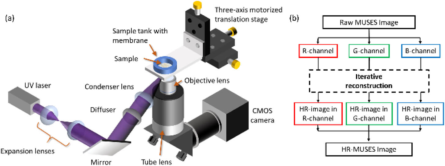
    Fig. 1. (a) System configuration of MUSES. (b) An iterative algorithm for the R, G, and B channels.
    (a)–(c) Comparison of 4X MUSE, 4X MUSES, and 10X MUSE images of blue fluorescent beads with a diameter of 500 nm. (d) and (e), (f) and (g), (h) and (i) Zoomed-in images of the bead inside the yellow dashed boxes in G- and B-channels under 4X MUSE, 4X MUSES, and 10X MUSE images, respectively. (j), (k) The corresponding line profiles in G- and B-channels of this bead under 4X MUSE (orange line) and 4X MUSES (purple line).
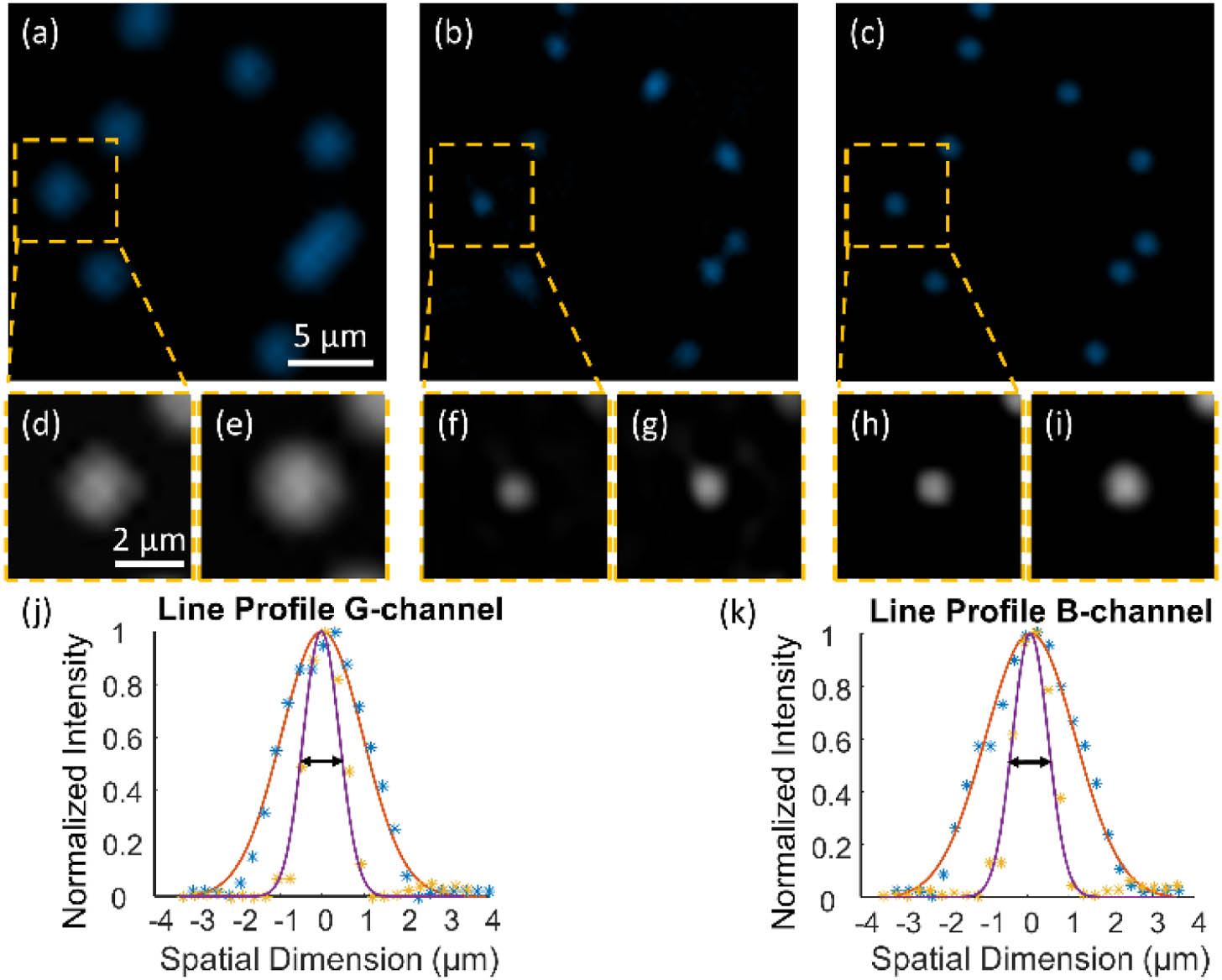
    Fig. 2. (a)–(c) Comparison of 4X MUSE, 4X MUSES, and 10X MUSE images of blue fluorescent beads with a diameter of 500 nm. (d) and (e), (f) and (g), (h) and (i) Zoomed-in images of the bead inside the yellow dashed boxes in G- and B-channels under 4X MUSE, 4X MUSES, and 10X MUSE images, respectively. (j), (k) The corresponding line profiles in G- and B-channels of this bead under 4X MUSE (orange line) and 4X MUSES (purple line).
    (a) 4X MUSE image of an FFPE mouse brain tissue slice that is stained with Rhodamine B and Hoechst 33342. (b) Corresponding H&E-stained FFPE slice. (c)–(e) Zoomed-in images of 4X MUSE, 4X MUSES, and corresponding H&E slice of the hippocampus region marked with an orange solid box in (a) and (b). (f)–(h) Zoomed-in images that correspond to the yellow dashed box regions marked in (c), (d), and (e), respectively. (i)–(k) Zoomed-in images that correspond to the blue dotted box regions marked in (c), (d), and (e), respectively.
    Fig. 3. (a) 4X MUSE image of an FFPE mouse brain tissue slice that is stained with Rhodamine B and Hoechst 33342. (b) Corresponding H&E-stained FFPE slice. (c)–(e) Zoomed-in images of 4X MUSE, 4X MUSES, and corresponding H&E slice of the hippocampus region marked with an orange solid box in (a) and (b). (f)–(h) Zoomed-in images that correspond to the yellow dashed box regions marked in (c), (d), and (e), respectively. (i)–(k) Zoomed-in images that correspond to the blue dotted box regions marked in (c), (d), and (e), respectively.
    (a) 4X MUSE image of formalin-fixed mouse brain tissue stained with Rhodamine B and Hoechst 33342. (b)–(d) Zoomed-in 4X MUSE, 4X MUSES, and its standard H&E (from adjacent layer) images of the orange solid box marked in (a), respectively. (e)–(g) Zoomed-in images that correspond to the green dashed box regions marked in (b), (c), and (d), respectively. (h)–(j) Zoomed-in 4X MUSE, 4X MUSES, and its standard H&E (from adjacent layer) images of the yellow dotted box marked in (a), respectively.
    Fig. 4. (a) 4X MUSE image of formalin-fixed mouse brain tissue stained with Rhodamine B and Hoechst 33342. (b)–(d) Zoomed-in 4X MUSE, 4X MUSES, and its standard H&E (from adjacent layer) images of the orange solid box marked in (a), respectively. (e)–(g) Zoomed-in images that correspond to the green dashed box regions marked in (b), (c), and (d), respectively. (h)–(j) Zoomed-in 4X MUSE, 4X MUSES, and its standard H&E (from adjacent layer) images of the yellow dotted box marked in (a), respectively.
    (a)–(c) 4X MUSE, 4X MUSES, and 10X MUSE images of fresh hand-cut mouse brain tissue stained with Rhodamine B and Hoechst 33342. (d)–(f) Virtual H&E-stained images of (a), (b), and (c), respectively, generated by CycleGAN. (g)–(i) 4X MUSE, 4X MUSES, and 10X MUSE images of another fresh mouse brain tissue stained with Hoechst 33342 and propidium iodide. Cell nuclei from other layers are clearly visualized only in the 4X MUSES image with improved resolution and long DOF (orange arrows). (j)–(l) Virtual H&E-stained images of (g), (h), and (i), respectively, generated by CycleGAN.
    Fig. 5. (a)–(c) 4X MUSE, 4X MUSES, and 10X MUSE images of fresh hand-cut mouse brain tissue stained with Rhodamine B and Hoechst 33342. (d)–(f) Virtual H&E-stained images of (a), (b), and (c), respectively, generated by CycleGAN. (g)–(i) 4X MUSE, 4X MUSES, and 10X MUSE images of another fresh mouse brain tissue stained with Hoechst 33342 and propidium iodide. Cell nuclei from other layers are clearly visualized only in the 4X MUSES image with improved resolution and long DOF (orange arrows). (j)–(l) Virtual H&E-stained images of (g), (h), and (i), respectively, generated by CycleGAN.
    Ivy H. M. Wong, Yan Zhang, Zhenghui Chen, Lei Kang, Terence T. W. Wong. Slide-free histological imaging by microscopy with ultraviolet surface excitation using speckle illumination[J]. Photonics Research, 2022, 10(1): 120
    Download Citation