• Opto-Electronic Advances
  • Vol. 1, Issue 9, 180015 (2018)
Yu Zheng1、2, Zhifang Wu2、3、*, Perry Ping Shum1、2, Zhilin Xu4, Gerd Keiser5, Georges Humbert6, Hailiang Zhang1、2, Shuwen Zeng6, and Xuan Quyen Dinh2、7
Author Affiliations
  • 1COFT, School of EEE, Nanyang Technological University, Singapore 639798, Singapore
  • 2CINTRA, CNRS/NTU/Thales Research Alliance, Singapore 637553, Singapore
  • 3Fujian Key Laboratory of Light Propagation and Transformation, College of Information Science and Engineering, Huaqiao University, Xiamen 361021, China
  • 4Center for Gravitational Experiments, School of Physics, Huazhong University of Science and Technology, Wuhan 430074, China
  • 5Department of Electrical and Computer Engineering, Boston University, Boston 02215, USA
  • 6XLIM Research Institute, UMR 7252 CNRS/University of Limoges, Limoges 87060, France
  • 7R&T, Thales Solutions Asia Pte Ltd, Singapore 498755, Singapore
  • show less
    DOI: 10.29026/oea.2018.180015 Cite this Article
    Yu Zheng, Zhifang Wu, Perry Ping Shum, Zhilin Xu, Gerd Keiser, Georges Humbert, Hailiang Zhang, Shuwen Zeng, Xuan Quyen Dinh. Sensing and lasing applications of whispering gallery mode microresonators[J]. Opto-Electronic Advances, 2018, 1(9): 180015 Copy Citation Text show less
    Schematic of light trapping inside a WGM microcavity by frustrated total internal reflection.
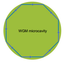
    Fig. 1. Schematic of light trapping inside a WGM microcavity by frustrated total internal reflection.
    Different methods of exciting WGMs.(a) Prism coupling. (b) Polished fiber coupling. (c) Tapered fiber coupling.
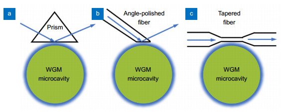
    Fig. 2. Different methods of exciting WGMs.(a) Prism coupling. (b) Polished fiber coupling. (c) Tapered fiber coupling.
    (a) Layout of the WGM sensing set-up. (b), (c) Transient interactions of single zinc or mercury ions with the NRs with the corresponding spectrum shifts. Figure reproduced from ref.5, Springer Nature.
    Fig. 3. (a) Layout of the WGM sensing set-up. (b), (c) Transient interactions of single zinc or mercury ions with the NRs with the corresponding spectrum shifts. Figure reproduced from ref.5, Springer Nature.
    The illustration of the coupled liquid-core laser.
    Fig. 4. The illustration of the coupled liquid-core laser.
    (a) Scanning electron micrograph of a polystyrene bead coated with ELNPs. (b) Left: wide-field image of a lasing microsphere. Right: simulated field distributions in the x–y plane. (c) Simulated NIR spectra of WGMs supported by a 5-µm polystyrene microsphere. Figure reproduced from ref.66, Springer Nature.
    Fig. 5. (a) Scanning electron micrograph of a polystyrene bead coated with ELNPs. (b) Left: wide-field image of a lasing microsphere. Right: simulated field distributions in the x–y plane. (c) Simulated NIR spectra of WGMs supported by a 5-µm polystyrene microsphere. Figure reproduced from ref.66, Springer Nature.
    (a) Schematic of an all-optical tunable microlaser. (b) Fabrication process of the erbium-doped hybrid microbottle cavity coated with iron oxide nanoparticles. Figure reproduced from ref.68, American Chemical Society.
    Fig. 6. (a) Schematic of an all-optical tunable microlaser. (b) Fabrication process of the erbium-doped hybrid microbottle cavity coated with iron oxide nanoparticles. Figure reproduced from ref.68, American Chemical Society.
    Illustrations of aqueous QDs (a) in solution inside an OFRR and (b) immobilized as a single layer on the inner surface of an OFRR. (c) Illustration of the experimental setup using confocal geometry. Figure reproduced from ref.8, American Chemical Society.
    Fig. 7. Illustrations of aqueous QDs (a) in solution inside an OFRR and (b) immobilized as a single layer on the inner surface of an OFRR. (c) Illustration of the experimental setup using confocal geometry. Figure reproduced from ref.8, American Chemical Society.
    Diagram and operation of the dual-microcavity narrowlinewidth laser.Figure reproduced with permission from ref.7, The Optical Society.
    Fig. 8. Diagram and operation of the dual-microcavity narrowlinewidth laser.Figure reproduced with permission from ref.7, The Optical Society.
    MicroresonatorSensitivity(nm/℃)
    PMMA microbubble380.039
    U-shaped optical fiber520.624
    Doped polystyrene microparticle530.122
    Silicon microring540.11
    Dye-doped CLC microdroplet550.96
    Holmium doped microsphere560.01
    Doped oil mirodroplet570.377
    Table 1. Sensitivities of temperature sensors based on WGM microresonator.
    Yu Zheng, Zhifang Wu, Perry Ping Shum, Zhilin Xu, Gerd Keiser, Georges Humbert, Hailiang Zhang, Shuwen Zeng, Xuan Quyen Dinh. Sensing and lasing applications of whispering gallery mode microresonators[J]. Opto-Electronic Advances, 2018, 1(9): 180015
    Download Citation