1COFT, School of EEE, Nanyang Technological University, Singapore 639798, Singapore
2CINTRA, CNRS/NTU/Thales Research Alliance, Singapore 637553, Singapore
3Fujian Key Laboratory of Light Propagation and Transformation, College of Information Science and Engineering, Huaqiao University, Xiamen 361021, China
4Center for Gravitational Experiments, School of Physics, Huazhong University of Science and Technology, Wuhan 430074, China
5Department of Electrical and Computer Engineering, Boston University, Boston 02215, USA
6XLIM Research Institute, UMR 7252 CNRS/University of Limoges, Limoges 87060, France
7R&T, Thales Solutions Asia Pte Ltd, Singapore 498755, Singapore
Optical whispering gallery mode (WGM) microresonators have attracted great attention due to their remarkable properties such as extremely high quality factor, small mode volume, tight confinement of modes, and strong evanescent field. All these properties of WGM microresonators have ensured their great potentials for applications, such as physical sensors, bio/chemical sensors and microlasers. In this mini-review, the key parameters and coupling conditions of WGM microresonators are firstly introduced. The geometries of WGM optical microcavities are presented based on their fabrication methods. This is followed by the discussion on the state-of-the-art applications of WGM microresonators in sensors and microlasers.
The whispering gallery mode (WGM) was first discovered by Lord Rayleigh in sound waves in 1910, when he studied the phenomenon of whispering gallery in St Paul's Cathedral1. Then, in 1961 Garrett et al. found that optical waves, which can undergo reflection, refraction and diffraction like a sound waves, can generate WGM in an optical resonator as well2. In WGM optical resonators, light with specific wavelengths is confined near a circular ring boundary via total internal reflection.
WGM microresonators have been extensively studied since they allow ultrahigh quality factor (Q factor), small mode volume, and a strong evanescent field. These remarkable properties have led to myriads of applications in extremely sensitive sensors. For example, the sensitivity of bio/chemical sensors based on WGM microresonators is as high as single atomic ions level3-5. Moreover, due to
extraordinarily high Q factor and small mode volume, WGM microresonators have another important application in microlasers. The WGM-based microlasers own some excellent properties and advantages such as ultralow lasing threshold6 and extremely narrow linewidth7. For instance, the pump threshold of an optofluidic microlaser based on WGM resonance could be down to 0.1 μJ/mm28. Compared with many good reviews on WGM6, 9-12, in this paper we will emphasize the current state of the art of WGM sensors and mcirolasers, especially in terms of the sensing sensitivity and the pump threshold respectively. In this paper, the key parameters and coupling conditions of WGM microresonators are discussed first. Then, we present some common geometries based on their fabrication processes. Finally, we focus on the recent progresses on applications of WGM microresonators in sensing and lasing fields.
Key parameters and coupling conditions of WGM microresonators
Resonant wavelength
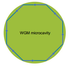
For preliminarily understanding how WGMs are formed in the microcavities, we can consider WGMs in a 2D model, as shown in Fig. 1. Supposing the system is lossless, the constructive interference will occur among the propagating light waves when the total propagation distance is equal to integer multiples of the resonant wavelength. These wavelengths will satisfy the following equation13:
where m, an integer number, is the azimuthal mode number, λ is the resonant wavelength, R is the radius of the microcavity, and neff is the effective refractive index of the microcavity.
WGM microcavities can propagate simultaneously transverse magnetic (TM) modes and transverse electric (TE) modes. A numerical approximation of WGM resonances was developed by Lam et al. in 199214. The more accurate resonant wavelengths are expressed as15:
where r is the radial mode number, nr=n1/n2, n1 and n2 are the refractive indices of microcavity and ambient materials, respectively, η(r) is the Airy function solution and equals 2.338 if r=1, L is the polarization characteristic coefficient and L=1/nr for TM modes and L=nr for TE modes.
Quality factor
WGM microcavities with different geometries can obtain significantly high Q factors. To the best of our knowledge, the maximal Q factor has exceeded 1011 achieved with fluoride crystalline materials16. The Q factor is defined by the resonator's capability to store energy at a specific frequency, and can be measured in the frequency or time domain, as shown in Equation (3).
where Q0 is the resonator's intrinsic Q factor, ω is the resonance frequency, Estored is the energy stored in the resonator, Pdiss is the power dissipated from the resonator, τ is the photon life time (or cavity ring-down time), and Δω is the resonance linewidth as measured from the frequency spectrum. Theoretically, the intrinsic Q factor of an independent WGM microcavity is determined by several loss factors as expressed by17:
where Qmat is the material loss factor, Qrad is the radiative loss factor, Qsca is the scattering loss factor and Qcont is the contaminant loss factor.
Generally, Qmat caused by material absorption and Rayleigh scattering, is dominant among these loss factors. Qmat can be calculated as:
where α is the linear absorption coefficient, n is the refractive index of the microcavity material.
Qrad, the radiative loss factor, is inevitable for all circular geometries and is determined by the curvature of the microcavity boundary. Generally, Qrad can be defined as the ratio between the real and imaginary parts of the wave number. When the angular mode number l= m, Qrad can be calculated approximately as follows18:
where σrms is the root-mean-squared size and B is the correlation length of roughness. From this equation, it is clear to see that Qsca is proportional to the cavity radius R under the same surface inhomogeneities, which indicates that bigger microcavities can confine light better than smaller ones. Meanwhile, by optimizing the size and length of inhomogeneity, the scattering loss can be reduced.
Qcont defines the influence from the surface contaminants, such as water molecules from the atmosphere. The contaminants can form nanolayers or nanoclusters on the microcavity surface and cause additional absorption and scattering loss.
Mode volume
Another prominent advantage of WGM microcavities as mentioned in the introduction section is the small mode volume. The mode volume can be described as the electromagnetic field localization in a WGM microcavity, and it is an especially important parameter for nonlinear applications. The mode volume of a spherical WGM resonator can be calculated using Equations (9):
$
V = 3.4{{\rm{ \mathsf{ π} }}^{\frac{3}{2}}}{\left( {\frac{\lambda }{{2{\rm{ \mathsf{ π} }}n}}} \right)^3}{m^{\frac{{11}}{6}}}\sqrt {m - v + 1},
$
where λ is the wavelength of the pumping light and ν is the magnetic number.
Optical coupling approaches
Optical WGMs can be excited by using a free-space setup, which is composed of a microcavity and a coupling component. The geometries of such optical microcavities include microspheres19, 20, microrings21, microdisks4, 22, microbubbles23-25 and microcapillaries26, 27. The coupling component, like a prism, an angle-polished fiber, or a tapered fiber, is positioned close to the WGM microcavity, as shown in Fig. 2. The prism coupler is considered as one of the oldest optical couplers28. It is suitable for coupling evanescent waves into large-size WGM cavities by frustrated total internal reflection, typically in the diameter range of 1-2 cm29. The phase-matching condition and critical coupling can be adjusted by respectively changing the incident angle, and the gap between resonator and prism. To the best of our knowledge, the highest coupling efficiency of a prism coupler was reported around 80%30. The angle-polished fiber coupler was first proposed and demonstrated by Ichenko in 199931. The mechanism of such a coupler is basically the same as that of the prism coupler. An angle-polished fiber was shown to provide significant improvement on mode quality over the prism by confining the input beam within its core. However, the output beam of both angle-polished fiber and prism couplers still has to be collected in free space. This limits their applications for compact and integrated devices for in situ measurements. Comparatively, a tapered fiber is more commonly used, because the light is coupled and decoupled in-line within a same fiber. In order to increase the overlap of the evanescent fields between the fiber guided mode and the WGM, the diameter of the tapered fiber is usually less than 2 μm.
Phase matching is one of the critical conditions to obtain high efficiency for light power coupling from one device to another. For WGM coupling, phase matching means that the propagation constant of the proper mode in the coupler is equal to the propagation constant of the WGM in the microcavity. Considering the coupling between a tapered optical fiber and a microsphere as an example, when l = m is assumed, the propagation constant βms of a microsphere can be expressed as32:
$
{\beta _{{\rm{ms}}}} = kl/{x_{rlm}},
$
where k is the free space propagation constant and xrlm is the size parameter that corresponds to the r, l, and m resonance. For the tapered fiber, the fundamental mode will significantly extend out into surroundings, and its propagation constant can be calculated as:
where n' is the refractive index of the fiber and ρ is the diameter of the fiber taper. When βms=βft, the phase matching between the tapered fiber and the microsphere is obtained.
Critical coupling
For achieving optimal coupling, besides the satisfaction of phase matching, the distance between the WGM microcavity and the coupler should be properly adjusted in order to exactly compensate the microcavity loss by the amount of power delivered from the coupler. At this time, the coupler output hits zero at the resonant wavelength and the so-called 'critical coupling' is achieved. After the light propagating in the waveguide is coupled into a WGM microcavity with a photon lifetime τe, it will resonate along the circular ring boundary and experience intrinsic losses from the microcavity with an intrinsic photon lifetime τ033. Based on the variation of τ0 and τe, the different coupling regimes can be described as:
1) Undercoupled, τ0 < τe, the cavity intrinsic loss is higher than the input power.
2) Critical coupling, τ0=τe, the cavity intrinsic loss is equal to the input power.
3) Overcoupled, τ0 > τe, the cavity intrinsic loss is lower than the input power.
Geometries of WGM optical microcavities
Up to now, various geometries, including microspheres, microrings, microdisks, microtoroids, microbottles, microbubbles, and microcapillaries etc. have been demonstrated for exciting WGM. These geometries of microcavities are fabricated from the different materials and methodologies. In this section, these geometries are classified and presented based on their fabrication methods.
Microspheres
Microspheres were the most common microresonator used to generate WGMs in early studies. There are mainly two methods for the fabrication of microspheres, melting of glass materials and the sol-gel process. The former method can be used for a large range of glass materials, since its general principle is to melt the glass materials, which is able to fabricate self-assembly microspheres due to surface tension. One representative method is called fiber splicing. The fiber is first tapered into a proper diameter and then put into one of the arms of the splicer. The fiber tip is melted by the electric arcs and the spherical microcavity is produced by surface tension. By adjusting the power and discharge times of electric arcs generated by the splicer, the diameter of the microsphere can be controlled. The sol-gel process is a chemical synthesis method used to generate microspheres from the molecular level, which is relatively flexible and cheap. The fabrication of silica microspheres based on sol-gel process with the diameter from 150 nm to 2 μm has become a very mature method12.
Microrings, mirodisks and microtoroids
Due to possessing planar structures, microrings, microdisks and microtoroids are more suitable for photonic integration compared with microspheres. Meanwhile, the fabrication of these planar structures is more controllable. The fabrication methods generally include top-down and bottom-up approaches. Simply, the former one means a small device is produced by removing the useless parts from larger material and adding the other useful parts. The main techniques consist of electron-beam lithography, dry etching and wet chemical etching. For example, Duan et al. reported a simple and robust method to produce well-controlled and uniform MAPbBr3 perovskite-based mircodisk arrays by controlling the concentration of the perovskite precursor, pattern sizes, and hydrophilicity of the substrate34. The bottom- up approach normally refers to producing a larger device from smaller materials, such as individual molecules.
Microbottles and microbubbles
The fabrication method of microbottles and microbubbles are similar and more flexible than the first two kinds of geometries25, 38, 39. A simple process of fabricating a microbottle is to taper the optical fiber two times in two adjacent places. Compared with the tubular microcavities, the microbottles have the better ability of light confinement along the cylinder axis. In the early studies, microbubbles were updated from heating microcapillaries with a gas pressure. Recently, some novel methods are applied to fabricate microbubbles38-40. For instance, Wang et al. prepared microbubbles based on the drying process of the colloidal quantum dots (CQDs)/ polymethyl methacrylate (PMMA) nanocomposite droplets25.
Applications
Sensors
Generally, the operational mechanism of sensors based on WGM microresonators is to monitor variations of WGM resonance wavelengths induced by physical changes of the system, and the ultrahigh Q factor and the strong evanescent waves make the variations of WGM resonances very sensitive to the surroundings. Hence, WGM microresonators have been developed as a variety of highly sensitive sensors4, 19-21, 40, such as single molecules/atomic-ions sensor, refractive index (RI) sensor, temperature sensor and humidity sensor.
1. Bio/chemical sensor
When a molecule binds onto a WGM resonator surface, the resonance frequencies of the optical modes will be shifted since the resonator's evanescent field is required to polarize the molecule41. Based on this principle, Su et al. developed a single-molecule sensor called frequency locked optical whispering evanescent resonator that can be used to detect a wide range of nanoscale objects with radii from 100 to 2.5 nm, including exosomes, ribosomes, mouse immunoglobulin G and human interleukin-241. Besides detecting the nanoscale objects directly, WGM resonators can be applied to sensing a specific analyte by surface functionalization approaches. For example, Ghali et al. developed a biosensor by functionalizing the WGM microdisk with LysK for real-time detecting of Staphylococcus aureus, and the limit of detection is 5 pg/mL42.
Swaim et al. built a theoretical model, in which WGM resonance frequency shifts can be enhanced through the combination of a microtoroid and a single Au nanorod, using the boundary element method in 201143. After that, the sensing applications based on the combination between Au nanorod and microcavities have been widely studied. Recently, Baaske and Vollmer built a system utilizing a WGM sensing method to detect single atomic ions in an aqueous medium5. Gold nanorods (NRs) were bound onto the surface of a silica microsphere for exciting the NR's LSPR and generating local intensity hot spots at the tips of the NRs, and the hot spots can increase the resonance wavelength shift when single atomic ions interacted with plasmonic NRs, as shown in Fig. 3. In this work, the ion-NR interaction under different solution's ionic strength of both zinc and mercury ions was studied and a clear distinction of their behaviors was found.
Figure 3.(a) Layout of the WGM sensing set-up. (b), (c) Transient interactions of single zinc or mercury ions with the NRs with the corresponding spectrum shifts. Figure reproduced from ref.5, Springer Nature.
Besides detecting the resonance wavelength shifts, there are also some intensity based WGM sensors. For instance, Heylman et al. demonstrated a single-particle photothermal absorption spectrometer by measuring the shift of the WGM energy44. This spectrometer applies on-chip optical WGM microtoroids as ultrasensitive thermmeters and is a convergence of narrow Fano resonances and WGMs, which makes it possess ultrahigh sensitivity up to sub-100 Hz resonance shift.
2. RI sensor
The resonant wavelengths of a microresonator have direct relationships with the RI difference between a microcavity and the surrounding medium. Therefore, microresonators with high Q factor can be applied as highly sensitive RI sensors, and these microresonators can be classified into passive microresonators and active microresonators. Compared with the RI sensors with passive microresonators45, 46, for example, Zhu et al. developed a RI detector utilizing liquid core optical ring resonators (LCORRs), and the sensitivity is 20 nm/RIU (refractive index units)47, recently applying active microresonators to build RI sensors has been receiving more attention, since they can achieve the practical advantages of free space interrogation. By applying a high RI glue onto half of an active microsphere, Kang et al. demonstrated a RI sensor based on mode-splitting by the free space excitation48. This method is able to discriminate surrounding RI changes and temperature changes. Wan et al. developed a robust RI sensor using the dye-doped polymer microring laser based on the free-space optics measurement setup, which possessed the sensitivity of 75 nm/RIU49. Krämmer et al. utilized free-space optics to demonstrate that the RI sensitivity can be improved from 60 nm/RIU to 150 nm/RIU by decreasing the thickness of a dye-doped polymeric microdisk from 0.9 μm to 0.21 μm50. Ren et al. proposed a coupled liquid-core laser by the parallel placing of two thin-wall silica capillaries as shown in Fig. 451. One of the capillaries was filled with a dye solution, and the other one was injected with a sample. The sensitivity was up to 3874 nm/RIU by taking advantage of the Vernier effect.
Figure 4.The illustration of the coupled liquid-core laser.
Temperature variation can lead to the shift of resonant wavelengths of a microresonator, because it will induce a change of the microcavity refractive index and the microcavity size that are determined by the thermal optic coefficient and the thermal expansion coefficient of the microcavity. Hence, many temperature sensors based on WGM microresonators of different geometries and different materials were developed. The geometries included microspheres, microrings and microbubbles, and the materials included polymethyl methacrylate (PMMA), cholesteric liquid crystal (CLC), doped silicate glass, and doped oil. The sensitivities of these temperature sensors are shown in Table 1.
Microresonator
Sensitivity(nm/℃)
PMMA microbubble38
0.039
U-shaped optical fiber52
0.624
Doped polystyrene microparticle53
0.122
Silicon microring54
0.11
Dye-doped CLC microdroplet55
0.96
Holmium doped microsphere56
0.01
Doped oil mirodroplet57
0.377
Table 1. Sensitivities of temperature sensors based on WGM microresonator.
Since the RI and size of some materials will vary when they absorb water vapor, the WGM microresonators consisting of these materials can be applied as humidity sensors. For example, Eryürek et al. presented an optical humidity sensor based on an on-chip SU-8 polymer microdisk, and demonstrated that the RI change of the microdisk was dominant when SU-8 interacted with water vapor58. Since SU-8 has the strong ability of interaction with water vapor, this sensor has a high sensitivity of 108 pm/% RH in the 0-1% RH range. Labrador-Páez et al. demonstrated a WGM microdroplet resonator that is made out of a 160 μm droplet of glycerol and rhodamine 6G adhered at the tip of an optical fiber59. Huang et al. developed an active microdisk of dye-doped PEG-DA hydrogel as a humidity sensor with a sensitivity of 43.11 pm/% RH60. The operating mechanism is that the size of the microdisk will homogenously be modified when the surrounding relative humidity changes, resulting in a linear shift of WGM lasing peaks.
Microlasers
Since Garrett et al. demonstrated the first WGM lasers in 19612, WGM microresonators have been widely used in the field of microlasers because of the high Q factor and the small mode volumes8, 25, 53, 61-64. Due to these advantages, microlasers based on WGM microresonators can obtain an ultralow lasing threshold. The pump threshold is defined by the minimum pump power that is able to make cavity gain equal to loss in one round-trip. Moreover, microlasers based on WGM microresonators can naturally achieve very narrow linewidth due to the ultrahigh Q factor.
In 2017, Lim et al. fabricated a stabilized MgF2 resonator laser with a 32 Hz Allan deviation and a sub-25 Hz linewidth by compensating the intrinsic thermal expansion of the WGM resonator65. Coating an active material onto a microresonator is another approach to fabricate the WGM microlaser. Fernandez-Bravo et al. developed a continuous-wave (CW) upconverting nanoparticle (UCNP) laser with a threshold of 14 kW/cm2 by coating Tm3+-doped energy-looping nanoparticles (ELNPs) onto a polystyrene microsphere with a diameter of 5 μm, as shown in Fig. 566. This CW laser can work more than 5 hours at blue and near-infrared wavelengths.
Figure 5.(a) Scanning electron micrograph of a polystyrene bead coated with ELNPs. (b) Left: wide-field image of a lasing microsphere. Right: simulated field distributions in the x–y plane. (c) Simulated NIR spectra of WGMs supported by a 5-µm polystyrene microsphere. Figure reproduced from ref.66, Springer Nature.
Although light pumping is dominant in the field of WGM microlasers, electric current also can be applied as a pump source for a WGM microlaser. Moiseev et al. demonstrated a semiconductor microdisk laser that applied InGaAs quantum well-dots as an active area by photolithography and dry etching67. The pump threshold current density of this laser is 710 A/cm2 for the microdisk of diameter of 31 μm, and around 70% of total absolute power (18.5 mW) is concentrated within the lasing modes.
As mentioned in Section 2, the WGM resonance wavelength is closely related to microresonactor size, RI difference and so on. Hence, a tunable laser based on WGM resonance is feasible through cavity size modification. Zhu et al. developed an all-optical tunable microlaser based on an erbium-doped hybrid microbottle with a high Q factor of 5.2×10⁷ and a low lasing threshold of 1.65 mW, as shown in Fig. 668. This hybrid microbottle was generally fabricated as follows: first, attaching Er3+ ion solution onto the SMF surface; then, applying a CO2 laser to process the SMF into a microbottle shape with a spherical end; finally, coating with iron oxide nanoparticles that can convert control light energy into thermal energy on the spherical end for tuning the lasing wavelength.
Figure 6.(a) Schematic of an all-optical tunable microlaser. (b) Fabrication process of the erbium-doped hybrid microbottle cavity coated with iron oxide nanoparticles. Figure reproduced from ref.68, American Chemical Society.
Besides these microlasers based on solid microcavities, Kiraz et al. demonstrated two types of optofluidic microlasers by combining aqueous quantum dots (QDs) and high Q factor optofluidic ring resonator (OFRR), as shown in Fig. 78. One was achieved by filling 2 μM QDs with a bulk buffer solution into the OFRR cavity. The pump threshold of this microlaser could be down to 0.1 μJ/mm2. The other one was constructed by fixing a single layer of QDs onto the interface between the buffer solution and the OFRR inner wall. The surface density was from 3×109cm-2 to 3×1010 cm-2. In both cases, the laser emission persisted 5-10 min under uninterrupted pulsed excitation, which was a significant improvement compared with organic-dye-based lasers.
Figure 7.Illustrations of aqueous QDs (a) in solution inside an OFRR and (b) immobilized as a single layer on the inner surface of an OFRR. (c) Illustration of the experimental setup using confocal geometry. Figure reproduced from ref.8, American Chemical Society.
Since most WGM microcavities are circularly symmetric, the lasing emission is usually isotropic accordingly. In order to satisfy the requirement of unidirectional laser emission desired in many applications, some specially-designed microcavities have been proposed61, 69. Zhan et al. fabricated a 3D polymer microlaser consisting of a stacked circular ring and a spiral ring on a narrow bandpass filter substrate by the femtosecond laser processing method70. The characteristic of this microlaser includes the low threshold of 60 μJ/cm2 and the unidirectional and single mode laser output. By comparing a series of different structures, they found that the circular ring is treated as an oscillator to generate the low-threshold WGMs lasing emissions, and the spiral ring serves as a mode filter and output port.
In addition, WGM microresonators also can be used to stabilize the laser and make it obtain a very narrow linewidth through their ultrahigh Q factors. A dual-microcavity laser was demonstrated by Loh et al., in 20157. One on-chip silica microresonator was applied to generate tunable 1550 nm laser through stimulated Brillouin scattering (SBS) and another microresonator was used to stabilize the laser frequency, as shown in Fig. 8. The laser linewidth was approximately 87 Hz. The fractional frequency noise of this configuration was also reduced to 7.8×10⁻¹⁴ Hz-1/2 when the offset was 10 Hz, which was a remarkable noise performance for a microresonator laser.
Figure 8.Diagram and operation of the dual-microcavity narrowlinewidth laser.Figure reproduced with permission from ref.7, The Optical Society.
In this paper, we have reviewed the key parameters and coupling conditions of WGM microresonators, geometries of WGM optical microcavities, and various applications in sensors and microlasers. Since the high Q factor can be easily obtained for WGM microresonators and the evanescent field exists around the surface of WGM microcavities, WGM resonance is extremely sensitive to the environment. This property gives the microresonators excellent behaviors even in detection of single atomic ions. On the other hand, microlasers based on WGM microresonators can achieve ultralow pump threshold and very narrow linewidth due to the ultrahigh Q factor and ultrasmall mode volume. These features are highly desired for mass production and future commercialization of the microlaser devices.
Although there are many significant research achievements of WGM in photonics10, 11, 71-75, most of them are realized only in the laboratory with setups that require very careful alignment and positioning. For instance, sub-micrometer scale precision positioning is required to achieve effective coupling, as any variation in the distance between the microcavity and the coupler will result in a change of coupling efficiency and a change of the resonator performance. Furthermore, the coupler, such as a tapered fiber, with the diameter less than 2 μm, is fragile and is easily deteriorated by external air flow or dust. Hence, in our opinion, developing efficient, robust coupling methods and package approaches are crucial for practical applications of WGM microresonators in the future.
As mentioned in the Microlasers Section, since most WGM microcavities are circularly symmetric, the lasing emission is usually isotropic accordingly, but the requirement of unidirectional laser emission is highly desired in many applications. Although some specially-designed microcavities based on different unidirectional emission approaches such as scatter-induced unidirectional emission, chaos-induced unidirectional emission and grating-induced vertical emission, have been proposed61, 70, 76 (for example Kim et al. proposed the design of the deformed microresonators for emission directionality by tailoring WGM properties69), the performance of unidirectional emission is required to be improved with respect to traditional laser performance.
Tunable lasing is an important branch in the field of lasers. Although some tunable microlasers based on WGM microresonators have been demonstrated, realizing the large tunable range and long lifetime are still challenging.
Acknowledgements
This work is partially supported by National Natural Science Foundation of China (11774102), the Scientific Research Funds and Promotion Program for Young and Middle-aged Teacher in Science & Technology Research of Huaqiao University (ZQN-YX504, 17BS412), Open Fund of IPOC (BUPT), National Research Foundation Singapore (NRF) (NRF-CRP13-2014-05), European Union's Horizon 2020 Research and Innovation Programme under the Marie Sklodowska-Curie Grant Agreement (No. 798916) and Singapore Ministry of Education Academic Research Fund Tier 1 (RG89/16).
Competing interests
The authors declare no competing financial interests.
[18] TaV D Flexible whispering gallery mode optical microcavities for lasers and sensors (Nanyang Technological University, Singapore, 2014)Ta V D. Flexible whispering gallery mode optical microcavities for lasers and sensors (Nanyang Technological University, Singapore, 2014).