Microcantilever is one of the most popular miniaturized structures in micro-electromechanical systems (MEMS). Sensors based on microcantilever are ideal for biochemical detection, since they have high sensitivity, high throughput, good specification, fast response, thus have attracted extensive attentions. A number of devices that are based on static deflections or shifts of resonant frequency of the cantilevers responding to analyte attachment have been demonstrated. This review comprehensively presents state of art of microcantilever sensors working in gaseous and aqueous environments and highlights the challenges and opportunities of microcantilever biochemical sensors.Microcantilever is one of the most popular miniaturized structures in micro-electromechanical systems (MEMS). Sensors based on microcantilever are ideal for biochemical detection, since they have high sensitivity, high throughput, good specification, fast response, thus have attracted extensive attentions. A number of devices that are based on static deflections or shifts of resonant frequency of the cantilevers responding to analyte attachment have been demonstrated. This review comprehensively presents state of art of microcantilever sensors working in gaseous and aqueous environments and highlights the challenges and opportunities of microcantilever biochemical sensors.
1. Introduction
Micro-cantilever is one of the popular miniaturized devices in MEMS, and it is widely used in sensing, imaging, and so on[1]. Microcantilever sensing technology which can serve for highly sensitive, label-free biochemical molecular analysis and real-time detection, has been applied to detect toxic gas molecules[2-5], proteins[6,7], DNA/RNA[8] and microbiology for thirty years[9–11].
Fritzet al. from IBM Zurich reported static mode microcantilever sensors performing hybridization recognition of DNA molecular fragments[12]. Direct translation of biomolecular recognition into nanomechanics has been achieved, feasibility of microcantilever sensors for rapid DNA comparisons and potential early detection of the disease have been demonstrated.
Resonant microcantilever sensors have attracted considerable attention due to high-quality factor of 105, it can achieve trace detection in the order of 10–18 g[13]. Thundatet al. pioneered study[14] on dynamic detection of biomolecules theoretically and experimentally. Specific adsorption of biomolecules changes not only effective mass, but also stiffness coefficient of the microcantilever due to immune-binding induced surface stress which reduces sensitivity of cantilever biosensor. Ramoset al. in Spanish National Microelectronics Center systematically investigated change of resonance frequency while printing the bacterial solution at different positions on the cantilever and concluded that the highest mass detection sensitivity can be achieved when the biological reaction took place at the free end of the cantilever[15].
Nugaevaet al. from University of Basel used microcantilever arrays to study the specific immobilization of fungi and the growth process of yeast and Aspergillus niger[16]. Real-time monitoring of colony growth was achieved by testing changes of the dynamic resonant frequency and detecting fungal concentrations in the range of 103–106 CFU/mL.
Detection of biomolecules in complex media such as serum is challenging due to the nonspecific binding. The group of Manalis from MIT reports a highly sensitive, batch-fabricated microcantilever, the suspended microchannel resonators (SMRs), a device with embedded microchannels that enables direct quantification of the adsorbed mass by changes in resonant frequency[17]. This approach enables detection of activated leukocyte cell adhesion molecule (ALCAM) in undiluted serum with a detection limit of 10 ng/mL.
In addition to the application in the field of biological monitoring, microcantilever technology has been exceptionally suitable for explosive detection due to its advantages of small size, high sensitivity, low power consumption, and versatility to integrate[18-20]. Microcantilevers coated with chemically selective layers or receptors for the target molecule were used for explosive detection. 4-MBA (4-mercaptobenzoic acid), fluoroalcoholpolysiloxane(SXFA), and other receptors have been adopted for the most widely used explosives such as TNT, RDX, and PETN[21,22]. The functionalization process is based on the receptor and microcantilever surface chemistry.
This work will comprehensively present microcantilever-based biochemical sensors and their application in gaseous and aqueous environments, facing challenges and potential for exploring high-sensitive sensors are also involved.
2. Theoretical analysis of cantilever-based biochemical sensors
There are two working modes for microcantilevers, static mode and dynamic mode. The static mode is also known as the deflection mode. The difference between the stresses on the top and bottom surfaces of the microcantilever causes cantilever bending. The deflection of the cantilever can be expressed as[23]
where
is the length of the cantilever,
is Poisson’s ratio,
is the Young’s modulus of cantilever materials,
is the thickness of the cantilever,
is the deflection, and
is the differential surface stress between the top and bottom surfaces of the microcantilever. According to Eq. (1), there is a linear relation between cantilever bending and differential surface stress.
The dynamic working mode of the micro-cantilever is also called resonance mode. The resonant frequency of cantilever can be derived from the mass-spring-dashpot model
where
is the effectivemass of cantilever,c is the dashpot constant,k is the spring constant, and
is the external force. In case that there is negligible damping and no external force, when the analyte adsorbs on the surface of the microcantilever, the mass of cantilever increases, resulting in shift of the resonance frequency. The resonance frequency
of cantilever can be expressed as[24]
where
is the stiffness coefficient and
is the effective mass of the cantilever beam. For the fundamental mode of a rectangular cantilever,
=nmb where n = 0.24 andmb is the mass of the cantilever. Assuming that the variation ofk is negligible, the mass sensitivity
resulting for adsorption can be expressed as[25]
where
and
are the change in the effective mass of the cantilever beam and the change of the resonance frequency caused by adsorption,l andw are the length and width of the microcantilever, and
andE are the mass density and Young’s modulus of lever, respectively.
The absolute mass sensitivity is determined by the quality factor (Q) in the resonance mode. TheQ can be defined as
where
is the resonant frequency, and
is the full width at half maximum FWHM of the resonance peak. The geometry of the cantilever and the environment in which it works have a significant impact on the quality factor. The mass resolution
can be expressed as
where
denote the amplitude of oscillation, Boltzmann constant, absolute temperature, the bandwidth, and quality factor, respectively. It indicates that the higher quality factor the lever has, the smaller mass it can detect.
3. Detection in gaseous environments
3.1. Toxic gas
Huang's group[25] first reported a novel freon gas sensor, it has high sensitivity for determining Freon-12 as a special application for indoor air quality control. The piezoelectric microcantilever coated with zeolite was employed to detect Freon-12 in dynamic mode. The zeolite nano-channel system provides a size and shape selective matrix for absorbing molecules. The sensitivity for Freon gas response was –0.0024% ppm–1 and the minimum detectable mass was 3.5 × 10⁻⁹ g. Excellent accuracy and sensitivity have been demonstrated for gas detection.
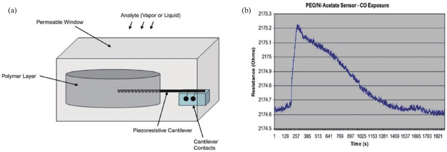
Small, portable, and low-cost sensors are currently attracting more attentions. Porter’s group detected carbon monoxide (CO) gas using embedded piezoresistive microcantilever (EPM) sensor[26]. The design of EPM sensors has facilitated improvements in measurement technology, as shown inFig. 1, where the piezoresistive micro-cantilever was fabricated and fully or partially embedded in the sensing material. As a result, the tiny (a few tens of microns in dimension) sensor element is extremely rigid and robust.
Figure 1.(Color online) (a) Schematic diagram of EPM sensor and (b) EPM sensor response to CO.
The resistance variation demonstrates that the EPM sensor can detect the presence of carbon monoxide gas and could be used for gas sensing.
EPM sensors have been widely reported due to small size, low cost, and robust. Timothyet al. verified that EPM sensors have fast response[27]. EPM sensor has been adopted to detect hydrogen cyanide gas (a colorless and odorless toxic gas) with sensitivity of approximately 4.7 Ω/µm and rapid response of less than 5 s to HCN levels.
3.2. Flammable and explosive gases
Flammable and explosive gases detection is important for security and environmental monitoring.
Thundatet al.[28] reported on the gas-phase detection of 2,4-dinitrotoluene (DNT) using polymer-coated micro-cantilever beams as early as 2004. A SXFA polymer-covered cantilever was developed and repeatedly exposed to DNT at approximately 45 ppb, resulting in an extremely sensitive and reversible response. The detection sensitivity was about 4.5 nm/ppb, and the polymer-coated microcantilever could detect about 300 ppt of DNT in 5 s. This result sets a precedent for explosive vapor detection with rapid response and high sensitivity.
Explosive vapor detection with low limit of detection (LOD), high selectivity, and rapid response remains a technical challenge due to extremely low explosive vapor pressure, complex interference, and various species. Metal nanoparticles[29] and self-assembled monomolecular layers (SAMs) of ligands, lipid membranes, and peptide receptors[30,31] have been developed as surface coating reagents to functionalize carbon nanotube sidewalls for selective detection of TNT. Microcantilever sensors with SAMs immobilized on a surface have been extensively studied for detection of explosive vapors by measuring bending deflection or resonance frequency.
As shown inFig. 2, Wanget al. developed a chemical sensor that consists of a suspended microcantilever and a carbon nanotube (CNT) networkin-situ synthesized[32]. The large surface-to-volume ratio of CNT enabled the chemical sensor to rapidly adsorb explosive vapors and improve the LOD. By grafting special sensing groups on the sidewalls of carbon nanotubes, the final equivalent LOD for TNT detection was 2.4 pg. High-performance CNT chemical sensors demonstrates substantial progress in LOD of gas detection.
Figure 2.(Color online) Schematic diagram of the sensor structure and operating principle. (a) Explosive vapor adsorbed on a carbon nanotube sensor. (b) Heating the explosive vapor to make it micro-detonate.
Rahimiet al. has demonstrated that Photothermal cantilever deflection spectroscopy (PCDS) can detect femtogram level physisorbed methane on a thermally sensitive microcantilever[33]. As shown inFig. 3, the selectivity of mid-IR spectroscopy is combined with the thermal sensitivity of a bi-material cantilever in this approach. The PCDS is capable of detecting methane adsorbed on the cantilever at the femtogram level (1.6 × 10⁻¹⁵ g for a gas flow rate of 2.5 vol% methane). Finally, the selectivity and sensitivity of photothermal cantilever deflection spectroscopy for physisorbed methane detection were demonstrated.
Figure 3.(Color online) Schematic diagram of the PCDS experimental gas sensing device. (a) Controlled vapor generation device. (b) Sensing section.
Conventional methods (such as broth macrodilution and agar dilution) for determining and characterizing bacterial resistance towards antibiotics require at least 24 h, so rapid and low-cost techniques for bacterial drug sensitivity assays are essential to fight infections worldwide. Longoet al. has reported on AFM cantilever as nanomechanical sensors for rapid detection of bacterial resistance to antibiotics[34].
The device is shown inFig. 4, a small group of live bacteria is immobilized on both sides of a cantilever sensor which is inserted into the analysis chamber. Cantilever deflection is monitored through an AFM illumination detection system. The movement of the bacteria causes an increase in the amplitude of the sensor fluctuations, which varies with the medium present in the analysis chamber to obtain information about the bacteria.
Figure 4.(Color online) Schematic diagram of the setup and floating cantilever. (a) Top: attached live bacteria. bottom: optical image of the cantilever. (b) Top indicates the acquisition chamber. Bottom: AFM illumination detection system. (c) Description of fluctuations generated by B adsorption on its surface.
Two bacteria, Escherichia coli and Staphylococcus aureus were detected with triangle cantilever, which were exposed to different concentrations of antibiotics. In summary, all the bacteria induced an increase in the 0.1–200 Hz fluctuations of the sensor.
The experimental results demonstrate that the cantilever sensor is capable of monitoring the nanoscale motion of live specimens exposed to different media much faster than conventional tools.
Thundatet al. has developed a platform of portable, high-throughput devices for real-time detection of bacteria and their response to antibiotics[35], as shown inFig. 5. A bimaterial microcantilever (BMC) embedded in a microfluidic channel with internal surface is chemically functionalized by receptors which selectively captures bacteria passing through the channel. Adsorption of bacteria inside the cantilever causes changes in resonance frequency and cantilever deflection. Excitation of capture bacteria using infrared radiation (IR) bendsthe cantilever in proportion to the IR absorption of the bacteria, which provided a nanomechanical IR spectrum for selective identification. The BMC platform was applied to in-site monitoring bacterial responses to antimicrobials
Figure 5.(Color online)Schematic diagram of the BMC and its multiple modes of operation. (a) BMC filled with bacteria on a silicon substrate. (b) Scanning electron microscope (SEM) image located at the bottom of the chip. (c) Cross-section of a cantilevered 32 mm wide microchannel. (d) Fluorescence image of the top of the BMC. (e) SEM image of the BMC tip. (f) Nanomechanical deflection of the BMC when the bacteria inside the BMC absorb infrared light. (g) The resonance frequency is sensitive to the mass increase caused by bacterial adsorption inside the BMC. (h) Nanomechanical deflection map of BMC when irradiated by a range of infrared light shows the wavelengths at which the bacteria absorb infrared light. This can provide excellent selectivity in complex mixtures.
Shekhawatet al. reported metal-oxide semiconductor field-effect transistor (MOSFET) embedded microcantilevers for measuring the deflection of biomolecular sensors[36] , as shown inFig. 6. Biomolecules are detected by immune binding to a fixed probe molecule on cantilever. Experiments were performed by detecting goat antibodies (secondary immunoglobulin G) with rabbit antibodies (primary immunoglobulin G) embedded in MOSFETs. When the goat anti-rabbit IgG antibody (0.1 mg/mL) was introduced, the current changed by almost two orders of magnitude, indicating that binding between antibodies to secondary antibodies resulted in microcantilever bending. Steady-state saturation was reached when the molecular and surface interactions were complete. The MOSFET detection method provides an extremely unique scheme for microcantilever sensor.
Figure 6.(Color online) (a) Schematic diagram of the interaction between the probe and target molecules in the embedded MOSFET cantilever system. (b) Schematic diagram of MOSFET drain current variation during probe-target bonding. (c) Variation of drain current with time.
Timurdoganet al. from KOC University in Turkey used a microcantilever sensor to detect hepatitis A (HAV) and hepatitis C viruses (HCV). It proposed a flow cell for antigen-antibody reaction detection in real time[37], as shown inFig. 7. Simultaneous detection of both HAV and HCN is achieved through the proposed method, and the minimum detection limit of 0.1 ng/mL (1.66 pM) is obtained. This is the first reported work on detection of hepatitis antigen through cantilevers resonating in undiluted serum.
Figure 7.(Color online) (a) Schematic diagram of the flow cell. (b) Schematic diagram of the closed-loop control system.
Yersinia, a type of infectious diseases that has ravaged human society for a long time, is the causative agent of plague. Our group developed a novel microcantilever sensor (as shown inFig. 8) that can be used for detection of Yersinia[38]. The microcantilever sensor with specific receptors immobilized on the surface can selectively capture the target bacteria or antigen, and then convert the binding signal to a mechanical signal. It increased the number of binding sites by introducing secondary antibodies and optimized the microcantilever surface modification method. The results showed that for the solution of 107 cells/mL bacteria, the whole reaction beam adsorbed a large number of Yersinia pestis. The frequency change reaches 216.3 Hz, the binding efficiency is greatly improved.
Figure 8.(Color online) (a) Schematic diagram of the micro-cantilever sensor and (b) scanning electron microscope (SEM) image of the cantilever.
The microcantilever sensing system has achieved good results for the earlier detection of hepatocellular carcinoma markers due to high sensitivity and low detection limit. In 2015, our group developed a novel structure of cantilever array sensor using circuit-compatible embedded MEMS technology, which was utilized to accurately detect liver cancer biomarkers[39]. A microcavity and pillar array were designed at the free end of the cantilever (as shown inFig. 9) for local antibody immobilization, and tumor markers adsorption only in the microcavity to reduce the effect of adsorption on the change of stiffness coefficient. The AFP antigen detection result (7.6 pg/mL) was very close to the calculated value (5.5 pg/mL) and two orders of magnitude higher than the fully antibody immobilized cantilever sensor.
Figure 9.(Color online) Illustration of the cantilever arrays (top) and enlarged view of the cantilever (bottom).
Our group proposed a joint detection scheme of multiple biomarkers using microcantilever array[40]. Simultaneous detection of multiple biomarkers is an effective and reliable method for the early diagnosis of cancer. The resonance frequency shift of the three liver cancer markers AFP, GGT-2, and HGF has a linear relationship with antigen concentration. Its high sensitivity and high detection accuracy are critical for early diagnosis of liver cancer, promoting the practical application of cantilever sensors in cancer detection.
4.3. Gene detection
Hegneret al. has improved the label-free cantilever array sensor (as shown inFig. 10) for detection of mRNA biomarker candidates in total cellular RNA[41].
Figure 10.(Color online) Setup showing sensor and reference cantilevers and the biofunctionalized cantilever array.
Normal intact human RNA samples were detected with sensitivity of 10 pM and LOD of 1–6 pM within minutes, which qualifies the technique as a rapid method for validating biomarkers that reveal disease risk, disease progression, or treatment response.
Tamayoet al. showed that adsorption of water by a self-assembled DNA monolayer functionlized on a silicon microcantilever can be detected by measuring the tension change of monolayer DNA due to hydration[42], which is mainly governed by the hydration force of the DNA intermolecular channels. A label-free DNA sensor that can detect single mutation was developed with the approximately constant response rate for target concentrations above 0.5 nM. The high grafting density of the ssDNA monolayer achieves sub-nanometer channels between molecules and contributes to the femtomolar sensitivity, at least two orders of magnitude higher than those of label-free nano-mechanical sensors and label-dependent microarrays.
5. Issues and solutions
The microcantilever biochemical sensors have been explored and investigated for decades, but some issues remain to be coped with for practical application. For sensors working in static mode, according to Eq. (1), the sensitivity of cantilever is inversely proportional to the Young’s module. Therefore, polymer-based cantilevers have drawn more attention, due to the lower Young’s module compared with silicon. In addition, the longer and thinner cantilever has the bigger bending deflection.
For dynamic mode, according to Eq. (6), the sensitivity of cantilever is proportional to the mass (m*), inversely proportional to
and
. The high signal-to-noise ratio of microcantilevers could be achieved with higher frequency in vacuum. Meantime, the longer and thicker lever can achieve higher sensitivity.
In addition, the steric hindrance effect leads to low molecular trapping efficiency of microcantilever sensors, which greatly reduces the signal-to-noise ratio. Therefore, amplification technology is needed to improve detection sensitivity of cantilever sensors.
5.1. Micro-nano structure of cantilever beam
The geometry and resonance modes greatly affect the resonant frequency, quality factor, and the sensitivity of the microcantilever sensor[43-46].
In static mode, Hawariet al.[47] analyzed different microcantilevers using ANSYS in 2014; the trapezoidal design microcantilever would have significant displacement compared to rectangular microcantilever. Limet al.[48] investigated the effect of the length, thickness, and material of the microcantilever on the sensitivity of the microcantilever sensor and demonstrated the beam with the larger length and the smaller thickness and stiffness are susceptible to larger displacements.
In dynamic mode, Ansariet al.[44] numerically analyzed the amplitude, resonance frequency of rectangular, triangular, and step profile microcantilevers, triangular and step profile cantilevers have better resonant characteristics than the rectangular one. Using analytical and finite element methods, Wang's group[46] investigated the resonance frequency of microcantilevers with various shapes, placing the detecting region close to the free end can enhance the sensor's performance. When the target gas is absorbed, microcantilevers with smaller mass near the free end can generate a larger frequency shift. A new stepped microcantilever is developed, featuring a rectangular pad at the free end and a trapezoidal part at the clamped end, as shown inFig. 11, the optimized structure increases the sensitivity by about 316% compared with the rectangular cantilever.
Figure 11.(Color online) (a) Shift of resonant frequency and sensitivity versusw, (b) position dependence of the rectangular cantilever sensitivity and (c) novel stepped microcantilever.
Polymers have lower elastic constants than semiconductors and dielectrics, polymer-based microcantilevers have attracted more and more attentions in recent years.
A piezoresistive microcantilever sensor made of polydimethylsiloxane (PDMS) was reported by Kimet al. in 2017, as shown inFig. 12. Cardiomyocytes are detectable by the microcantilever sensor at concentrations between 0.05 and 1μM[46]. Zhaoet al. has developed a polymeric microcantilever with the surface passivation layer of parylene-C and the strain resistor of poly(3,4-ethylenedioxythiophene)/poly (styrene sulfonate) (PEDOT/PSS) and demonstrated for immunoassays. The detection limit for IgG is 10 ng/mL, and the elastic constant is 17 nN/m[49]. Yenet al. reported a complementary metal–oxide–semiconductor piezoresistive microcantilever sensor with a minimum detection concentration of 9.44 g/mL in 2019[50].
Figure 12.(Color online) (a) The SEM top view of the MCL. (b) The SEM side view of MCL. (c) The schematic of immobilizing the modification process. (d) The entity experiment diagram.
Pathogens range in size from 0.1 to 10μm, and the capture area of the microcantilever is small, thus the steric hindrance effect leads to low molecular capture efficiency and a low signal-to-noise ratio. As a result, signal amplification technology is critical for improving the detection sensitivity of cantilever sensors. So far, three techniques for amplification of sensor signals have been reported: gold nanoparticle amplification, magnetic field amplification, and electric field amplification.
Etayashet al.[51] used gold nanoparticle amplification to achieve 0.1 pg/mL sensitivity for breast cancer biomarkers. Kimet al. used magnetic field amplification to detect human papillomavirus and achieved a 1000-fold increase in electrical signal output[52]. However, the above two methods require labeling procedure, which complicates and extends the detection process. Leahyet al. integrated the electrode structure and microfluidic unit into the cantilever to improve capture efficiency, and the output signal-to-noise ratio was 82 and 26 at concentrations of 107 and 105 CFU/mL, respectively[53]. Our group integrated a glass plate based on alternating current electrothermal technology (ACET) and a sensor chip to enhance the sensitivity of a microcantilever, the detection limit for Vibrio parahaemolyticus (Vp) was as low as 5 × 10⁵ CFU/mL. The detection time is reduced to 40 min[54], as shown inFig. 13.
Figure 13.(Color online) (a) Schematic diagram of reaction module. (b) Schematic diagram of electrode and cantilever chip position. (c) Partial enlarged view of ACET electrode. (d) PDMS microfluidic channel chip.
There are still numerous intricate issues and potential opportunities for microcantilever sensors. Sensor noise has yet been optimized. As uninterrupted internet technology and gradual popularization of internet application, combination with AI technology is essential.
6.1. Noise
Resistance for the piezoresistive sensors is apt to fluctuate in response to cantilever movement[10]. In comparison to optical detection, piezoresistive detection is more portable and requires no extra equipment. The disadvantage is that temperature-depended on resistor variations in the cantilever caused by current flowing might lead to parasitic deflection and piezoresistive changes[55].
Resonators or oscillators often undergo variations in amplitude and frequency/phase response due to changes in surrounding environment[56] and leads to measurement uncertainty[1,23,57-59]. Tang's group have demonstrated experimentally a strong dissipative acousto-optic interaction in a microfiber-microcavity coupling system[60]. The detection sensitivity shows the 2-orders-of-magnitude improvement over the dispersive mechanism in the same device. The detection sensitivity is demonstrated to be insensitive to modeQ factors and does not rely on the mechanical properties of cavity materials. A noise equivalent pressure as low as 0.81 Pa at 140 kHz in air is demonstrated experimentally, insensitive to cavityQ factors and does not rely on mechanical resonances.
6.2. Portability
Currently, optical lever is popular technology. The laser beam reflected from the vibrating or bending beam surface was detected by the PSD position sensor. However, this approach is difficult to be miniaturized or integrated for portable application. Efforts have been made to simplify optical detection methods. Kimet al. simplified the detection arrangement by placing a microcantilever in front of a photodetector and concentrating a light in the in-plane direction to the side of the cantilever. The out-of-plane vibration of the beam modulates the intensity of light reaching the photodetector[61]. There is a growing trend toward miniaturization of sensor arrays for multiplexed detection, and the integration of electronic, mechanical, and fluidic units must be improved further in comparison to currently available diagnostic platforms such as ELISA (enzyme linked immunosorbent assay) or nucleic acid microarrays.
6.3. Combining with AI
The addition of artificial intelligence (AI) expands the capabilities of MEMS sensors and opens up new opportunities in automation, intelligent industrial manufacturing, medical devices and life sciences. MEMS sensors using machine learning methods have significantly advanced the development of next-generation intelligent detection systems with an emphasis on big data and ultra-sensitive detection. With the rapid development of the 5G information industry and human-machine interface technology, AI is being applied more to human-computer interaction and interconnection. This interactive system with future sensor characteristics has numerous potential applications in a variety of scenarios, including sports training simulation, medical rehabilitation, even in entertainment.
MEMS microcantilever biochemical sensors should be capable of a wider range of human-computer interactions and interconnections, as well as connectivity to the Internet. Deep data processing, like data processing and data fusion using neural algorithms, may benefit AI applications in sensors. With the rapid development of the Internet today, examples of biochemical sensors for man-computer interaction device of smart wristbands can be found everywhere. MEMS microcantilever sensors based on artificial intelligence technology and neural algorithms can be used in medical treatment, industrial production, environmental protection, and other areas in the near future.
7. Conclusion
Microcantilever biochemical sensors and their application in gaseous and aqueous environments are discussed. Microcantilever biochemical sensors have advantages of high sensitivity, fast response, small volume, and so on, however, anti-interference capability must be enhanced via signal amplification technology for noise-resistant cantilever sensor. In addition, the combination of AI and microcantilever sensors will explore more potential applications for the sensors.