• Laser & Optoelectronics Progress
  • Vol. 59, Issue 6, 0617027 (2022)
Yingjie Qu1, Shufang Chang2, and Xiaorong Xu3、*
Author Affiliations
  • 1School of Artificial Intelligence, Anhui Polytechnic University, Wuhu , Anhui 241000, China
  • 2Department of Obstetrics and Gynecology, The Second Affiliated Hospital of Chongqing Medical University, Chongqing 400010, China
  • 3Suzhou Institute for Advanced Research, University of Science and Technology of China, Suzhou , Jiangsu 215009, China
  • show less
    DOI: 10.3788/LOP202259.0617027 Cite this Article Set citation alerts
    Yingjie Qu, Shufang Chang, Xiaorong Xu. Preliminary Study of Vulvar Lichen Sclerosis Diagnosis Based on Hyperspectral Imaging[J]. Laser & Optoelectronics Progress, 2022, 59(6): 0617027 Copy Citation Text show less
    Schematic of the hyperspectral imaging system
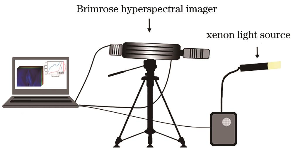
    Fig. 1. Schematic of the hyperspectral imaging system
    Flowchart of the clinical data collection process
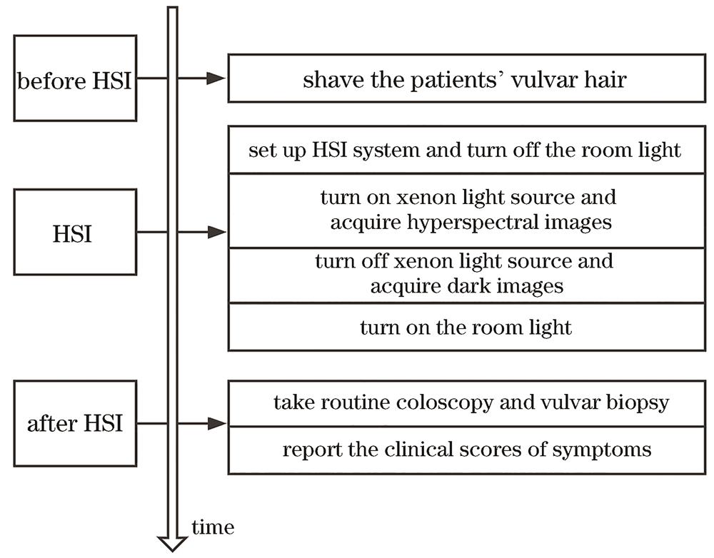
    Fig. 2. Flowchart of the clinical data collection process
    Process for calculating melanin content index and StO2 index in vulvar skin tissue. (a)-(c) Hyperspectral images of the vulva, PTFE board, and dark background; (d) relative reflectance image of the vulva at 550 nm wavelength; (e) the raw intensity curve and the relative reflectance curve retrieved from the dot area in Fig.3 (d); (f) corresponding melanin content index; (g) corresponding StO2 index
    Fig. 3. Process for calculating melanin content index and StO2 index in vulvar skin tissue. (a)-(c) Hyperspectral images of the vulva, PTFE board, and dark background; (d) relative reflectance image of the vulva at 550 nm wavelength; (e) the raw intensity curve and the relative reflectance curve retrieved from the dot area in Fig.3 (d); (f) corresponding melanin content index; (g) corresponding StO2 index
    Hyperspectral analysis results of two VLS patients. (a) Colposcopy images of the vulva, circles are the suspicious disease areas identified by the clinician; (b) corresponding relative reflectance images at 550-nm wavelength; (c) absorption spectra between normal tissues and VLS tissues; (d) melanin content index maps; (e) StO2 index maps
    Fig. 4. Hyperspectral analysis results of two VLS patients. (a) Colposcopy images of the vulva, circles are the suspicious disease areas identified by the clinician; (b) corresponding relative reflectance images at 550-nm wavelength; (c) absorption spectra between normal tissues and VLS tissues; (d) melanin content index maps; (e) StO2 index maps
    Statistical comparison of melanin content index and StO2 index of 40 VLS patients
    Fig. 5. Statistical comparison of melanin content index and StO2 index of 40 VLS patients
    Yingjie Qu, Shufang Chang, Xiaorong Xu. Preliminary Study of Vulvar Lichen Sclerosis Diagnosis Based on Hyperspectral Imaging[J]. Laser & Optoelectronics Progress, 2022, 59(6): 0617027
    Download Citation