Journals >Laser & Optoelectronics Progress
Contents


2022
Volume: 59 Issue 6
32 Article(s)
Export citation format
Medical Optics and Biotechnology
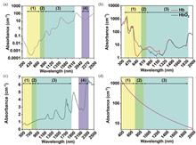
Advances on in vivo Fluorescence Bioimaging in the Second Near-Infrared Window
Zhe Feng, and Jun Qian
Near-infrared-II (NIR-II) fluorescence visualization is an in vivo optical imaging technique with large imaging depth and high resolution. Compared with visible (360?760 nm) and near-infrared-I (760?900 nm) windows, the light absorption of bio-tissues in the second near-infrared window (900?1880 nm) improves significanNear-infrared-II (NIR-II) fluorescence visualization is an in vivo optical imaging technique with large imaging depth and high resolution. Compared with visible (360?760 nm) and near-infrared-I (760?900 nm) windows, the light absorption of bio-tissues in the second near-infrared window (900?1880 nm) improves significantly, suppressing the scattering background in two-dimensional array detection, thus remarkably improving imaging quality. Currently, scientists have developed various luminescent probes, imaging systems, and theranostics pathways corresponding to the NIR-II window. The experimental animals cover from rodents to non-human primates. The clinical surgery of human liver tumors guided by NIR-II fluorescence imaging has also been initially evaluated. With light absorption and photon scattering in the visible and near-infrared regions in biological tissues, this study expounds on the mechanism of imaging quality improvement on NIR-II fluorescence imaging. It briefly introduces several typical NIR-II luminophores and reviews the developed multimodal NIR-II optical imaging methods. The multi-functional theranostics platform guided by precious NIR-II optical imaging is also discussed. These aim to provide references for further development and accelerate clinical translation..
Laser & Optoelectronics Progress
- Publication Date: Mar. 25, 2022
- Vol. 59, Issue 6, 0617001 (2022)

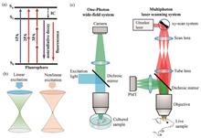
Near Infrared-Ⅱ Excited Multiphoton Fluorescence Imaging
Shaowei Wang, and Ming Lei
Multiphoton fluorescence imaging, which uses near-infrared light excited multiphoton absorption process, has been widely applied in biological imaging, especially in in vivo deep-tissue imaging due to its deep penetration, high spatiotemporal resolution, high signal-to-noise ratio, and low phototoxicity. Compared with Multiphoton fluorescence imaging, which uses near-infrared light excited multiphoton absorption process, has been widely applied in biological imaging, especially in in vivo deep-tissue imaging due to its deep penetration, high spatiotemporal resolution, high signal-to-noise ratio, and low phototoxicity. Compared with UV-vis light used in traditional one-photon bioimaging, near-infrared light greatly improved the penetration depth of multiphoton fluorescence imaging. Recently, the light in near infrared-II region (1000-1700 nm) has attracted tremendous attention in one-photon and multiphoton fluorescence imaging, which further improves the imaging depth due to its lower scattering and better penetration capability in biological tissues. In this review, we introduce the mechanism of multiphoton fluorescence imaging and the properties of near-infrared light in biological tissues. Furthermore, we introduce and discuss recent advances in near infrared-II light excited multiphoton fluorescence imaging. Finally, we present challenges facing this technique and future developments..
Laser & Optoelectronics Progress
- Publication Date: Mar. 25, 2022
- Vol. 59, Issue 6, 0617002 (2022)
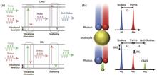
Hyperspectral Coherent Raman Scattering and Its Applications
Fan Wu, Shangyu Li, Weili Hong, Shuhua Yue, and Pu Wang
There is abundant material information in biological samples, and each material has its unique properties and functions. Research on the production, transportation, and distribution of these materials in organisms can give us a deep understanding of biological systems. Optical microscopes with chemical specificity are There is abundant material information in biological samples, and each material has its unique properties and functions. Research on the production, transportation, and distribution of these materials in organisms can give us a deep understanding of biological systems. Optical microscopes with chemical specificity are powerful and reliable tools for these studies. Raman scattering technology has greatly promoted the development of biomedicine because it solves the problems caused by the fluorescence microscope. Especially, hyperspectral coherent Raman scattering (HS-CRS) microscope can perform label-free chemical imaging of substances with different components. By providing image and spectral information at the same time, the properties, content and distribution of substances in the sample can be determined. It provides scientific researchers with more dimensional material information. In recent decades, HS-CRS has been continuously updated and applied in the biomedical field. This paper introduces the main HS-CRS implementation and schemes and summarizes its application in the biomedical field..
Laser & Optoelectronics Progress
- Publication Date: Mar. 25, 2022
- Vol. 59, Issue 6, 0617003 (2022)
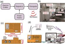
Research Progress and Prospect of Microwave-Induced Thermoacoustic Imaging Technology
Shanxiang Zhang, Xiaoyu Tang, and Huan Qin
Microwave-induced thermoacoustic imaging (MTAI) is a novel nondestructive physical medical imaging method that combines the advantages of microwave imaging's high contrast and ultrasound imaging's high resolution. Through the transformation of energy transfer from microwave to ultrasound, MTAI achieves nondestrMicrowave-induced thermoacoustic imaging (MTAI) is a novel nondestructive physical medical imaging method that combines the advantages of microwave imaging's high contrast and ultrasound imaging's high resolution. Through the transformation of energy transfer from microwave to ultrasound, MTAI achieves nondestructive and centimeter deep high-resolution imaging of biological tissues using the microwave as an excitation source and ultrasound as the information carrier. The contrast of MTAI depends on the difference in microwave absorption. In biological tissues, polar molecule such as water molecules (molecular polarization loss) and ions (ion polarization loss) are mainly used as signal sources, to obtain images of the structure and function of biological tissues. It has distinct advantages in the field of biomedical imaging and has piqued the interest of many researchers. This review discusses the principle of MTAI technology, microwave radiation module and data acquisition module and data processing module, biomedical applications of MTAI, and MTAI probes. Additionally, by combining MTAI with the current challenges, we forecast its development direction..
Laser & Optoelectronics Progress
- Publication Date: Mar. 25, 2022
- Vol. 59, Issue 6, 0617004 (2022)

Creating the sound of life by light: a discussion about photoacoustic imaging
Changhui Li
Photoacoustic imaging (PAI) is a very unique existence in biomedical imaging methods owing to the very different physical signals for excitation and detection. This "mixed" imaging mode breaks through the barrier in high-resolution optical imaging for deep tissue, providing a powerful platform technology for Photoacoustic imaging (PAI) is a very unique existence in biomedical imaging methods owing to the very different physical signals for excitation and detection. This "mixed" imaging mode breaks through the barrier in high-resolution optical imaging for deep tissue, providing a powerful platform technology for both biomedical research and clinical applications. However, PAI has not been widely used thus far. On the one hand, it is limited by its own mechanism, and on the other hand, there are some challenges in its practical application. This study discusses PAI technology based on the author's long-term scientific research experience and thinking in the field of PAI. Unlike the general research review, it does not discuss some of the important related technologies and application achievements in detail, instead it focuses on the historical development, connotation, and main challenges of PAI to foster the understanding of this technique and accelerate its implementations..
Laser & Optoelectronics Progress
- Publication Date: Mar. 25, 2022
- Vol. 59, Issue 6, 0617005 (2022)

Diffusion Correlation Spectroscopy for Tissue Blood Flow Monitoring and Its Clinical Applications
Zhe Li, Jinchao Feng, and Kebin Jia
Diffusion correlation spectroscopy (DCS) is a relatively new methodology that has been extensively used for the noninvasive monitoring of tissue blood flow. This technology irradiates the tissue surface with near-infrared light, calculates the light intensity autocorrelation function of the scattered spot on the tissueDiffusion correlation spectroscopy (DCS) is a relatively new methodology that has been extensively used for the noninvasive monitoring of tissue blood flow. This technology irradiates the tissue surface with near-infrared light, calculates the light intensity autocorrelation function of the scattered spot on the tissue surface, and computes the movement of red blood cells to realize the quantitative detection of blood flow changes in tissues. DCS measurements show more promise for the noninvasive, radiation-free, continuous and real-time monitoring, wide application range, and low detection requirements of tissue blood flow than the other blood flow monitoring methods such as laser Doppler flowmetry (LDF), magnetic resonance imaging (MRI), and positron emission tomography (PET). Moreover, DCS technology can be utilized for the bedside monitoring of tissue blood flow. The DCS technique is mainly introduced in which its theoretical background, instrumentation, progress, and clinical applications are included, as well as its future development prospects are discussed..
Laser & Optoelectronics Progress
- Publication Date: Mar. 25, 2022
- Vol. 59, Issue 6, 0617006 (2022)
Application of Synthetic Aperture Technology in Photoacoustic Imaging
Tiancheng Lei, Rongkang Gao, and Chengbo Liu
Photoacoustic imaging is a hybrid imaging method that integrates optical contrast and ultrasound penetration, reserving the advantages of both the optical imaging and the ultrasonic imaging. In acoustic-resolution photoacoustic imaging (AR-PAM), focused transducers with large numerical are usually applied to AR-PAM to Photoacoustic imaging is a hybrid imaging method that integrates optical contrast and ultrasound penetration, reserving the advantages of both the optical imaging and the ultrasonic imaging. In acoustic-resolution photoacoustic imaging (AR-PAM), focused transducers with large numerical are usually applied to AR-PAM to achieve high image quality. However, the lateral resolution and signal-to-noise ratio of photoacoustic images degrade significantly in the out-of-focus region due to the limited focal depth of transducer. To address this challenge, algorithms of synthetic aperture focusing technology (SAFT) based on virtual point concept have been developed. These include one-dimensional-SAFT, two-dimensional-SAFT, adaptive-SAFT, spatial impulse response-based scheme, and delay-multiplication-sum solution. This review introduces the advantages and limitations of these algorithms, and discusses their outlooks for future developments in AR-PAM..
Laser & Optoelectronics Progress
- Publication Date: Mar. 25, 2022
- Vol. 59, Issue 6, 0617007 (2022)
Research Progress of Multi-Modal Contrast Agent in Optical Coherence Tomography
Jie Hu, Yongwei Guo, and Haomiao Zhu
Recently optical coherence tomography (OCT) has obtained a fast development and application in clinic due to its "optical biopsy" properties. In addition, OCT could also provide molecular information after using contrast agents. However, single modal OCT technique has its disadvantages, such as poor penetratiRecently optical coherence tomography (OCT) has obtained a fast development and application in clinic due to its "optical biopsy" properties. In addition, OCT could also provide molecular information after using contrast agents. However, single modal OCT technique has its disadvantages, such as poor penetration depth. These can be avoided by multi-modal imaging technique, which will also help to make full use of OCT system and to get a full understanding of organism from different aspect. In this review, we summarized the development of multi-modal contrast agents for OCT, fluorescence imaging, photoacoustic imaging and magnetic resonance imaging, and discussed the future of multi-modal contrast agents..
Laser & Optoelectronics Progress
- Publication Date: Mar. 25, 2022
- Vol. 59, Issue 6, 0617008 (2022)
Recent Advances in Structured Illumination Microscope Super-Resolution Image Reconstruction
Yujun Tang, Linbo Wang, Gang Wen, and Hui Li
Structured illumination microscopy (SIM) has become one of the most popular super-resolution (SR) instruments for dynamic imaging of live cells. However, the final SR images of SIM depends heavily on the image reconstruction algorithms, which could dramatically affect the image quality. In the past five years, nearly 1Structured illumination microscopy (SIM) has become one of the most popular super-resolution (SR) instruments for dynamic imaging of live cells. However, the final SR images of SIM depends heavily on the image reconstruction algorithms, which could dramatically affect the image quality. In the past five years, nearly 10 open-source software packages for SIM reconstruction have been developed with advantage on different situations. And deep learning based SIM reconstruction algorithms has also been reported. Understanding the principles and differences of each algorithm becomes a priority to select the appropriate algorithms for practical applications. This review firstly introduces the principle of SIM, and then presents the latest advances for the reference of SIM researchers and users from three aspects: estimation of structured illumination parameter, spectrum optimization, and deep learning based reconstruction. Finally, the remaining issues that need to be addressed further for high-quality SIM super-resolution image reconstruction are summarized..
Laser & Optoelectronics Progress
- Publication Date: Mar. 25, 2022
- Vol. 59, Issue 6, 0617009 (2022)
Activatable NIR-Ⅱ Small Molecules for Bioimaging
Hanming Dai, Xiaohong Ruan, Jinjun Shao, and Xiaochen Dong
The second near-infrared (NIR-Ⅱ, 1000-1700 nm) fluorescence imaging (FLI) can overcome the problems of strong tissue absorption, high scattering, and auto-fluorescence, significantly improve the spatiotemporal resolution, signal-to-noise ratio, and penetration depth, so as to play a vital role in the diagnosis and treaThe second near-infrared (NIR-Ⅱ, 1000-1700 nm) fluorescence imaging (FLI) can overcome the problems of strong tissue absorption, high scattering, and auto-fluorescence, significantly improve the spatiotemporal resolution, signal-to-noise ratio, and penetration depth, so as to play a vital role in the diagnosis and treatment of major diseases. Among NIR-Ⅱ fluorescent materials, small organic molecules present great potential for clinical transform by their good biocompatibility, fast metabolism, and high biosafety. At present, most always-on type NIR-Ⅱ small molecules lack accuracy in the detection of lesions, leading to uncertainty in diagnosis. Activatable NIR-Ⅱ small molecules can generate specific fluorescent signals under the activation of biomarkers, which greatly improves the accuracy and reliability of detection. This review systematically summarizes the recent progresses on activatable NIR-Ⅱ small molecules for bioimaging, focusing on their molecular design strategies and bioimaging performance. In addition, the current problems, challenges, and future development directions are discussed..
Laser & Optoelectronics Progress
- Publication Date: Mar. 25, 2022
- Vol. 59, Issue 6, 0617010 (2022)
Single Molecule Counting Immunoassay
Yuting Su, and Hongwei Gai
Immunoassay is an indispensable analytical method in modern medicine. At present, most immune technologies have the problems such as high detection limit, low sensitivity, and poor reliability in complex systems, which are difficult to meet the requirements of precision medicine. Single molecule immunoassay, also knownImmunoassay is an indispensable analytical method in modern medicine. At present, most immune technologies have the problems such as high detection limit, low sensitivity, and poor reliability in complex systems, which are difficult to meet the requirements of precision medicine. Single molecule immunoassay, also known as digital immunoassay, can quantify by measuring the number of immune complexes. It is an ultrahigh sensitive immunoassay technology, which can meet the needs of high sensitive detection such as early detection of major diseases and detection of rare antigens in blood. This paper introduces the basic theory, classification methods and specific applications of single molecule immunoassay, focuses on the research status in the field of single molecule counting immunoassay at home and abroad, and discusses the prospects of the applications of this immunoassay technology in medical clinical diagnosis..
Laser & Optoelectronics Progress
- Publication Date: Mar. 25, 2022
- Vol. 59, Issue 6, 0617011 (2022)
Label-Free Optical Microscopy Technique and Its Biomedical Applications
Yitao Cao, Xue Wang, Xinchao Lu, and Chengjun Huang
Currently, microscopic imaging methods based on fluorescent labeling are the mainstream approaches in biomedical research. However, fluorescent labeling faces bottlenecks, such as photobleaching, photo quenching, specific labeling difficulty, and fluorescence disturbance. Therefore, the development of label-free microsCurrently, microscopic imaging methods based on fluorescent labeling are the mainstream approaches in biomedical research. However, fluorescent labeling faces bottlenecks, such as photobleaching, photo quenching, specific labeling difficulty, and fluorescence disturbance. Therefore, the development of label-free microscopic imaging techniques has attracted numerous attentions. In this review, four label-free optical microscopy techniques are described: coherent Raman scattering imaging, photothermal imaging, surface plasmon resonance imaging, and interference scattering imaging. The basic principles of these techniques are illustrated, and their applications in biomedical fields, including morphology of biomolecules, viruses, and cells, as well as biodynamics, are described. Finally, the performance of the four imaging techniques is compared, and the advantages, limitations, and application prospects are summarized..
Laser & Optoelectronics Progress
- Publication Date: Mar. 25, 2022
- Vol. 59, Issue 6, 0617012 (2022)
Application Progress of Multiphoton Microscopy in Prognostic Prediction of Breast Tumor Microenvironments
Shunwu Xu, Jiajia He, Gangqin Xi, Lianhuang Li, Xiahui Han, Liqin Zheng, and Jianxin Chen
Brest cancer has historically ranked number one in the global incidence of malignant tumors in women. The latest International Agency for Research on Cancer by the World Health Organization shows that breast cancer has surpassed lung cancer as the leading type of cancer worldwide. Multiphoton microscopy (MPM) is an advBrest cancer has historically ranked number one in the global incidence of malignant tumors in women. The latest International Agency for Research on Cancer by the World Health Organization shows that breast cancer has surpassed lung cancer as the leading type of cancer worldwide. Multiphoton microscopy (MPM) is an advanced medical imaging technique based on nonlinear optical effects, such as multiphoton excited fluorescence and harmonic generation, occurring owing to laser interactions with biological tissues. MPM offers the advantage of a resolution comparable to digital histopathology as well as requires no labeling or sectioning while providing three-dimensional optical imaging that reflects the metabolic function information of cells, three-dimensional targeted ablation capability, low photobleaching and photodamage, and a high imaging contrast. Therefore, MPM has garnered considerable research interest locally and globally in the field of basic and applied research in medical pathological diagnosis, particularly in the clinical translation of tumor pathological diagnosis. Herein, the application progress of MPM in the prognostic prediction of breast tumor microenvironments is introduced in detail and its future development prospects are presented..
Laser & Optoelectronics Progress
- Publication Date: Mar. 25, 2022
- Vol. 59, Issue 6, 0617013 (2022)
Advances on SERS Based on Shifting of Raman Characteristic Peaks
Jianming Yang, Guojun Weng, Jianjun Li, Jian Zhu, and Junwu Zhao
Surface-enhanced Raman scattering (SERS) is a type of molecular vibration spectroscopy with numerous applications in materials, biomedicine, and the military due to its high sensitivity, good selectivity, and nondestructive nature. At present, the main strategy for SERS detection is using the intensity change of characSurface-enhanced Raman scattering (SERS) is a type of molecular vibration spectroscopy with numerous applications in materials, biomedicine, and the military due to its high sensitivity, good selectivity, and nondestructive nature. At present, the main strategy for SERS detection is using the intensity change of characteristic peaks of Raman reporters. The characteristic peaks of the Raman reporters would shift, resulting from the influence of various factors. SERS applications involving the shifting of Raman characteristic peaks have gradually gained popularity due to their higher stability and more reliable reproducibility when compared to intensity changes. In this review, we summarized the mechanism of Raman characteristic peak shifting and discussed recent advances in SERS detection based on Raman characteristic peak shifting in cancer and disease diagnosis, environmental monitoring, and food safety detection. These examples will provide new strategies and ideas for the development of high-stability SERS methods..
Laser & Optoelectronics Progress
- Publication Date: Mar. 25, 2022
- Vol. 59, Issue 6, 0617015 (2022)
Application of Raman Spectroscopy in Cancer Diagnosis
Fengxiang Liu, Lihao Zhang, and Xia Huang
Raman spectroscopy is a nondestructive photon spectrum detection technology that can directly detect the changes in cell molecular composition and structure, providing rapid and precise identification for cancerous cells. Because cancer is the leading cause of mortality in humans, early tumor detection has become a focRaman spectroscopy is a nondestructive photon spectrum detection technology that can directly detect the changes in cell molecular composition and structure, providing rapid and precise identification for cancerous cells. Because cancer is the leading cause of mortality in humans, early tumor detection has become a focus of medical research in recent years. This study introduced the concepts of various Raman detection techniques, and their diagnostic applications on clinic samples including tumor tissue, tumor pathological sections, patient serum, and cancerous single cells. It also presented the Raman spectroscopy technology's recent research advances concerning breast cancer, digestive system cancer, skin cancer, and glioma. Cancerous cells differ from normal cells in terms of the structure and quantity of biomacromolecules. Raman spectroscopy can quickly and precisely detect these differences, making its ideal for early cancer diagnosis..
Laser & Optoelectronics Progress
- Publication Date: Mar. 25, 2022
- Vol. 59, Issue 6, 0617016 (2022)
Automatic Detection of Retinal Diseases Based on Lightweight Convolutional Neural Network
Lingxiao Wang, Jun Yang, Wensai Wang, and Ting Li
One major method for detecting retinopathy in clinics is optical coherence tomography. However, this manual diagnostic model is affected by strong subjectivity and low efficiency. Therefore, this paper proposes a lightweight convolutional neural network for the automatic detection of retinopathy. The proposed network cOne major method for detecting retinopathy in clinics is optical coherence tomography. However, this manual diagnostic model is affected by strong subjectivity and low efficiency. Therefore, this paper proposes a lightweight convolutional neural network for the automatic detection of retinopathy. The proposed network consists of two modules. The first module combines atrous convolutions and depth wise separable convolutions to reduce the number of parameters; the second module uses the decomposition convolution method to extend the depth by decomposing the conventional convolution layer into multilayer asymmetric convolution. Both modules are combined to form a feature extractor, and the Softmax function is used as the classifier to obtain a lightweight model with 44 layers deep and 9.2 MB parameters. The accuracy, sensitivity, specificity, and area under the receiver operating characteristic curve of the proposed network on the test set are 0.980, 0.954, 0.987, and 0.997, respectively. The visualization results show that the diagnostic basis of the model is consistent with that of ophthalmologists. These results show that the proposed network can accurately automate retinal disease detection..
Laser & Optoelectronics Progress
- Publication Date: Mar. 25, 2022
- Vol. 59, Issue 6, 0617017 (2022)
Inversion Algorithm for Optical Properties of Double-Layer Tissue Based on Convolutional Neural Network
Rui Bao, Qingwen Liu, Yuanyuan Liu, and Zuyuan He
The absorption and scattering coefficients of biological tissue are related to the physiological state of the tissue, which are important parameters for the detection of human health. The prediction accuracy of absorption and scattering coefficients in the current optical property inversion method of the double-layer bThe absorption and scattering coefficients of biological tissue are related to the physiological state of the tissue, which are important parameters for the detection of human health. The prediction accuracy of absorption and scattering coefficients in the current optical property inversion method of the double-layer biological tissue model is greatly affected by parameters such as the thickness of the upper layer tissue, which limits the scope of application. In this study, an inversion method of absorption and scattering coefficients is proposed, which is insensitive to parameters such as the thickness of upper layer tissue, in which the spatial and temporal distribution information of diffuse reflectance is collected; then, convolution neural network algorithm is applied to predict the absorption and scattering coefficients of double-layer biological tissue. The inversion accuracy of absorption and scattering coefficients is high under the random parameters of thickness and refractive index of upper layer tissue. In the simulation experiment, using a modified Monte-Carlo simulation, the diffuse reflectance of the double-layer skin model at different space detection positions and different time is obtained, and the convolution neural network is trained and tested using the simulation data to predict the absorption and scattering coefficients of two-layer skin tissue. Results show that the mean relative errors of absorption and scattering coefficients are less than 4% when the upper layer tissue thickness and refractive index are constant, whereas when the upper layer tissue thickness and refractive index change randomly, the mean relative errors of absorption and scattering coefficients are still less than 8%. Compared with other methods, the measurement scheme and inversion algorithm proposed in this study improve the prediction accuracy and expand the practical application prospect and provide a new method for the noninvasive measurement of biological tissue optical properties..
Laser & Optoelectronics Progress
- Publication Date: Mar. 25, 2022
- Vol. 59, Issue 6, 0617018 (2022)
Automated Analysis Methods for Autofluorescence Lifetime Microscopic Images of Yeast
Jiahui Zhong, Junxin Wu, Yawei Kong, Wenhua Su, Jiong Ma, and Lan Mi
Saccharomyces cerevisiae is one of the most attractive microorganisms, and monitoring changes in its metabolic state at different growth periods is significant for both basic biology and industrial research. In this paper, yeast cells are cultured for different periods based on the yeast generation curve rule, autofluoSaccharomyces cerevisiae is one of the most attractive microorganisms, and monitoring changes in its metabolic state at different growth periods is significant for both basic biology and industrial research. In this paper, yeast cells are cultured for different periods based on the yeast generation curve rule, autofluorescence lifetime images are collected using fluorescence lifetime imaging microscopy (FLIM), and an automatic analysis method based on machine learning is proposed, which can rapidly identify young and senile yeast cells without markers. First, a deep-supervised U-Net is applied to automatically segment yeast cells. Then, the features of fluorescence lifetime and morphology of each yeast cell are extracted. Finally, the classification is achieved using the unsupervised clustering method. The experimental results reveal that yeast senescence is accompanied by changes in metabolism. FLIM, as a label-free imaging technique, can be used for the metabolic analysis of yeast cells. When combined with the automated analysis process, it can swiftly and accurately distinguish cells with different metabolic differences, laying the foundation for subsequent screening of single cells..
Laser & Optoelectronics Progress
- Publication Date: Mar. 25, 2022
- Vol. 59, Issue 6, 0617019 (2022)
Hybrid Registration Method of Narrow-Band Blood Vessel Image for Vascular Targeted Photodynamic Therapy
Ruijie Chen, Yanhui Niu, Tianlong Chen, Haixia Qiu, Ying Gu, Yi Shen, and Buhong Li
Automatic quantitative assessment of vascular damage in mice's dorsal skin-fold window chamber (DSWC) plays an important role in personalized and precise vascular targeted photodynamic therapy (V-PDT). The narrow-band light blood vessel image of DSWC, however, will produce nonrigid deformations in varying degrees, Automatic quantitative assessment of vascular damage in mice's dorsal skin-fold window chamber (DSWC) plays an important role in personalized and precise vascular targeted photodynamic therapy (V-PDT). The narrow-band light blood vessel image of DSWC, however, will produce nonrigid deformations in varying degrees, which is caused by breathing, heartbeat, and involuntary movement of mice during V-PDT, and this leads to quantification errors of vascular damage. A judgment hybrid registration method combining the advantages of feature-based and gray-scale registration methods was proposed. The slight and severe deformations were first distinguished using the mean square difference (MSD) of registration groups. For slight deformation, the differential homeomorphism Demons (Log-Demons) algorithm was directly adopted for image registration. For severe deformation, the speeded up robust features thin-plate splines (SURF-TPS) algorithm was used for global registration, and then the Log-Demons algorithm was used to realize local registration. The experimental results show that the proposed algorithm has better registration accuracy and higher processing efficiency than the SURF-TPS, Log-Demons, and SURF-TPS+Log-Demons algorithm. Furthermore, this algorithm could effectively correct the inaccuracy assessment of vascular injury caused by severe deformation..
Laser & Optoelectronics Progress
- Publication Date: Mar. 25, 2022
- Vol. 59, Issue 6, 0617020 (2022)
Numerical Simulation and Experimental Confirmation on Reconstruction of Bessel Beam
Jiajun Xie, Hangshi Xu, Wenhui Yu, Rui Hu, Junle Qu, and Liwei Liu
The Bessel beam has the characteristics of non-diffraction and reconstruction, and has important application value in the fields such as optical tweezers and biological imaging. Based on the angular spectrum theory, the reconstruction behavior of the Bessel beam blocked by obstacles is analyzed, and the cross-correlatiThe Bessel beam has the characteristics of non-diffraction and reconstruction, and has important application value in the fields such as optical tweezers and biological imaging. Based on the angular spectrum theory, the reconstruction behavior of the Bessel beam blocked by obstacles is analyzed, and the cross-correlation coefficient is used to characterize the similarity of the cross-section intensity distributions of the reconstructed Bessel beam and the original one, thus to determine the recovery distance in the simulation, and get the influence of different obstacles on Bessel beam reconstruction. The simulation results show that the relationship between the recovery distance and the obstacle size is not simply linear. The recovery distance will only increase when the obstacle blocks more sidelobes and the off-axis obstacle causes longer recovery distance than that of the on-axis obstacle. For on-axis obstacles, when their sizes are smaller than the hollow areas of the high-order Bessel beam, the high-order Bessel beam has a stronger self-healing ability. In the experiment, the spatial light modulator is used to generate a flexible Bessel beam to verify the simulation results. The experimental results are in good agreement with the simulation results, which indicates that the cross-correlation method based on the angular spectrum theory can accurately simulate the reconstruction characteristics of Bessel beam..
Laser & Optoelectronics Progress
- Publication Date: Mar. 25, 2022
- Vol. 59, Issue 6, 0617021 (2022)
Sparse-View Photoacoustic Image Quality Enhancement Based on a Modified U-Net
Tong Wang, Wende Dong, Kang Shen, Songde Liu, Wen Liu, and Chao Tian
In photoacoustic tomography, an ultrasonic transducer array is usually used to receive photoacoustic signals, which is expensive to manufacture, and the number of array elements has an important impact on the final imaging quality. To improve photoacoustic image quality reconstructed under sparse view conditoin, this sIn photoacoustic tomography, an ultrasonic transducer array is usually used to receive photoacoustic signals, which is expensive to manufacture, and the number of array elements has an important impact on the final imaging quality. To improve photoacoustic image quality reconstructed under sparse view conditoin, this study proposes a modified U-Net based on the replacement of the skip connection in a conventional U-Net with continuous convolutional layers, thereby increasing the matching degree of features transferred from the encoder to the decoder. Furthermore, the loss function based on the structural similarity index measure is used to train the network. Experimental results based on simulation and in vivo dataset show that compared with the conventional U-Net, the modified U-Net achieves more image details and the quality of the reconstructed image is significantly better..
Laser & Optoelectronics Progress
- Publication Date: Mar. 25, 2022
- Vol. 59, Issue 6, 0617022 (2022)
Modified Beer-Lambert Law for Nd∶ YAG Laser Transmission in Blood
Xiaoxi Dong, Yaqun Kong, Huijuan Yin, Weinan Dong, Jizhi Zhao, and Jichun Yang
To acquire the exact expression of the modified Beer-Lambert law for the transmission of Nd∶YAG laser in blood, the relationship between a geometry-dependent factor and blood depth must be clarified. In this study, we built a laser energy detecting system that can measure the transmitted Nd∶YAG laser energy using in viTo acquire the exact expression of the modified Beer-Lambert law for the transmission of Nd∶YAG laser in blood, the relationship between a geometry-dependent factor and blood depth must be clarified. In this study, we built a laser energy detecting system that can measure the transmitted Nd∶YAG laser energy using in vitro blood samples. Laser decay was measured in cylinder-shaped blood layer with a thickness of 1.0-2.5 mm, which is the typical blood layer thickness in clinical vascular diseases. Statistical methods and the R language were used to build linear, log, index, and power regression models of the geometry-dependent factor with blood thickness as an independent variable. The highest R2 value among these four models is 0.9219, corresponding to the linear regression model. Based on the abovementioned results, the exact expression of the specially modified Beer-Lambert law has been built for Nd∶YAG laser transmission in blood..
Laser & Optoelectronics Progress
- Publication Date: Mar. 25, 2022
- Vol. 59, Issue 6, 0617023 (2022)
Preliminary Exploration of Plasmon-Enhanced Four-Wave Mixing Imaging and Its Possible Application in Antibody-Drug Metabolism in the Body
Yijia Geng, Lili Cong, Xiumian Cao, Xin Guan, Zepeng Huo, Gang Chen, Yu Liu, Weiqing Xu, Chongyang Liang, and Shuping Xu
In this study, several imaging functions, such as two-photon imaging and multi-mode four wave mixing(FWM) imaging, were realized on a single multiphoton spectrometer by the combination of double-output picosecond pulse laser and laser scanning microscope platform. Different nonlinear effects were tested to prove that tIn this study, several imaging functions, such as two-photon imaging and multi-mode four wave mixing(FWM) imaging, were realized on a single multiphoton spectrometer by the combination of double-output picosecond pulse laser and laser scanning microscope platform. Different nonlinear effects were tested to prove that this system could achieve spectroscopic recording and have excellent imaging performance. We explored the possible application of plasmon-enhanced FWM imaging for the metabolism residues of antibody drugs in a mouse's liver and kidney using 5 nm gold nanoparticles as a probe. This method acts as a tool for conducting the metabolism evaluation of protein drugs in the body..
Laser & Optoelectronics Progress
- Publication Date: Mar. 25, 2022
- Vol. 59, Issue 6, 0617024 (2022)
Multiphoton Technique for Visualization of Angiomatous Meningiomas
Na Fang, Zanyi Wu, Xingfu Wang, Yuanxiang Lin, and Jianxin Chen
Angiomatous meningiomas comprise numerous blood vessels. The ability to clearly diagnose angiomatous meningiomas preoperatively or intraoperatively is crucial for developing surgical plans and prognosis. In this study, we combined the second harmonic, two-photon excited fluorescence, and spectroscopic techniques to reaAngiomatous meningiomas comprise numerous blood vessels. The ability to clearly diagnose angiomatous meningiomas preoperatively or intraoperatively is crucial for developing surgical plans and prognosis. In this study, we combined the second harmonic, two-photon excited fluorescence, and spectroscopic techniques to realize a label-free study of angiomatous meningiomas. Results show that this multiphoton technique can identify the endogenous components of angiomatous meningioma and visualize its microstructure. Furthermore, combined with image analysis techniques, the multiphoton technique can further automatically locate vessels and calculate number of vessels. With the rapid development in fiber optic and multiphoton endoscopic technologies, multiphoton technique poses as a new technique for the clinical diagnosis of angiomatous meningiomas..
Laser & Optoelectronics Progress
- Publication Date: Mar. 25, 2022
- Vol. 59, Issue 6, 0617025 (2022)
Automatic Phase Recognition Method Based on Convolutional Neural Network
Ying Ji, Lingran Gong, Shuang Fu, and Yawei Wang
Aiming at the problem that the extraction of sample morphological information in quantitative phase imaging technology is cumbersome and not conducive to automatic detection and analysis, the feasibility and training strategy of an accurate recognition of phase objects with similar contour based on small-scale datasetsAiming at the problem that the extraction of sample morphological information in quantitative phase imaging technology is cumbersome and not conducive to automatic detection and analysis, the feasibility and training strategy of an accurate recognition of phase objects with similar contour based on small-scale datasets are explored. The phase distribution and interference fringe datasets of four types of samples, including polystyrene microspheres and red blood cells are established accordingly. A convolution neural network (CNN) model is constructed to recognize the phase diagram successfully, and then the phase values of different samples are transformed to increase recognition difficulty. All sample types are successfully recognized on the verification set by improving the network model. To simplify the detection, the interference fringes corresponding to four types of samples are identified. The residual module is used to improve the network degradation of CNN model and realize an accurate classification. According to the actual situation of complex and changeable fringe visibility and carrier frequency, the impact on the recognition accuracy is investigated, respectively. The recognition efficiency of the model is improved via optimizing the training set, which shows the potential of machine learning technology in phase information recognition..
Laser & Optoelectronics Progress
- Publication Date: Mar. 25, 2022
- Vol. 59, Issue 6, 0617026 (2022)
Preliminary Study of Vulvar Lichen Sclerosis Diagnosis Based on Hyperspectral Imaging
Yingjie Qu, Shufang Chang, and Xiaorong Xu
To compare the hyperspectral differences of the vulvar lichen sclerosis (VLS) tissue and normal vulvar tissue and extract their respective features, the melanin content and tissue oxygen saturation (StO2) in the vulvar skin tissue of 40 patients with VLS were quantitatively analyzed via hyperspectral imaging. Our experTo compare the hyperspectral differences of the vulvar lichen sclerosis (VLS) tissue and normal vulvar tissue and extract their respective features, the melanin content and tissue oxygen saturation (StO2) in the vulvar skin tissue of 40 patients with VLS were quantitatively analyzed via hyperspectral imaging. Our experimental results suggest that the melanin content index and StO2 index in lesions are statistically lower than those in the normal vulvar tissues. Therefore, by quantifying the difference between melanin content index and StO2 index in normal vulvar tissue and VLS tissue, hyperspectral imaging technology provides the possibility of in vivo assisted diagnosis of VLS..
Laser & Optoelectronics Progress
- Publication Date: Mar. 25, 2022
- Vol. 59, Issue 6, 0617027 (2022)
BiT-Based Early Gastric Cancer Classification Using Endoscopic Images
Hongxiao Li, Shu Li, Xiafei Shi, Xiaoxi Dong, Ge Jin, Lanping Zhu, Yingxin Li, and Huijuan Yin
Gastric cancer is one of the significant lethal cancers in China. Most patients are diagnosed at an advanced stage, and if gastric cancer can be detected at an early stage through large-scale screening, patient survival can be considerably improved. In China, there are two obstacles toward the large-scale screening of Gastric cancer is one of the significant lethal cancers in China. Most patients are diagnosed at an advanced stage, and if gastric cancer can be detected at an early stage through large-scale screening, patient survival can be considerably improved. In China, there are two obstacles toward the large-scale screening of early gastric cancer. One is that endoscopy is overly invasive, resulting in low patient acceptance, and the other is that the number of endoscopists is too small compared with China's large population. A capsule endoscopic robot can alleviate the first obstacle, and the second obstacle is expected to be solved using artificial intelligence. We transferred the state-of-the-art Big Transfer (BiT) to a small dataset of early gastric cancer endoscopic images and built an early gastric cancer classification model based on white-light endoscopic images. We customized the BiT hyperparameter rules in transfer learning based on local situations. The batch size was selected according to the GPU memory limit, and based on the batch size, the linear scale rules were used to adjust the optimizer's initial learning rate dynamically. The total number of training images for the small dataset was set at 256000, on which other hyperparameters of the transfer learning were set. This study experimented with multiple models having the same structure of ResNet-v2 but different depths and widths. The best model has a depth of 101 and a width three times the original one. It has an accuracy of 97.14%, an F1 score of 94.77%, a sensitivity of 90.67%, and a specificity of 99.73% on the test set. Furthermore, the results show that the effect of batch size on the model training is statistically insignificant. This paper transferred a large model to a small dataset of endoscopic images with the BiT customization. This will promote the use of large-scale models in the field of endoscopic image analysis, which can help realize a large-scale screening of early gastric cancer..
Laser & Optoelectronics Progress
- Publication Date: Mar. 25, 2022
- Vol. 59, Issue 6, 0617028 (2022)
Medical Image Fusion Based on Multi-Scale Feature Learning and Edge Enhancement
Wanxin Xiao, Huafeng Li, Yafei Zhang, Minghong Xie, and Fan Li
As an effective method for integrating the information in different forms of medical images, medical image fusion has been commonly used in various clinical applications, such as disease diagnosis and treatment planning. However, the existing medical image fusion methods do not effectively solve the problem of blurred As an effective method for integrating the information in different forms of medical images, medical image fusion has been commonly used in various clinical applications, such as disease diagnosis and treatment planning. However, the existing medical image fusion methods do not effectively solve the problem of blurred boundaries between different organs, making the fused images more difficult to understand. Therefore, to solve this problem, this paper proposes a medical image fusion model based on multi-scale feature learning and edge enhancement. First, the receptive field is expanded using multiple dilated convolutions with different dilate rates to enable the model to learn more discriminative multi-scale features of the source images. Then, according to the maximum fusion strategy, the source image features are fused to obtain the fused feature. The convolutional layer is used to reconstruct it to obtain the fused image. Further, the edge enhancement module is introduced to enhance the edge information in the fused image to better solve the problem of blurred boundaries between different organs in medical image fusion. The experimental results show that the results obtained by using the proposed method are superior to comparison methods in terms of subjective visual effects and objective quantitative evaluation..
Laser & Optoelectronics Progress
- Publication Date: Mar. 25, 2022
- Vol. 59, Issue 6, 0617029 (2022)
Experimental Sampling Strategy for Rapid Extraction of Morphological Features Based on Phase Edge Detection
Ying Ji, Xinyu Wei, Mingming Zhang, and Yawei Wang
To address the problem of how to select appropriate edge sampling points in the phase distribution map, in this paper, we analyze the advantages and disadvantages of different processing schemes and propose a sampling strategy based on phase imaging theory and experiments. Combined with the phase imaging mechanism, sevTo address the problem of how to select appropriate edge sampling points in the phase distribution map, in this paper, we analyze the advantages and disadvantages of different processing schemes and propose a sampling strategy based on phase imaging theory and experiments. Combined with the phase imaging mechanism, several gradient-based classical edge detection operators were used to analyze the source of uncertainty in the position of sampling feature points and its influence on the detection results. Actual samples showing a morphological structure with different complexities were selected to conduct experimental research. Furthermore, the influence of different sampling points on the sample size calculation results was compared and analyzed. Subsequently, the causes of various errors were investigated and their theoretical correlation and explanation were presented. Based on the results of a comparative analysis of multiple groups of experimental data, a morphological structure feature extraction scheme suitable for samples with different complexities was proposed. The processing efficiency of this scheme can meet the needs of a rapid real-time detection..
Laser & Optoelectronics Progress
- Publication Date: Mar. 25, 2022
- Vol. 59, Issue 6, 0617030 (2022)
Local-Connection-Network-Based Reconstruction Method for Cerenkov Luminescence Tomography
Weitong Li, Haolin Wang, Kang Li, Guohua Geng, Mingquan Zhou, and Xin Cao
The traditional reconstruction method of Cerenkov luminescence tomography (CLT) has a relatively low reconstruction accuracy. In this paper, we propose a finite-element-subdivision-based local connection network (FES-LCN). The network consists of a fully connected subnetwork and a node-connected subnetwork, in which thThe traditional reconstruction method of Cerenkov luminescence tomography (CLT) has a relatively low reconstruction accuracy. In this paper, we propose a finite-element-subdivision-based local connection network (FES-LCN). The network consists of a fully connected subnetwork and a node-connected subnetwork, in which the latter is based on finite element subdivision and used to predict the residual error between the initial reconstruction result and real light source. The physical simulation experiment and digital mouse experiment are designed to test the performance of the proposed network. The results show that the FES-LCN has good stability and accuracy..
Laser & Optoelectronics Progress
- Publication Date: Mar. 25, 2022
- Vol. 59, Issue 6, 0617031 (2022)
Reviews
[in Chinese]
Zhenxi Zhang, Xunbin Wei, Minbiao Ji, and Ke Si
Laser & Optoelectronics Progress
- Publication Date: Mar. 25, 2022
- Vol. 59, Issue 6, 0600000 (2022)
Label-Free Optical Microscopy Technique and Its Biomedical Applications
Yitao Cao, Xue Wang, Xinchao Lu, and Chengjun Huang
Currently, microscopic imaging methods based on fluorescent labeling are the mainstream approaches in biomedical research. However, fluorescent labeling faces bottlenecks, such as photobleaching, photo quenching, specific labeling difficulty, and fluorescence disturbance. Therefore, the development of label-free microsCurrently, microscopic imaging methods based on fluorescent labeling are the mainstream approaches in biomedical research. However, fluorescent labeling faces bottlenecks, such as photobleaching, photo quenching, specific labeling difficulty, and fluorescence disturbance. Therefore, the development of label-free microscopic imaging techniques has attracted numerous attentions. In this review, four label-free optical microscopy techniques are described: coherent Raman scattering imaging, photothermal imaging, surface plasmon resonance imaging, and interference scattering imaging. The basic principles of these techniques are illustrated, and their applications in biomedical fields, including morphology of biomolecules, viruses, and cells, as well as biodynamics, are described. Finally, the performance of the four imaging techniques is compared, and the advantages, limitations, and application prospects are summarized..
Laser & Optoelectronics Progress
- Publication Date: Mar. 25, 2022
- Vol. 59, Issue 6, 0600012 (2022)

© Copyright 2018-2021 | Chinese Laser Press. All Rights Reserved 沪ICP备15018463号-20