
Zhiguo Sun, Bo Cai, Xi Chen, Wenxian Wei, Xiaoming Li, Dandan Yang, Cuifang Meng, Ye Wu, Haibo Zeng. Prediction and observation of defect-induced room-temperature ferromagnetism in halide perovskites[J]. Journal of Semiconductors, 2020, 41(12): 122501
- Journal of Semiconductors
- Vol. 41, Issue 12, 122501 (2020)
Abstract
1. Introduction
Modern logic and memory devices work through electrical control of n- or p-type charge carriers in semiconductor transistors. However, their further miniaturization has reached a bottleneck due to the quantum tunneling effect and also due to energy-consumption issues. Making use of another property of electrons (i.e., the spin property) to develop semiconductor spintronic devices is key to overcome these barriers[
However, there is an increasing number of studies which have reported that the vacancy defects that are responsible for the d0 ferromagnetism; for example, the oxygen vacancy in ZnO, TiO2, and SnO2, the zinc and titanium vacancies in ZnO and TiO2, and the gallium vacancy in GaN are deep in energy levels[
Recently, a class of lead halide perovskite (LHP) semiconductors with the chemical formula ABX3 (A = Cs+, CH3NH3+et al., B = Pb2+, and X = Cl−, Br−, or I−) has attracted extensive studies in the fields of photovoltaics and optoelectronics[
Here, we demonstrate, through both theoretical and experimental approaches, that even without partially-filled d or f orbitals, defective LHPs can show ferromagnetic states, due to their lifted spin degeneracy by halogen vacancy. The vacancy-induced d0 ferromagnetism is robust at RT, and can be improved by incorporating a tiny fraction of 3d ions into Pb sites. Our results may boost LHPs with excellent optical properties for applications in novel spintronic devices, such as spin light-emitting diodes and spin field-effect transistors.
2. Experimental section
2.1. Materials
PbBr2 (99.99%), CsBr (99.5%), PbCl2 (99.99%), PbI2 (99.9%), CsCl (99.99%), CsI (99.9%), manganese(II) acetylacetonate [Mn(acac)2, 97%], Fe(acac)2 (98%), Co(acac)2 (99%), Ni(acac)2 (96%), Cu(acac)2 (99%), Zn(acac)2 (98%), hydrobromic acid (HBr, 40%), hydroiodic acid (HI, 47%), hydrochloric acid (HCl, 36.5%), 1-octadecene (ODE, 90%), and diethyl ether were purchased from Macklin. Cs2CO3 (99.99%), oleylamine (OAm, 90%), oleic acid (OA, 85%), dimethylsulfoxide (DMSO, 99.8%), methanol (MeOH, 99.9%), ethyl acetate (99%), and dimethylformamide (DMF, 99.9%) were purchased from Aladdin. Toluene (99.5%) was purchased from Sinopharm Chemical Reagent Corp., China. Methylammonium bromide (MABr, 99.5%) was purchased from Xi'an Polymer Light Technology Corp., China. All materials were used without further purification.
2.2. Preparation of OAmBr (Cl or I)
We take OAmBr as an example to show how OAmBr (Cl or I) was made. OAmBr was synthesized by reaction of the OAm with HBr. 20 mmol OAm in absolute ethanol was stirred and cooled in ice-bath, and then 20 mmol HBr was added drop by drop. The reaction solution was stirred for 12 h until all OAm was reacted. Then rotary evaporation was applied to obtain OAmBr pulp at 70 °C. After three times washing with diethyl ether, white powder was obtained and dried under vacuum at 40 °C overnight for future use. Similarly, OAmCl and OAmI were synthesized by reaction of the OAm with HCl and HI, respectively.
2.3. Synthesis and surface passivation of pure and 3d ion-doped LHPs at room temperature
We take CsPbBr3 as an example to show how LHPs were made. Firstly, a mixture of PbBr2 (1.2 mmol), CsBr (1.0 mmol), and Ni(acac)2 at a designated Ni/Pb molar ratio from 0 to 20 mol% were dissolved in dimethylsulfoxide (DMSO, 10 mL). The insoluble residues were removed by using a filter with 22 μm pore size, and then the precursor solution was obtained. Next, oleylammonium (OAm, 20 μL), oleic acid (OA, 20 μL), and the precursor solution (0.2 mL) were then loaded into a 20 mL vial. Then, toluene (15 mL) was quickly added into the vial under vigorous stirring. After 5 min, the solution was centrifuged by 8000 rpm and then CsPbBr3 nanocrystals were obtained. Finally, the obtained nanocrystals were washed by 8 mL toluene twice. After washing, the nanocrystals were redispersed in 4 mL toluene for further use. OAmBr (0.025 mg/mL) was added into the toluene-dispersed nanocrystals to situ passivate the nanocrystal surfaces. The whole synthetic process was carried out at room temperature.
2.4. Preparation of Cs-oleate
Cs2CO3 (0.36 g), OA (1.5 mL) and ODE (15 mL) were added to a 100 mL three-neck round-bottom flask and degassed under an Ar flow at room temperature for 15 min, and then heated at 120 °C under an Ar flow with constant stirring for 15 min to remove the moisture from the raw materials. Thereafter, the mixture was heated to 150 °C for 15 min under an Ar flow with constant stirring and lowered to 110 °C until further use.
2.5. Synthesis of pure and 3d ion-doped LHPs via hot-injection
We take CsPbBr3 as an example to show how hot-injection LHPs were made. A mixture of 0.54 mmol of PbBr2, Ni(acac)2 at a designated Ni/Pb mole ratio from 0 to 20 mol% and 15 mL of ODE, 1.5 mL of OA, 1.5 mL of OAm was first added to a 100 mL three-neck flask, dried under vacuum for 1 h at 120 °C with constant stirring to remove the moisture from the raw materials, and then heated at 120 °C under a Ar flow. After 5 min, the mixture was heated to 170 °C, the Cs-oleate precursor (1.5 mL) were subsequently quickly injected. After reacting for 5 s, the reaction mixture was cooled to 20 °C rapidly by using an ice-water bath. The obtained quantum dots were inject 40 mL ethyl acetate collected by centrifugation at 10 000 rpm for 1 min.
2.6. Preparation of precursors for single crystal growth
2 mmol CsBr and 3 mmol PbBr2 were dissolved by 5 mL DMSO with continuous stirring for 1 h at room temperature. Then, the solution was filtered using 45 μm-sized filter to remove the precipitate, and clear solution was obtained. After that, MeOH was titrated into the clear solution until the orange precipitates no longer dissolved. Finally, the orange precipitates were filtered and the clear precursor was collected for further crystal growth.
2.7. Growth of CsPbBr3 single crystals
CsPbBr3 single crystals were grown by the antisolvent vapor-assisted crystallization method. About 10 mL clear precursors obtained above were put in a 20 mL container, and 15 mL MeOH was then added into an outer petri dish before sealing. MeOH was volatilized from the outer container to the inner one, forming saffron yellow CsPbBr3 crystals. This growing process took 2 days. Finally, the obtained CsPbBr3 crystals were washed with 110 °C DMF solution to remove the precursors attached to the crystal surfaces.
2.8. Composition, structural, magnetic, and optical characterizations
The real molar ratios of TM ions relative to Pb in TM-doped CsPbBr3 were determined by using an inductively coupled plasma mass spectrometry (iCAP TQ, Thermo Scientific). The crystalline structures were characterized by powder X-ray diffraction (Bruker-AXS D8 Advance). The microstructure was characterized by using a Tecnai G20 transmission electron microscopy with operation voltage of 200 kV. The samples used for magnetic properties measurements were dried in a vacuum drying oven at vacuum of 2 × 10⁻² Pa and at temperature of 40 °C. Then, the obtained powders were loaded into a capsule, and the magnetic properties were measured using a physical property measurement system (Quantum Design). The elemental valence states were investigated using an X-ray photoelectron spectrometer (Thermo Scientific ESCALAB 250Xi). Before recording the spectra, the surface contaminants of samples were removed by Ar ion etching with etching time of 60 s. To eliminate the charge effect, all binding energies were calibrated by the C 1s line at 284.6 eV. The electron spin resonance was carried out on a JEOL FA-200 instrument at X-band. The photoluminescence spectra were detected on Varian Cary Eclipse instrument.
2.9. First-principle calculation details
The theoretical derivation was carried out using first-principle density functional theory implemented in the Vienna Ab-initio Simulation Package (VASP)[
3. Results
3.1. Structural and optical properties characterizations
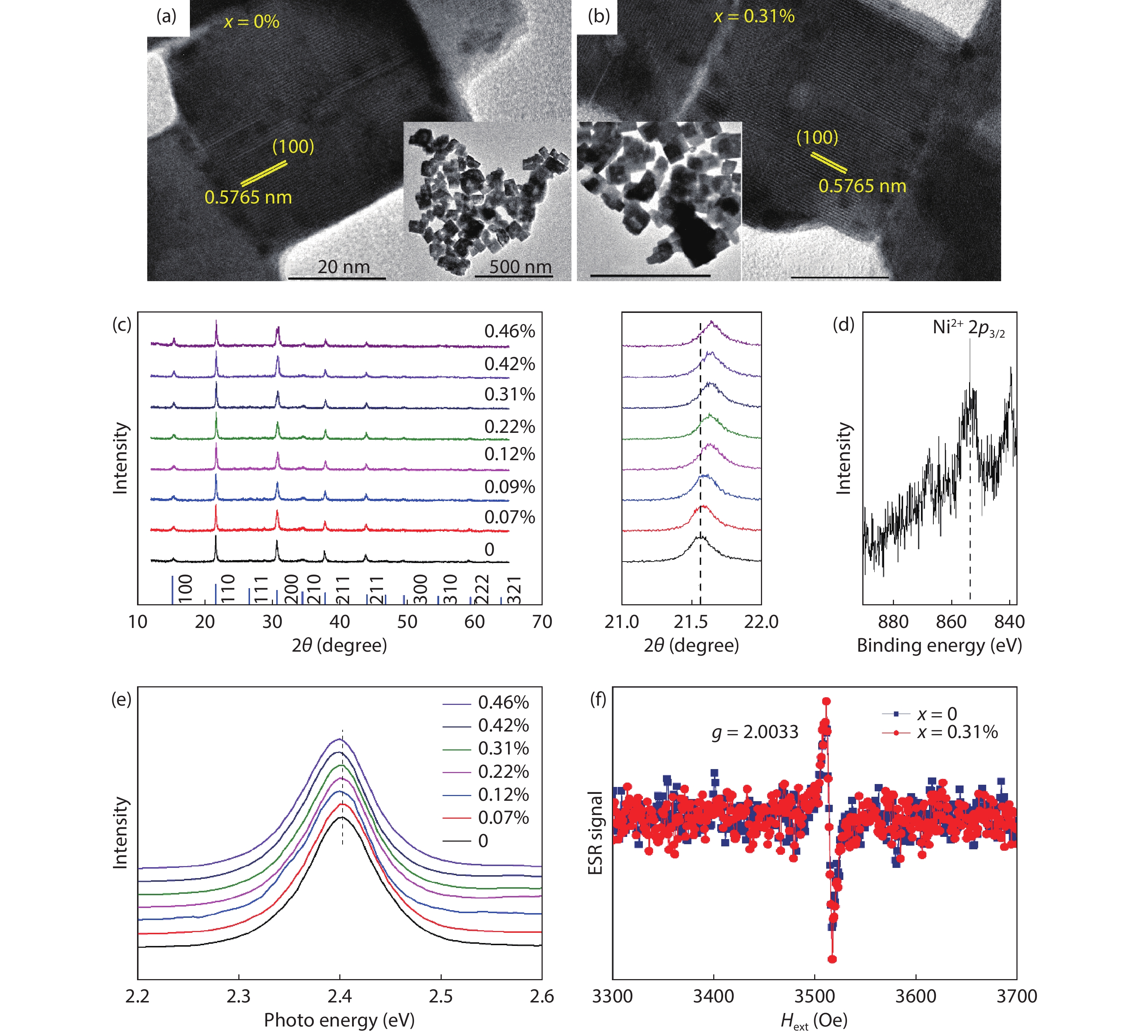
Before showing the magnetic results, we would like to present some structural and optical characterizations of Ni-doped CsPbBr3 as a representative. The samples are named as CsPb1–xNixBr3, where x is the real molar ratio of Ni relative to Pb, as determined from inductively coupled plasma mass spectrometry (Table S1, Supporting Information), and x = 0 represents pure CsPbBr3. The samples were synthesized at RT through solution processing (see Experimental section). Figs. 1(a) and 1(b) display high-resolution transmission electron microscopy (TEM) images of CsPb1–xNixBr3 with x = 0 and 0.31%, respectively. Clear lattice fringes can be observed, indicating that our samples are well crystalized. The fringe spacing determined by using fast Fourier transform patterns was found to be 0.5765 nm for both samples, which we attribute to the (100) plane. The low-resolution TEM images shown in insets reveal that both samples have typical square nanocrystal morphology, with an average size of ~ 55.2 nm. Fig. 1(c) shows X-ray diffraction (XRD) patters of CsPb1–xNixBr3 with different x values. All samples can be indexed as CsPbBr3 with cubic-phase structure. Close examination of the XRD patterns near 2θ = 21.5º reveals that the Bragg angle θ shifted slightly but systematically to higher positions as x increased, suggesting that the lattice shrank. Fig. 1(d) shows the X-ray photoelectron spectrum of the Ni 2p level in CsPb1–xNixBr3 with x = 0.31%. The peak located at around 855.42 eV is assigned to the Ni2+ 2p3/2 level. As the ion radius of Ni2+ (~0.83 Å for an octahedral site) is smaller than that of Pb2+ (~0.1 Å for an octahedral site), the lattice shrink confirms that the Ni2+ dopants were incorporated into the Pb2+ sites. Fig. 1(e) shows normalized photoluminescence spectra of CsPb1–xNixBr3 nanocrystals with different x values measured at RT. The optical gap (i.e., the energy value at the photoluminescence peak) was 2.4 eV for x = 0, which exhibits a slight red-shift with increasing x.
![]()
Figure 1.(Color online) Crystal and electronic structure characterizations. High-resolution TEM images of CsPb1–
We adopted electron spin resonance (ESR, also known as electron paramagnetic resonance) to determine the type of defects that is predominant in our samples, as ESR is a defect-sensitive technique that is widely used to study defect physics. Fig. 1(f) shows the ESR first derivative signals as a function of the external magnetic field (Hext), obtained from CsPb1–xNixBr3 nanocrystals with x = 0 and 0.31% at RT. The two samples had the same weight (10.2 mg) for these ESR measurements. We can see that, although they are weak, clear resonance signals can be observed. Both samples exhibit the same resonance peak, same line shape, and same line width, indicating that the signals share the same origin. Using the formula g = hγ/μBHext, where h is the Planck’s constant, γ is the microwave frequency (9.85 GHz), and μB is the Bohr magnetron, the g factor was calculated to be 2.0033 for both samples, which can be assigned to the Br vacancy (VBr) donor defect[
3.2. First-principle prediction of vacancy-induced magnetism
We next elucidated the defect-induced magnetic states in CsPbBr3 through first-principle density functional theory (DFT) calculations. Based on the above ESR results, we focused only on VBr and its effects on the electronic structures of CsPbBr3. Fig. 2(a) shows the perfect 3 × 3 × 3 cubic-phase CsPbBr3 supercells (left-hand column) used in the DFT calculations. As for defective CsPbBr3 (right-hand column), a Br atom (indicated by the blue arrow) was removed from the perfect supercell to create a VBr, and the lattice was then fully relaxed to a stable state for study; see Experimental section for calculation details. Fig. 2(b) displays the slice of deformation charge density (DCD) of the Pb−Br layer from the CsPbBr3 (200) plane, without and with VBr. This permits us to study the effects of VBr on charge transfer after forming chemical bonds. The Pb−Br bonds showed ionic character, where the Br atoms gained electrons and the Pb atoms contributed electrons. The charge density distribution in perfect CsPbBr3 was highly symmetric. When VBr was introduced, it became asymmetric, particularly in the vicinity of VBr. The VBr site exhibited a charge-accumulation environment, indicating that there was strong bonding between the Pb atoms around VBr.
![]()
Figure 2.(Color online) First-principle calculations. (a) The 3 × 3 × 3 CsPbBr3 supercells used in DFT calculations. Left: perfect lattice, where the Br atom indicated by the blue arrow will be removed to create a VBr. Right: relaxed lattice with the presence of a VBr. (b) DCD of the perfect (left) and defective (right) CsPbBr3. High (low) charge density corresponds to charge accumulation (depletion) regimes. (c) Spin-resolved total and partial DOSs of the perfect and defective CsPbBr3 supercells.
To study whether or not the charge distribution asymmetry could induce magnetic states, calculations on spin-resolved density of states (DOSs) were carried out; the results are shown in Fig. 2(c). As expected, the total DOSs of the perfect CsPbBr3 showed high spin-degeneracy, that is, the distribution of the spin-up and spin-down electrons was completely symmetrical, indicating the nonmagnetic nature of perfect CsPbBr3. The total DOSs of the perfect CsPbBr3 were discrete and sharp, revealing that the electronic states were rather localized. No states were present inside the bandgap of perfect CsPbBr3. Regarding defective CsPbBr3, clear spin splitting can be seen from its total DOSs (i.e., the spin degeneracy has lifted). The magnitude of the spin splitting near the valence band maximum was ~ 38 meV. The net magnetic moment of the defective supercell was calculated to be 6μB. Moreover, the defective CsPbBr3 exhibited extended DOSs, indicating that the electronic states in defective CsPbBr3 were much more delocalized than those in perfect CsPbBr3. Particularly, some impurity states were present inside the bandgap of defective CsPbBr3. Analysis of the partial DOSs of the defective CsPbBr3 revealed that: (1) the conduction band consisted of Pb 6p orbitals (predominant) and Br 4s and 4p orbitals; (2) the valence bands were formed by Br 4p orbitals (predominant) and Pb 6s and 6p orbitals; (3) Pb 6s and 6p orbitals exhibited strong hybridization with Br 4s and 4p orbitals; and (4) the impurity states were mainly composed of Pb 6p orbitals, whereas the Br 4p orbital also contributed a small part of the impurity states, due to its hybridization with the Pb 6p orbital. The exchange interaction between vacancies was studied by calculating the total energy of a 3 × 3 × 3 CsPbBr3 supercell containing two VBr and comparing the energy for the ferromagnetic (EFM) and antiferromagnetic (EAFM) states. It was found that EFM was lower than EAFM, with an energy of 3.73 meV, suggesting that the bivacancy system had a ferromagnetic ground state.
3.3. Experimental demonstration of vacancy-induced ferromagnetism
In experiment, we measured the magnetic properties of a number of LHPs including pure CsPbCl3, CsPbBr3, CsPbI3, and CH3NH3PbBr3, using a vibrating sample magnetometer (see Experimental section). Fig. 3(a) displays the magnetization versus Hex curves of RT-synthesized CsPbBr3 nanocrystals at measuring temperatures of 4, 100, 200, 300, and 400 K. The linear diamagnetic backgrounds have already been subtracted. All curves show a clear ferromagnetic behavior with S-shape signals. That is, the magnetization increased with Hex before then becoming saturated at a certain Hex value. The curves show little or no hysteresis (inset of Fig. 3(a)), and the saturation magnetization (Ms) does not change much with temperature (Fig. 3(b)), features of d0 ferromagnetism[
![]()
Figure 3.(Color online) Magnetic properties of RT-synthesized LHPs. (a) Magnetization versus
Moreover, as shown in Fig. 3(c), the ferromagnetism of the CsPbBr3 nanocrystal could be tuned by treating the nanocrystal surfaces with oleylammonium bromide (OAmBr): the Ms decreased from 0.99 memu/g before the treatment to 0.57 memu/g after the treatment. We also studied the magnetic properties of CsPbBr3 quantum dots synthesized by hot injection at 170 ºC and CsPbBr3 single crystals (see Experimental section for synthesis details). We found that both samples only showed diamagnetic background signals at 300 K (Fig. S1, Supporting Information), which indicates that they were nonmagnetic at 300 K. Surface treatment with OAmBr can passivate the VBr and thus decrease VBr concentration near the surfaces. Hot-injection synthesized and single-crystal CsPbBr3 also have reduced VBr, as indicated by our ESR measurements (Fig. S2, Supporting Information). Together with the first-principle calculation results, the magnetic results of the surface-passivation and high-quality CsPbBr3 samples confirmed the VBr origin of the ferromagnetism observed in the RT-synthesized CsPbBr3 nanocrystals.
Figs. 3(d)–3(f) present the magnetic properties of RT-synthesized tetragonal-phase CsPbCl3, orthorhombic-phase CsPbI3, and cubic-phase CH3NH3PbBr3 (structure and optical characterizations are shown in Fig. S3, Supporting Information), respectively. They all exhibited clear d0 ferromagnetism at 300 K, and surface passivation suppressed it. Moreover, similar to the case of CsPbBr3, no ferromagnetism was observed in the hot-injection synthetized CsPbCl3, CsPbI3, and CH3NH3PbBr3 at a measuring temperature of 300 K (Fig. S4, Supporting Information). Accordingly, we conclude that vacancy-induced d0 ferromagnetism should be universal in LHP materials.
3.4. Ferromagnetism enhancement with 3d ion doping
For practical device applications, the ferromagnetism should be as strong as possible, to stabilize the spins against external thermal fluctuations. As it has a defect-origin nature, d0 ferromagnetism can in principle be enhanced by increasing the defect concentration. However, having too many vacancies in LHPs is potentially hazardous to their structural stability, due to the vacancy-mediated ionic migration effect[
Fig. 4(a) presents the magnetization versus Hex curves of RT-synthesized CsPbBr3 nanocrystals doped with Mn, Fe, Co, Ni, Cu, and Zn, measured at 300 K (The XRD study confirmed the successful incorporation of these 3d dopants into the Pb site; see Fig. S5, Supporting Information). Even a tiny fraction (< 1%) of 3d dopants led to a significant modulation of the ferromagnetism of the CsPbBr3 nanocrystals. Compared with pure CsPbBr3, as shown in Fig. 4(b), doping with 0.54% Fe, 0.87% Co, and 0.31% Ni enhanced the Ms by a factor of three, two, and four, respectively, while doping with 0.35% Cu and 0.86% Zn impaired the Ms. The remarkable variation of the Ms indicates that the exchange coupling is sensitive to 3d ions doped, which have variable electron configuration in the 3d orbitals. As a representative, Fig. 4(c) shows the Ni2+ dopant concentration dependence of magnetic properties in CsPb1–xNixBr3 (see Fig. 1 for structural characterizations). The Ms roughly increased as increased from 0 to 0.46% (inset of Fig. 4(c)). Due to solubility limitation, we were unable to investigate the effects of higher x on the Ms. Fig. 4(d) displays the magnetization curves of CsPb1–xNixBr3 with x = 0.31% at measuring temperatures of 4, 100, 200, 300, and 400 K. In contrast to pure CsPbBr3, where the Ms did not vary significantly with temperature (Figs. 3(a) and 3(b)), the Ms of Ni-doped CsPbBr3 exhibited strong temperature dependence behavior: it decreased by approximately 42% as the temperature increased from 4 to 400 K. Nevertheless, all of the magnetization curves presented in Figs. 4(c) and 4(d) showed little or no hysteresis, indicating that the ferromagnetism of Ni-doped CsPbBr3 should also originate from VBr – the same as for pure CsPbBr3. The dramatic temperature dependence of the Ms reflects that there was strong coupling between the VBr and Ni2+ dopants. Only paramagnetism was found in the hot-injection-synthesized Ni-doped CsPbBr3 quantum dots (Fig. S6, Supporting Information), further confirming the VBr origin of the ferromagnetism.
![]()
Figure 4.(Color online) 3
4. Discussions
Here we discuss VBr-induced ferromagnetism and its enhancement with 3d dopants in an exchange coupling mechanism based on a magnetic polaron model[
![]()
Figure 5.(Color online) Schematic of vacancy-induced ferromagnetism. A donor electron associated with a VBr polarizes the surrounding lattices within its hydrogenic orbital, leading to the formation of a magnetic polaron (gray circles). Due to the shallow nature of the VBr, the magnetic polarons have extended wave functions. As a result, the overlap of the magnetic polarons aligns the spins of the VBr (red arrows) via exchange coupling, producing long-range spin ordering (
It is worthy to point out that, because of the large magnetic polarons, even a very low doping level of Ni2+ ions (up to 0.46%) can lead to the significant enhancement of the RT ferromagnetism in CsPbBr3. In contrast, previously widely studied oxide semiconductors had much smaller magnetic polaron than CsPbBr3; the D values for ZnO, TiO2, and SnO2 were 1.52, 0.96, and 1.72 nm, respectively[
5. Conclusions
In summary, we have reported a universal observation of vacancy-induced ferromagnetism in nominally nonmagnetic LHP semiconductors, including CsPbCl3, CsPbBr3, CsPbI3, and CH3NH3PbBr3. We have documented that this phenomenon is stable at temperatures well above 300 K, and that it is enhanced by doping LHPs with 3d ions. Our first-principle calculations suggest that the vacancy-induced ferromagnetism arises from spin-splitting states produced by halide vacancy. Our results are expounded within an exchange-coupled magnetic polaron model, and provide new physical insights for comprehensive understanding of defect physics in LHPs. Given that growth-related vacancies in LHP materials are unavoidable, making use of the vacancy-induced ferromagnetic properties will extend the functionalities of LHP-based devices, for example, for spintronic applications in spin light-emitting diodes and spin transistors. It would also be interesting to study more dopants, such as 4d TM ions and 4f rare-earth metal ions, and investigate whether they can lead to an even stronger ferromagnetism in LHPs, which would be crucial for practical applications.
Acknowledgements
Z. Sun and B. Cai contributed equally to this work. This work was financially supported by NSFC (61725402) and the Natural Science Foundation of Jiangsu Province (BK20190475).
References
[1] I Žutić, J Fabian, Sarma S Das. Spintronics: Fundamentals and applications. Rev Mod Phys, 76, 323(2004).
[2] B Qi, S Ólafsson, H P Gíslason. Vacancy defect-induced d0 ferromagnetism in undoped ZnO nanostructures: Controversial origin and challenges. Prog Mater Sci, 90, 45(2017).
[3] Y Matsumoto, M Murakami, T Shono et al. Room-temperature ferromagnetism in transparent transition metal-doped titanium dioxide. Science, 291, 854(2001).
[4] P Sharma, A Gupta, K V Rao et al. Ferromagnetism above room temperature in bulk and transparent thin films of Mn-doped ZnO. Nat Mater, 2, 673(2003).
[5] S B Ogale, R J Choudhary, J Buban et al. High temperature ferromagnetism with a giant magnetic moment in transparent Co-doped SnO2−
[6] V Fernandes, J J Klein, o N Mattoso et al. Room temperature ferromagnetism in Co-doped CeO2 films on Si(001). Phys Rev B, 75, 121304(R)(2007).
[7] M L Reed, N A El-Masry, H H Stadelmaier et al. Room temperature ferromagnetic properties of (Ga, Mn)N. Appl Phys Lett, 79, 3473(2001).
[8] J M D Coey.
[9] A Sundaresan, R Bhargavi, N Rangarajan et al. Ferromagnetism as a universal feature of nanoparticles of the otherwise nonmagnetic oxides. Phys Rev B, 74, 161306(2006).
[10] A K Rumaiz, B Ali, A Ceylan et al. Experimental studies on vacancy induced ferromagnetism in undoped TiO2. Solid State Commun, 144, 334(2007).
[11] P Zhan, W P Wang, C Liu et al. Oxygen vacancy–induced ferromagnetism in un-doped ZnO thin films. J Appl Phys, 111, 033501(2012).
[12] G Niu, E Hildebrandt, M A Schubert et al. Oxygen vacancy induced room temperature ferromagnetism in Pr-doped CeO2 thin films on silicon. ACS Appl Mater Interfaces, 6, 17496(2014).
[13] B Roul, M K Rajpalke, T N Bhat et al. Experimental evidence of Ga-vacancy induced room temperature ferromagnetic behavior in GaN films. Appl Phys Lett, 99, 162512(2011).
[14] H X Wang, Z C Zong, Y Yan. Mechanism of multi-defect induced ferromagnetism in undoped rutile TiO2. J Appl Phys, 115, 233909(2014).
[15] X P Han, J Lee, H I Yoo. Oxygen-vacancy-induced ferromagnetism in CeO2 from first principles. Phys Rev B, 79, 100403(2009).
[16] P Dev, Y Xue, P H Zhang. Defect-Induced intrinsic magnetism in wide-gap III nitrides. Phys Rev Lett, 100, 117204(2008).
[17] Y R Wang, J Y Piao, G Z Xing et al. Zn vacancy induced ferromagnetism in K doped ZnO. J Mater Chem C, 3, 11953(2015).
[18] C H Ahn, Y Y Kim, D C Kim et al. Erratum: “A comparative analysisof deep level emission in ZnO layers deposited by various methods” [J. Appl. Phys. 105, 013502 (2009)]. J Appl Phys, 105, 089902(2009).
[19] F Fabbri, M Villani, A Catellani et al. Zn vacancy induced green luminescence on non-polar surfaces in ZnO nanostructures. Sci Rep, 4, 5158(2014).
[20] B J Morgan, G W Watson. Polaronic trapping of electrons and holes by native defects in anatase TiO2. Phys Rev B, 80, 233102(2009).
[21] J L Lyons, C G van de Walle. Computationally predicted energiesand properties of defects in GaN. npj Comput Mater, 3, 12(2017).
[22] Q A Akkerman, G Rainò, M V Kovalenko et al. Genesis, challenges and opportunities for colloidal lead halide perovskite nanocrystals. Nat Mater, 17, 394(2018).
[23] A Walsh, D O Scanlon, S Y Chen et al. Self-regulation mechanism for charged point defects in hybrid halide perovskites. Angew Chem Int Ed, 54, 1791(2015).
[24] Q F Dong, Y J Fang, Y C Shao et al. Solar cells. Electron-hole diffusion lengths > 175
[25] A Dutta, R K Behera, P Pal et al. Near-unity photoluminescence quantum efficiency for all CsPbX3 (X = Cl, Br, and I) perovskite nanocrystals: A generic synthesis approach. Angew Chem Int Ed, 58, 5552(2019).
[26] N J Jeon, H Na, E H Jung et al. A fluorene-terminated hole-transporting material for highly efficient and stable perovskite solar cells. Nat Energy, 3, 682(2018).
[27] K Lin, J Xing, L N Quan et al. Perovskite light-emitting diodes with external quantum efficiency exceeding 20 per cent. Nature, 562, 245(2018).
[28] Y Cao, N N Wang, H Tian et al. Perovskite light-emitting diodes based on spontaneously formed submicrometre-scale structures. Nature, 562, 249(2018).
[29] W J Yin, T T Shi, Y F Yan. Unusual defect physics in CH3NH3PbI3 perovskite solar cell absorber. Appl Phys Lett, 104, 063903(2014).
[30] J Kang, L W Wang. High defect tolerance in lead halide perovskite CsPbBr3. J Phys Chem Lett, 8, 489(2017).
[31] Y X Zhai, S Baniya, C Zhang et al. Giant Rashba splitting in 2D organic-inorganic halide perovskites measured by transient spectroscopies. Sci Adv, 3, e1700704(2017).
[32] C Zhang, D L Sun, Z G Yu et al. Field-induced spin splitting and anomalous photoluminescence circular polarization in CH3NH3PbI3 films at high magnetic field. Phys Rev B, 97, 134412(2018).
[33] D L Sun, C Zhang, M Kavand et al. Surface-enhanced spin current to charge current conversion efficiency in CH3NH3PbBr3-based devices. J Chem Phys, 151, 174709(2019).
[34] J Wang, C Zhang, H Liu et al. Spin-optoelectronic devices basedon hybrid organic-inorganic trihalide perovskites. Nat Commun, 10, 129(2019).
[35] . Efficient iterative schemes for ab initio total-energy calculations using a plane-wave basis set. Phys Rev B, 54, 11169(1996).
[36] J P Perdew, A Ruzsinszky, G I Csonka et al. Restoring the density-gradient expansion for exchange in solids and surfaces. Phys Rev Lett, 100, 136406(2008).
[37] P Jakes, J Zimmermann, H von Seggern et al. Eu2+-doped CsBr photostimulable X-ray storage phosphors: Analysis of defect structure by high-frequency EPR. Funct Mater Lett, 7, 1350073(2014).
[38] M Coey, K Ackland, M Venkatesan et al. Collective magnetic response of CeO2 nanoparticles. Nat Phys, 12, 694(2016).
[39] J M D Coey. Magnetism in
[40] Y B Yuan, J S Huang. Ion migration in organometal trihalide perovskite and its impact on photovoltaic efficiency and stability. Acc Chem Res, 49, 286(2016).
[41] Z J Yong, S Q Guo, J P Ma et al. Doping-enhanced short-range order of perovskite nanocrystals for near-unity violet luminescence quantum yield. J Am Chem Soc, 140, 9942(2018).
[42] A Kaminski, S Das Sarma. Polaron percolation in diluted magnetic semiconductors. Phys Rev Lett, 88, 247202(2002).
[43] J M D Coey, M Venkatesan, C B Fitzgerald. Donor impurity band exchange in dilute ferromagnetic oxides. Nat Mater, 4, 173(2005).
[44] Y Kang, S Han. Intrinsic carrier mobility of cesium lead halide perovskites. Phys Rev Appl, 10, 044013(2018).
[45] F Pan, C Song, X J Liu et al. Ferromagnetism and possible application in spintronics of transition-metal-doped ZnO films. Mater Sci Eng R, 62, 1(2008).


Set citation alerts for the article
Please enter your email address

© Copyright 2018-2021 | Chinese Laser Press. All Rights Reserved 沪ICP备15018463号-20