1Laboratory of Thin Film Optics, Key Laboratory of Materials for High Power Laser, Shanghai Institute of Optics and Fine Mechanics, Chinese Academy of Sciences, Shanghai 201800, China
2Center of Materials Science and Optoelectronics Engineering, University of Chinese Academy of Sciences, Beijing 100049, China
3Hangzhou Institute for Advanced Study, University of Chinese Academy of Sciences, Hangzhou 310024, China
4CREOL, The College of Optics and Photonics, University of Central Florida, Orlando, Florida 32816, USA
5CAS Center for Excellence in Ultra-intense Laser Science, Shanghai 201800, China
The requirements for dichroic laser mirrors continue to increase with the development of laser technology. The challenge of a dichroic laser mirror coating is to simultaneously obtain spectral performance with significantly different reflection or transmission properties as well as a high laser-induced damage threshold (LIDT) at two different wavelengths. Traditional dichroic laser mirrors composed of alternating high- and low-refractive-index pure materials often has difficulty achieving excellent spectral performance and high LIDTs at two wavelengths simultaneously. We propose to use a new design with mixture layers and sandwich-like-structure interfaces to meet the challenging requirements. An mixture-based dichroic laser mirror, which can be used as a harmonic separator in a fusion-class laser or a pump/signal beam separator in a petawatt-class Ti-sapphire laser system, is experimentally demonstrated using e-beam deposition. The mixture-based dichroic mirror coating shows good spectral performance, fine mechanical property, low absorption, and high LIDT. For the -polarized 7.7 ns pulses at a wavelength of 532 nm and the -polarized 12 ns pulses at a wavelength of 1064 nm, the LIDTs are almost doubled. The excellent performance of this new design strategy with mixture layers and sandwich-like-structure interfaces suggests its wide applicability in high-performance laser coating.
1. INTRODUCTION
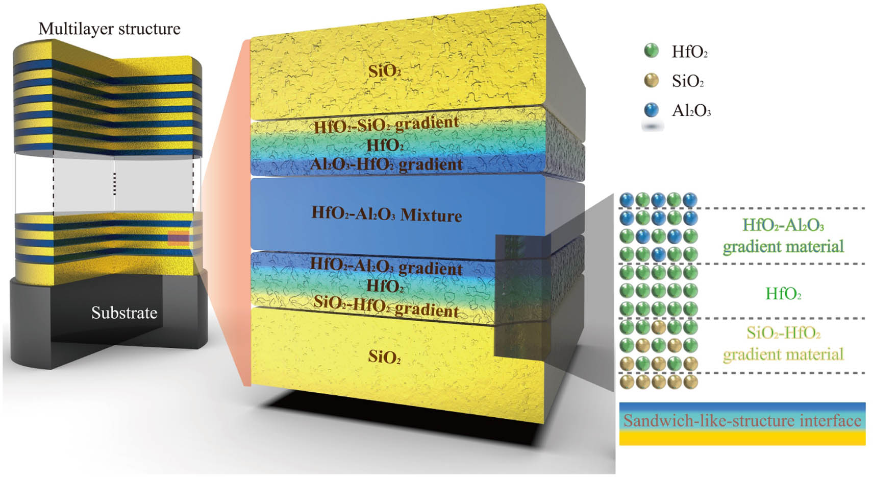
Dichroic laser mirrors are usually used as harmonic separators [1,2], beam combiners [3], or beam splitters [4] and play an important role in many laser applications, including inertial confinement fusion (ICF) lasers [5], petawatt femtosecond lasers [6], high power fiber lasers [3,7], compact -switched or mode-locked lasers [8–10], and other emerging lasers [11]. The ideal dichroic laser mirror for high-power lasers requires a significantly different reflection or transmission property and a high laser-induced damage threshold (LIDT) at two different wavelengths of interest. Unfortunately, a traditional dichroic laser mirror (TDLM) composed of alternating high- and low-refractive-index () pure materials often has difficulty achieving excellent spectral performance and high LIDTs at two wavelengths simultaneously [4]. Generally, TDLM for UV-NIR laser applications is achieved by alternately e-beam deposited layers and layers [12]. Sometimes, is chosen instead of as the high- material, which shows improved LIDT but requires a relatively large total number of coating layers [13,14]. There is a trade-off between the required optical performance and LIDT because suitable candidate coating materials are limited. In recent years, the library of available coating materials is expanded by co-evaporated or co-sputtered oxide mixtures [15–17]. The mixture materials provide us with adjustable optical gap values and optical constants, show superior properties over pure materials [16,18], and are attractive for many applications [19–21]. In addition to the coating material itself, it is also necessary to consider the interface-related issues of the traditional discrete interface, which is one of the key factors affecting LIDT. The co-evaporated interface with a graded-refractive index shows a significant increase in LIDT compared with the traditional discrete interface [22,23]. Therefore, by appropriately designing mixture materials and optimizing the interface, it is expected to realize an ideal dichroic laser mirror suitable for high-power lasers.
Figure 1.Schematic diagram of the proposed MDLM design.
All coatings are deposited on fused silica and BK7 substrates using e-beam evaporation, in which the mixture layer and the sandwich-like-structure interface are obtained by dual e-beam co-evaporation [18]. BK7 and fused silica substrates are used for stress measurement and other measurements, respectively. Before deposition, the coating chamber is heated to 473 K and evacuated to a base pressure of ; then, the substrate is cleaned with plasma ions at a bias voltage of 80 V for 120 s. The deposition rates for and in monolayer and nanolaminate coatings and the deposition rates for in multilayer coatings are 0.1, 0.1, and 0.2 nm/s, respectively. The deposition rates for and in the mixture coating are 0.028 and 0.072 nm/s, respectively. The deposition rates for and in the mixture layer of the MDLM coating are 0.05 and 0.05 nm/s, respectively. The oxygen pressures of the layer and other layers are and , respectively.
Figure 2.Schematic diagram of the deposition process of MDLM coating.
X-ray diffraction (XRD) (PANalytical Empyrean) is used to characterize the structure information of the coating. A VUV spectrometer (LZH ML 6500) and a UV-VIS-NIR spectrometer (Perkin Elmer Lambda 1050) are employed to measure the transmittance spectra in the ranges of 150–200 nm and 200–1200 nm, respectively. The reflectance spectra in the VIS region are calculated from the transmission data neglecting absorption. The refractive indices and optical bandgaps are estimated using the commercial thin film software (Essential Macleod) and the Tauc equation [24], respectively. The laser-induced temperature rise in the multilayer coating is obtained from the finite-element method (FEM) simulation.
Sign up for Photonics Research TOC. Get the latest issue of Photonics Research delivered right to you!Sign up now
The elemental composition profiles are determined by X-ray photoelectron spectroscopy (XPS, Thermo Scientific K-Alpha) using a monochromatic Al α (1486.6 eV) X-ray source. The spectra are recorded after every 20 s of etching with 1 keV Ar+ ions.
A 632.8 nm wavelength interferometer (ZYGO Mark III-GPI) is employed to inspect the sample surfaces before (substrates) and 60 days after the deposition (coatings) in a controlled environment with a temperature of and relative humidity of . The coating stress is obtained from Stoney’s formula.
The absorption of the coating at 1064 nm is measured by a home-made system based on the surface thermal lensing technique. The interfacial adhesion is characterized by a scratch test using a nano indenter (KLA Tencor). A load is gradually increased from 20 μN to 50 mN for the scratch test.
The 1-on-1 LIDT is tested according to ISO 21254. An -polarized Nd:YAG laser with a 7.7 ns pulse width and a -polarized Nd:YAG laser with a 12 ns pulse width are used for 532 and 1064 nm LIDT measurements, respectively. The test is performed at an angle of incidence of 45°. The effective beam sizes on the sample surface for 532 and 1064 nm LIDT measurements are approximately and , respectively. Fifteen sites are tested for each energy fluence. The damage morphology is characterized by a focused ion beam scanning electron microscope (FIB-SEM, Carl Zeiss AURIGA CrossBeam). The chemical composition of the damaged area is analyzed by energy dispersive spectroscopy (EDS, Oxford X-Max, ).
3. EXPERIMENTAL RESULTS AND DISCUSSION
A. Properties of the Pure Monolayer, Nanolaminate, and Mixture Coatings
Figure 3.Microstructure and optical property of the pure monolayer, nanolaminate, and mixture coatings. (a) XRD spectra, (b) transmittance, and (c) optical bandgap versus of monolayer, monolayer, nanolaminate, and mixture coatings. (d) Surface figure change () caused by the nanolaminate and mixture coatings.
The mixture layer has two advantages over the nanolaminate layers: the deposition process is simpler, and the bandgap is larger when is close. The mixture is therefore chosen as the high- material for MDLM coating in this work. This allows one to develop MDLM coatings with excellent optical and LIDT properties.
B. Mixture-Based Dichroic Laser Mirrors with Sandwich-like-Structure Interfaces
Figure 5.Elemental percentage profiles from the high- layer to the low- layer.
Figure 6.Microstructure and optical property of the TDLM and MDLM coatings. (a) XRD spectra. (b) Transmittance (left: incident angle at 0°; middle: incident angle at 45°, -polarized light) and reflectance spectra (right: incident angle at 45°, -polarized light). (c) Surface figures of the samples before and after coating. (d) Surface and cross-section morphologies after the scratch test. (e) Single-pulse damage probability as a function of the input fluence.
Figure 7.Damage morphology imaged by SEM and the depth profile of the marked area measured by FIB. (a)–(d) Damaged sites and (e)–(h) schematic diagram of the damage morphologies after irradiation of an -polarized 532 nm laser. (i) and (j) Plasma scald induced by -polarized 532 nm laser in MDLM coating.
Figure 8.Damage morphology imaged by SEM and the depth profile of the marked area measured by FIB. (a) and (b) Full field-of-view of the damaged area. (c)–(i) Typical damaged sites marked with asterisks. (j)–(m) Schematic diagram of the damage morphologies after irradiation of a -polarized 1064 nm laser.
An FEM simulation is used to investigate the 1064 nm -polarized laser-induced temperature rise in the two coatings. The extinction coefficients () of the layer () and mixture layer () are calculated by using Eq. (1) [31], based on the measured absorption, neglecting the absorption of the layers: where and are the absorption and the transmittance of coating, is the coating thickness (only the layers with absorption are taken into account), and is the wavelength. The extinction coefficient of dielectric materials typically increases with the decrease of wavelength; in this work, the mixture layer suggests a lower extinction coefficient over the whole wavelength range of interest than the layer.
Figure 10.Simulated laser-induced temperature rises in TDLM and MDLM coatings.
In summary, we have proposed and experimentally demonstrated a new MDLM coating with mixture layers and sandwich-like-structure interfaces. The proposed MDLM coating shows excellent spectral performance, and an LIDT that is almost twice of that of the TDLM coating at the two wavelengths of interest for the following reasons. First, the mixture layer in the MDLM coating has a larger optical bandgap and a lower absorption, resulting in a smaller temperature rise under the same fluence laser irradiation; second, the sandwich-like-structure interface allows the MDLM coating to exhibit enhanced mechanical properties. We believe that the described concept opens new avenues for improved dichroic mirror coatings and other laser coatings and can benefit many areas of laser technology that rely on high-quality laser coatings.
Acknowledgment
Acknowledgment. The authors express their appreciation to Prof. Wolfgang Rudolph for the fruitful discussions. The authors thank Ziyuan Xu and Yun Cui for the LIDT and FIB measurements, respectively.