
Jingzhou Zhang, Feng Chen, Jiale Yong, Qing Yang, Xun Hou. Research Progress on Bioinspired Superhydrophobic Surface Induced by Femtosecond Laser[J]. Laser & Optoelectronics Progress, 2018, 55(11): 110001
- Laser & Optoelectronics Progress
- Vol. 55, Issue 11, 110001 (2018)
![Creatures in nature with superhydrophobic surface microstructures. (a) Lotus leaf[1]; (b) rose petal[3]; (c) leg of water strider[4]; (d) mosquito eye[6]; (e) rice leaf[7]; (f) butterfly wing[8]](/richHtml/lop/2018/55/11/110001/img_1.jpg)
Fig. 1. Creatures in nature with superhydrophobic surface microstructures. (a) Lotus leaf[1]; (b) rose petal[3]; (c) leg of water strider[4]; (d) mosquito eye[6]; (e) rice leaf[7]; (f) butterfly wing[8]
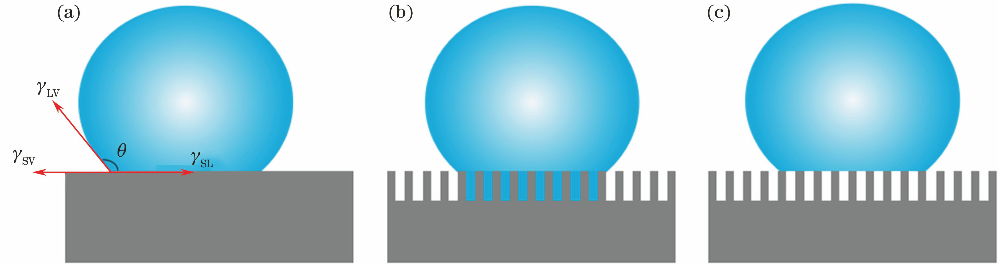
Fig. 2. Different wetting states of water droplets. (a) Young state; (b) Wenzel state; (c) Cassie/Baxter state
Fig. 3. Applications of femtosecond laser direct writing technique. (a) Treatment and correction of myopia[60]; (b) precision cutting[60]; (c) fabrication of microlens array[61]; (d) fabrication of micro-nano hierarchical rough structures[2]; (e)-(j) various patterned microstructures[62]
Fig. 4. Fabricated superhydrophobic rough surfaces by femtosecond laser and surface silanization treatment. (a) Top-view scanning electron micrograph (SEM) image, (b) 45° side-view SEM image, and (c) projection of water droplet contact angle under effect of femtosecond laser irradiation on silicon in SF6 environment[51]; (d) 45° side-view SEM image, (e) top-view SEM image, and (f) water droplet morphology picture on silicon surface under effect of femtosecond laser irradiation on silicon in air environm
Fig. 5. Process flowcharts and SEM images. (a) Process flowcharts of femtosecond laser fabrication and mask replication of PDMS; (b) microscopic and (c) nanoscopic SEM images of PDMS after femtosecond laser scanning; (d) microscopic and (e) nanoscopic SEM images of PDMS after mask replication[86]
Fig. 6. Femtosecond laser scanning of stainless steel. (a) SEM images of stainless steel after femtosecond laser scanning with power density of (a) 2.83 J·cm-2 and (b) 5.16 J·cm-2; (c) contact angle versus standing time of sample; XPS spectra of (d) intrinsic stainless steel, (e) stainless steel after laser scanning, and (f) stainless steel after standing of 52 d in air[92]
Fig. 7. Effect of femtosecond laser irradiation on titanium surface. (a) Microscopic SEM image of titanium surface after femtosecond laser irradiation; (b) SEM image with high magnification, and top-right is projection of water droplet contact angle; (c) schematic of laser fabrication; EDXS results (d) before and (e) after laser scanning[89]
Fig. 8. Methods for changing viscosity of superhydrophobic materials. (a) Distance control of well-shaped array on PDMS[87]; (b) distance control of pulse ablated craters on PDMS[102]
Fig. 9. Hierarchical rough structures of rice leaves. (a) Rice leaf surface and its anisotropic sliding behavior of water droplet; (b) top-view SEM image of rice leaf surface; (c) model of hierarchical rough structure of rice leaf; (d) schematic of femtosecond laser imitation of rice leaf
Fig. 10. Fabrication of graphene thin film with superhydrophobic structural color[116]. (a) Flowchart of fabrication; (b) hydrophobicity of graphene versus laser power; (c) structural color transferred from graphene film to glass substrate after laser scanning with power of 200 mW
Fig. 11. Fabrication of superhydrophobic metals with high light absorptivity. (a) Light absorptance and superhydrophobicity of metal material surface after femtosecond laser scanning[117]; (b) high transmittance and superhydrophobicity of PDMS surface with special micro/nano-structures[120]
Fig. 12. Laser ablation of zinc. (a) 3D profile of zinc surface after laser ablation; (b) composition change before and after fabrication; (c) reversible transformation between superhydrophobicity and superhydrophilicity[97]
Fig. 13. Schematic of microdroplet control by femtosecond laser. (a) Positioning of droplets; (b) no-loss droplet transportation[87]
Fig. 14. Interaction between living cells and biomaterials controlled by femtosecond micromachining technique. (a) SEM images of intrinsic Si and Si microstructures after femtosecond laser ablation with energy density of 0.34, 0.56, 0.9, and 1.69 J·cm-2 under smooth case; (b) higher magnification; (c) projection morphologies of water droplets on different samples; (d) confocal laser microscopy pictures of fibroblast cells cultured for three days[136]
Fig. 15. Oil-water separation. (a) SEM images of femtosecond laser induced rough PTFE after mechanical perforating; (b) photographs of water droplets and colored oil droplets on rough PTFE surface; effect images (c) before and (d) after oil-water separation in acid solution; effect images (e) before and (f) after oil-water separation in alkaline solution[138]; (g) schematic of another oil-water separation device; (h) mixtures of oil and water were successfully separated in 28 s based on this device[139]
Fig. 16. Microfluidic device. (a) SEM image of superhydrophobic copper substrate induced by femtosecond laser; (b) SEM image of superhydrophobic PDMS obtained after mask replication; (c) microfluidic device based on superhydrophobic PDMS[140]
Fig. 17. Effect image and schematic of water fog collection. (a) Collection by superhydrophilic film; (b) collection by superhydrophobic film; (c) collection by Janus film; (d) effect images by above three collection systems at beginning and after 15 min[141]
Fig. 18. Fabrication of hybrid rough structures by femtosecond laser ablation of silicon. (a) Projection of water droplet contact angle on substrate under three different laser powers and solid-liquid contact surface area versus time; (b) Raman scattering spectra obtained for different types of substrates[143]

Set citation alerts for the article
Please enter your email address
© Copyright 2018-2021 | Chinese Laser Press. All Rights Reserved 沪ICP备15018463号-20