
Bao-Kai MA, Mian LI, Ling-Zhi CHEONG, Xin-Chu WENG, Cai SHEN, Qing HUANG. Enzyme-MXene Nanosheets: Fabrication and Application in Electrochemical Detection of H2O2 [J]. Journal of Inorganic Materials, 2020, 35(1): 131
- Journal of Inorganic Materials
- Vol. 35, Issue 1, 131 (2020)
Abstract
Hydrogen peroxide (H2O2) is widely used as antimicrobial, oxidizing, reducing and bleaching agents in many fields including pharmaceutical, medical, textile, paper, and food processing[
Electrochemical biosensing technique has generated much interest due to its advantages of simple instrumentation, easy miniaturization, high sensitivity and selectivity, as well as rapid response[
Two-dimensional (2D) transition metal carbides, nitrides and carbonitrides (MXene) are produced by etching layers of sp elements (specifically groups 13 and 14) from their corresponding three-dimensional (3D) MAX phases which correspond to the general formula Mn+1AXn (n=1, 2, 3) where M represents early d-block transition metals (Ti, Sc, V, Cr, Ta, Nb, Zr, Mo, Hf), A represents main group sp elements and X is either C or N atom[
Present study aims to fabricate a horse radish peroxidase@MXene electrochemical biosensor for detection of H2O2 in food. HRP, a heme-containing enzyme, has been widely used to catalyze oxidation of a wide variety of substrates including hydrogen peroxide[
1 Experimental
1.1 Materials and chemicals
Horseradish peroxide (HRP, activity units•mg-1) was purchased from Sigma Aldrich. Natural flake graphite (48 μm), Ti powders (48 μm, purity of 99.9%), Al powders (48 μm, purity of 99.9%), hydrogen peroxide solution (30wt%), hydroquinone (HQ), chitosan (deacetylation 95%), potassium chloride, acetic acid were obtained from Aladdin, China. Other reagents including NaCl, KCl, sodium hydroxide (NaOH), K3[Fe(CN)6], K4[Fe(CN)6]• 3H2O were obtained from Sinoreagent, China. 0.1 mol•L-1 phosphate buffer solutions (PBS, pH 7.0) comprising NaH2PO4 and Na2HPO4 were used as electrolyte. All aqueous solutions were freshly prepared with ultra-pure water (18 MW•cm).
1.2 Synthesis of MXene (Graphite/TiC/Ti3C2)
G(graphite)/TiC/Ti3AlC2 were fabricated according to a previously reported method with slight modifications[
1.3 Characterization of MXene
MXene was characterized using XRD, FT-IR and SEM. XRD analysis were conducted at room temperature using Bruker D8 Discover XRD (Cu radiation, λ=0.l540596 nm) over the 2θ range of 5°~70° at room temperature. FT-IR spectra was obtained in the range of 500 to 4000 cm-1 by using a Fourier-transform infrared (FT-IR) spectroscopy (Nicolet 6700, Thermo, USA).
The microstructures of the powders were examined by a field emission scanning electron microscopy (FEI Quanta FEG 250) equipped with an EDS system and a TEM instrument (FEI Tecnai F20).
1.4 Fabrication of the HRP@MXene/chitosan/ GCE biosensor
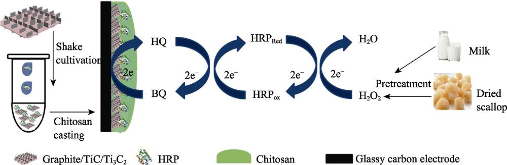
Fabrication of the HRP@MXene electrochemical biosensor is illustrated in Fig. 1. Glassy carbon electrodes (GCE, 3 mm) was firstly polished using Al2O3 (1.0, 0.3, 0.05 μm), cleaned by ethanol and water for three times, and finally dried under gentle N2 stream. Ten microliter of HRP solution [10 mg•mL-1, PBS (0.1 mol•L-1, pH 6.0)] and 20 μL of MXene aqueous solution (5 mg•mL-1) were mixed and shaked at 200 r•min-1 for 10 h at low temperature.
Following that, 10 μL chitosan solution (6 mg•mL-1, adjusted to pH 6.0 by 10 mg•mL-1 NaOH) was added to the mixture and vibrated for 3 min. Chitosan solution has been previously reported to be positively charged and have good electrical conductivity at pH 6.0 due to the protonation of amino groups[
![]()
Figure .Schematic illustration for fabrication of HRP@MXene (Graphite/TiC/Ti3C2)/chitosan/GCE and H2O2 sensing principle of HRP@MXene/chitosan/GCE
1.5 Electrochemical behavior of the HRP@MXene/chitosan/GCE biosensor
All electrochemical experiments were carried out using CHI760E electrochemical workstation (Chenhua, Shanghai) with GCE as working electrode, platinum wire as counter electrode and saturated calomel electrode (SCE) as reference electrode. The electrochemical impedance spectroscopy (EIS) and cyclic voltammograms (CVs) of electrodes fabricated using chitosan of different pH was conducted in N2-saturated 0.1 mol•L-1 KCl solution containing 5.0 mmol•L-1 Fe(CN)63-/4- at open circuit potential in the frequency range from 0.1 Hz to 105 Hz with the amplitude 5 mV. The EIS data were analyzed using ZVIEW software.
1.6 Electrochemical biosensing of H2O2 by HRP@MXene/chitosan/GCE biosensor
CVs were carried out in N2-saturated 0.1 mol•L-1 PBS (pH 7.5) in the presence of 2.0 mmol•L-1 H2O2 and 1 mmol•L-1 HQ (dissolved in methanol) at a scanning rate of 50 mV•s-1. Differential pulse voltammetry (DPV) was performed in N2-saturated 0.1 mol•L-1 PBS (pH 7.5) containing 2 mmol•L-1 H2O2 and 1 mmol•L-1 HQ (dissolved in methanol) with amplitude of 5 mV and pulse width of 0.2 s after five times of CV at a scanning rate of 50 mV•s-1 ranging from 0.8 V to -0.8 V. The effects of electrolyte PBS buffer pH (5.5 to 8) and the concentration of MXene were evaluated and optimized in terms of CV and DPV signal.
1.7 Electrochemical detection of H2O2 in spiked dried scallop and milk
Amperometric current-time curves for H2O2 were carried out to construct a calibration curve of current response at different H2O2 concentration. Measurements were performed in 10 mL of stirring 0.1 mol•L-1 PBS (pH 7.5) in the presence of 1 mmol•L-1 HQ with successive addition of H2O2 at room temperature under an applied peak potential value of -0.1 V. LOD was determined according to the following equation:
whereby SD refers to the standard deviation of the control measurement, and K refers to slope of the calibration curve.
Milk and dried scallop were chosen as model of liquid and solid food. Milk sample was used directly for H2O2 detection. Dried scallop was pre-treated according to the following procedure to extract H2O2 residual. Briefly, 2 g of dried scallop was immersed in 5 mL of H2O2 aqueous solution (3%) for 1 h. Following that, the scallop was immersed in 5 mL of water for 0.5 h to extract H2O2 residue. H2O2 concentration in spiked dried scallop test solution and milk solution (12.5, 50 and 125 μmol•L-1 H2O2) were detected using the amperometric current- time curves for H2O2. Recovery of the HRP@MXene/ Chitosan/GCE was calculated.
1.8 Selectivity of the biosensor
Selectivity of the fabricated HRP@MXene/chitosan/ GCE biosensor was evaluated using potentially interfering substances including uric acid, glucose and ascorbic acid [100 μmol•L-1 in 0.1 mol•L-1 PBS (pH 7.5)].
1.9 Storage stability of the biosensor
Storage stability of the HRP@MXene/GCE was evaluated by monitoring reduction peak in CVs in 0.1 mol•L-1 PBS with 1 mmol•L-1 HQ and 2 mmol•L-1 H2O2 during electrodes storage in 0.05 mol•L-1 PBS at 4 ℃.
2 Results and discussion
2.1 Characterization of the synthesized MXene and HRP@MXene
XRD patterns of the synthesized MXene (G/TiC/Ti3C2) and G/TiC/Ti3AlC2 are showed in Fig. 2(A). G/TiC/Ti3C2 demonstrates a dominant phase of graphite (peak at ~26°) and TiC (peaks at 35.9°, 41.8°). This is in agreement with previously reported finding[
![]()
Figure .XRD patterns of G/TiC/Ti3AlC2 and G/TiC/Ti3C2 (A); FT-IR spectra of the MXene, HRP and HRP@MXene (B); SEM images of the MXene G/TiC (C) and Ti3C2 (D)
As shown in Fig 2(B), FT-IR spectra of MXene do not display any absorption peaks from 3800 to 400 cm-1. Meanwhile, HRP demonstrates characteristic peaks at 2961, 1647, 1541, and 1080 cm-1. The amide I band (1700-1600 cm-1) can be assigned to the α-helical conformation of the HRP; meanwhile, the amide II band can be assigned to the β-sheet structure of the HRP[
SEM analysis shows a two dimensional multilayered structured of Ti3C2 (<1 μm) standing perpendicular to the plane of G/TiC forming interfacial junctions (Fig. 1(C)). The multilayer Ti3C2 also demonstrated typical MXene morphology of two-dimension structure (Fig. 1(D)). This two-dimensional multilayered interfacial junctions structure provides a large specific surface area for efficient enzyme immobilization/entrapment.
2.2 Electrochemical behavior of the fabricated GCE biosensor
Chitosan, a natural film-forming agent, is commonly used in fabrication of enzyme electrodes. It is positively charged at pH<6.3 due to protonation of amino groups[
![]()
Figure .EIS of various electrodes in 0.1 mol•L-1 KCL aqueous solution containing 5 mmol•L-1 [Fe(CN)6]3-/4-: Chit (pH 5.0)/GCE (curve b, red line), Chit (pH 6.0)/GCE (curve c, blue line) , Chit (pH 6.5)/GCE (curve d, green line), Chit (pH 7.0)/GCE (curve e, pink line) (A); CV curves of Chit (pH 5.0)/GCE (curve b, red line), Chit (pH 6.0)/GCE (curve c, blue line) , Chit (pH 6.5)/GCE (curve d, green line) , Chit (pH 7.0)/GCE (curve e, pink line) electrodes cycled in 0.1 mol•L-1 KCL aqueous solution containing 5 mmol•L-1 [Fe(CN)6]3-/4-: (potential window: -0.1-0.5 V
In addition, according to Fig. S1(B), redox peaks current decreased with increased pH, and peak separation (DEp) became bigger when pH from 6.0 to 7.0. The increasing Rct reflected the degressive electrical conductivity of chitosan because of protonation of amino groups, and the increasing DEp indicated the declined ability of electronic transfer. Considering the film-forming and electrical conductivity of chitosan, in addition, HRP was reported to be most active at nearly neutral[
![]()
Figure .EIS of Chit(chitosan)/GCE(a), MXene/Chit/GCE(b), HRP@MXene/Chit/GCE (c) electrodes cycled in 0.1 mol•L-1 KCL aqueous solution containing 5 mmol•L-1 [Fe(CN)6]3-/4- (A); CV curves of Chit/GCE (a), MXene/Chit/GCE (b), HRP@MXene/Chit/GCE (c) electrodes cycled in 0.1 mol•L-1 KCL aqueous solution containing 5 mmol•L-1 [Fe(CN)6]3-/4-: (potential window: -0.1-0.5 V
In comparison with Chitosan/GCE (curve a), MXene/ chitosan/GCE (curve b) demonstrated an increase in current response and similar ΔEp value (differences between anodic and cathodic peaks potential) indicating MXene is an excellent electric conducting material. Meanwhile, HRP@MXene/chitosan/GCE (curve c) demonstrated a decrease in current response and an increase in ΔEp value indicating HRP hindered the electron conductivity.
2.3 Electrochemical biosensing of H2O2 by the biosensor
Fig. 4 shows the CV of chitosan/GCE, MXene/chitosan/GCE, HRP@chitosan/GCE, and HRP@MXene/chitosan/ GCE electrodes obtained in 0.1 mol•L-1 N2-saturated PBS (pH 7.5) containing 1 mmol•L-1 HQ and 2 mmol•L-1 H2O2. Chitosan/GCE electrode demonstrated a pair of well-defined redox peaks with potentials at about 0.14 and -0.07 V which is characteristic of redox process of HQ and H2O2[
![]()
Figure .CV curves of Chit/GCE (curve a, black line), MXene/ Chit/GCE (curve b, red line), HRP/Chit/GCE (curve c, pink line), HRP@MXene/Chit/GCE (curve d, blue line) electrodes cycled in N2-saturated 0.1 mol•L-1 PBS (pH 7.5) containing 1.0 mmol•L-1 HQ and 2.0 mmol•L-1 H2O2 at a scanning rate of 50 mV•s-1 (potential window: -0.8-0.8 V
Fig. S2(A) shows the CV of HRP@MXene/chitosan/GCE obtained in 0.1 mol•L-1 N2-saturated PBS (pH 7.5) containing 1 mmol•L-1 HQ and 2 mmol•L-1 H2O2 at various scan rates. The redox peaks of HRP@MXene/chitosan/GCE increased linearly versus the square root of scanning rates from 20 to 500 mV•s-1 (Fig. S2(B)). The electrochemical behaviors were in accordance with a diffusion-controlled process occurring at the surface of the biosensor[
![]()
Figure .CV curves of HRP@MXene/Chit/GCE electrodes cycled in N2-saturated 0.1 mol•L-1 PBS (pH 7.5) containing 1.0 mmol•L-1 HQ and 2.0 mmol•L-1 H2O2 at a different scanning rates (20-500 mV•s-1) (A); Plot of cathodic and anodic peak current for HRP@MXene/Chit/GCE
Based on aforementioned findings, PBS buffer’s pH of 7.5 and MXene concentration of 5 mg•mL-1 were used for fabrication of HRP@MXene/chitosan/GCE in the subsequent analysis.
Electrochemical biosensing of H2O2 by HRP@MXene/ chitosan/GCE was optimized in terms of electrolyte PBS buffer’s pH (pH 5.5-8.0) and concentration of MXene (0.5-10 mg•mL-1). The pH value of the electrolyte is important for the performance of enzyme electrode as HRP activity is greatly affected by pH. Fig. S3(A) shows that the peak current of HRP@MXene/chitosan/GCE increased from pH 5.5 and reached maximum at pH 7.5. The value of pH was chosen for further study and was also in agreement with previous observations reported[
![]()
Figure .Effects of PBS buffer’s pH (A) and concentration of MXene (B) on the cathodic peak current of enzyme biosensor cycled in N2-saturated 0.1 mol•L-1 PBS ( pH 7.5) containing 1.0 mmol•L-1 HQ and 2.0 mmol•L-1 H2O2; Effects of PBS buffer’s pH (C) and concentration of MXene (D) on the DPV response of enzyme biosensor cycledin N2-saturated 0.1 mol•L-1 PBS (pH 7.5) containing 1.0 mmol•L-1 HQ and 2.0 mmol•L-1 H2O2
2.4 Electrochemical detection of H2O2 in spiked dried scallop and milk
The current-time curve which is a potential-controlled electrochemical analysis method was used to build a calibration curve of amperometric response at a series of H2O2 concentration. Fig. 5(A) shows the amperometric response of HRP@MXene/chitosan/GCE following successive additions of H2O2 to PBS buffer (Potential = -0.1 V). The corresponding calibration curves of HRP@MXene/ chitosan/GCE biosensor were presented in Fig. 5(B), which was linear at two concentration ranges (5~190 and 190~1650 μmol•L-1 H2O2) with a linear regression equation of Y=0.02644X+0.55914(R2=0.999) and Y=0.01959X +1.84114 (R2=0.996). Moreover, the fabricated biosensor also showed very low detection limit of 0.74 μmol•L-1. A comparison of linear range and detection limit for H2O2 with other H2O2 sensors reported in literature are summarized in Table S1. The data demonstrated that both the linear range and detection limit for H2O2 are comparable or even better than those detected using sensors recently reported. The excellent biosensing performance of HRP@MXene/chitosan/GCE can be ascribed to the unique vertical junction structure of the two dimensional MXene nanosheets which provided a suitable matrix for HRP immobilization and also platform for H2O2 and HQ redox reactions.
![]()
Figure .Amperometric responses of HRP@MXene/Chit/ GCE at -0.1 V upon successive additions of H2O2 in astirred 0.1 mol•L-1 PBS (pH 7.5) (A); Calibration curve of amperometric responses at different H2O2 concentrations (B); Amperometric responses of HRP@MXene/Chit/ GCE at -0.1 V upon successive additions of solutions extracted from milk sample (C) and dried scallop (D) spiked with different H2O2 under stirred 0.1 mol•L-1 PBS (pH 7.5)
| Sample | Added H2O2/ | Found H2O2/ | Recovery | RSD |
|---|---|---|---|---|
| Milk | 12.5 | 13.037 | 104.30 | 5.88 |
| Milk | 50 | 52.57 | 105.14 | 1.12 |
| Milk | 125 | 136.5 | 109.20 | 3.33 |
| Dried scallop | 0 | 66.56 | - | - |
| Dried scallop | 12.5 | 77.84 | 90.24 | 6.97 |
| Dried scallop | 50 | 120.08 | 107.04 | 1.46 |
| Dried scallop | 125 | 189.11 | 98.04 | 8.39 |
Table 2.
Detection of hydrogen peroxide in real food sample
Present work used dried scallop and milk as representative of solid and liquid food system to explore the application of HRP@MXene/chitosan/GCE biosensor in detection of H2O2 in food samples. Fig. 5(C, D) shows the amperometric response of HRP@MXene/chitosan/GCE following additions of solutions extracted from milk and dried scallop with different concentration of H2O2. The curves show HRP@MXene/chitosan/GCE is a rapid and sensitive method to detect H2O2 at different concentraitons. The recovery of H2O2 in food samples at different concentrations ranged from (90.24±6.97)% to (109.20± 3.33)% (Table 1). The results indicated that the fabricated biosensor is a reliable tool for detection of residual H2O2 in food samples.
| Electrode | Linear range/(mmol•L-1) | LOD/(mmol•L-1) | Ref. |
|---|---|---|---|
| HRP-CTAB-Au/GCE | 0.50-105 | 0.23 | [1] |
| HRP/GO/GCE | 0.002-0.5 | 1.6 | [2] |
| HRP/TB/CCB | 0.429-455 | 0.17 | [3] |
| HRP-BMIM·BF4/SWCNTs | 0.49 to 10.2 | 0.13 | [4] |
| HRP/PGN/GCE | 2.77-835 | 2.67 ×10⁻⁴ | [5] |
| Hb-MXene-GO/Au foil | 2-1×10³ | 1.95 | [6] |
| MXene/GCE | - | 0.7×10⁻³ | [7] |
| Hb-naf-MXene/GCE | 0.1-260 | 0.02 | [8] |
| TiO2-Hb-naf-MXene/GCE | 0.1-380 | 1.4×10⁻² | [9] |
| HRP@MXene/Chitosan/GCE | 5-1.65×10³ | 0.74 | This work |
Table 1.
Comparison of the performance of present work with other published electrodes for hydrogen peroxide detection
2.5 Selectivity and stability of the HRP@MXene/chitosan/GCE
The anti-interference performance of HRP@MXene/ chitosan/GCE biosensor was evaluated by detecting 100 μmol•L-1 H2O2 in the presence of the same concentration of ascorbic acid, glucose and uric acid as interfering substances. As shown in Fig. S4(A), there were no noticeable amperometric responses from glucose and uric acid. However, amperometric responses can be detected by ascorbic acid (34% H2O2) indicating ascorbic acid has the capability to participate in the redox process of HQ and H2O2; hence, interfering with the measurement of H2O2.
![]()
Figure .Amperometric response of HRP@MXene/Chit/GCE in 0.1 mol•L-1 pH 7.5 PBS containing 100 mmol•L-1 of ascorbic acid, glucose, uric acid and H2O2 (Applied potential: -0.1 V) (A); Reduction peak currents of HRP@MXene/Chit/GCE stored in 50 mmol•L-1 PBS (pH 7.5) at 4 for 10 d (B)
HRP@MXene/chitosan/GCE demonstrated good storage and operational stability. When stored in 0.05 mol•L-1 PBS (pH 7.5) at 4 ℃, HRP@MXene/chitosan/GCE was able to retain 84.8% of its initial response to H2O2 after a period of 10 d (Fig. S4(B)). This indicated that the vertical junction structure of the MXene (Graphite/ TiC/Ti3C2) were able to act as an effective and stable platform for entrapment enzyme HRP.
3 Conclusion
In summary, we have explored a new type of supporting material for immobilizing HRP and fabricated an electrochemical H2O2 biosensor for in situ detection of H2O2 in food products. The synthesized MXene exhibited large specific area, biocompatibility, excellent electronic conductivity, and good dispersion in aqueous phase. HRP enzymes molecules immobilized on MXene/chitosan/GCE electrode showed good electrochemical behaviors and electrocatalytic activity toward reduction of H2O2. The fabricated HRP@MXene/chitosan/GCE biosensor exhibited a wide linear range from 5 μmol•L-1 to 1.650 mmol•L-1 and a low detection limit of 0.74 μmol•L-1 with long-term stability, good reproducibility and high selectivity. The fabricated biosensor has also been successfully employed for detection of trace level of H2O2 in real food products (both solid and liquid food). The study provides a good concept for construction of electrochemical H2O2 biosensor based on MXene.
Supporting materials
Supporting materials related to this article can be found at
Enzyme-MXene Nanosheets: Fabrication and Application in Electrochemical Detection of H2O2
MA Bao-Kai1,2,3, LI Mian3, CHEONG Ling-Zhi2, WENG Xin-Chu1, SHEN Cai3, HUANG Qing3
(1. School of Life and Sciences, Shanghai University , Shanghai, 200444, China; 2. Department of Food Science and Engineering, College of Food and Pharmaceutical Sciences, Ningbo University, Ningbo 315211,China; 3. Institute of Materials Technology & Engineering, Chinese Academy of Sciences, Ningbo, 315201, China)
* HRP: Horseradish Peroxidase; CTAB: cetyltrimethylammonium bromide; GO: graphene oxide; TB: Toluidine blue; CCB: ceramic composite biosensor; BMIM·BF4: 1-butyl-3-methylimidazolium tetrafluoroborat; SWCNTs: Single-walled carbon nanotubes; PGN: porous grapheme; Hb: hemoglobin; naf: nafion.
References:
[1]YANG S, DING S, LI L, et al. One-step preparation of direct electrochemistry HRP biosensor via electrodeposition. Journal of The Electrochemical Society, 2017, 164(13): B710-B714.
[1]WANG Y, ZHAO K J, ZHANG Z Q, et al. Simple approach to fabricate a highly sensitive H2O2 biosensor by one-step of graphene oxide and horseradish peroxidase co-immobilized glassy carbon electrode. International Journal of Electrochemical Science, 2018, 13(3): 2921-2933.
[2]THENMOZHI K, NARAYANAN S S. Horseradish peroxidase and toluidine blue covalently immobilized leak-free Sol-Gel composite biosensor for hydrogen peroxide. Materials Science & Engineering C, Materials for Biological Applications, 2017, 70(Pt 1): 223-230.
[3]REN Q Q, WU J, ZHANG W C, et al. Real-time in vitro detection of cellular H2O2 under camptothecin stress using horseradish peroxidase, ionic liquid, and carbon nanotube-modified carbon fiber ultramicroelectrode. Sensors and Actuators B: Chemical, 2017, 245: 615-621.
[4]LIU Y, LIU X, GUO Z, et al. Horseradish peroxidase supported on porous graphene as a novel sensing platform for detection of hydrogen peroxide in living cells sensitively. Biosensors & Bioelectronics, 2017, 87: 101-107.
[5]ZHENG J, DIAO J, JIN Y, et al. An inkjet printed Ti3C2-GO electrode for the electrochemical sensing of hydrogen peroxide. Journal of The Electrochemical Society, 2018, 165(5): B227-B231.
[6]LORENCOVA L, BERTOK T, DOSEKOVA E, et al. Electrochemical performance of Ti3C2Tx MXene in aqueous media: towards ultrasensitive H2O2 sensing. Electrochimica Acta, 2017, 235: 471-479.
[7]WANG F, YANG C, DUAN C, et al. An organ-like titanium carbide material (MXene) with multilayer structure encapsulating hemoglobin for a mediator- free biosensor. Journal of The Electrochemical Society, 2014, 162(1): B16-B21.
[8]WANG F, YANG C, DUAN M, et al. TiO2 nanoparticle modified organ-like Ti3C2 MXene nanocomposite encapsulating hemoglobin for a mediator-free biosensor with excellent performances. Biosensors and Bioelectronics, 2015, 74: 1022-1028.
References
[2] D ADMINISTRATION F. Code of Federal Regulations, 21CFR184..
[4] Y WANG, J ZHAO K, Q ZHANG Z et al. Simple approach to fabricate a highly sensitive H2O2 biosensor by one-step of graphene oxide and horseradish peroxidase co-immobilized glassy carbon electrode. International Journal of Electrochemical, 13, 2921-2933(2018).

Set citation alerts for the article
Please enter your email address
© Copyright 2018-2021 | Chinese Laser Press. All Rights Reserved 沪ICP备15018463号-20