• Laser & Optoelectronics Progress
  • Vol. 57, Issue 3, 031402 (2020)
Peng Liu1, Zhikai Chen2、3、*, Quanming Jin1, and Qinghai Zhu2、3
Author Affiliations
  • 1Xuzhou Construction Machinery Group Hydraulics Business Division, Xuzhou, Jiangsu 221004, China
  • 2Jiangsu Xuzhou Construction Machinery Research Institute Co., Ltd., Xuzhou, Jiangsu 221004, China
  • 3State Key Laboratory of Intelligent Manufacturing of Advanced Construction Machinery, Xuzhou, Jiangsu 221004, China
  • show less
    DOI: 10.3788/LOP57.031402 Cite this Article Set citation alerts
    Peng Liu, Zhikai Chen, Quanming Jin, Qinghai Zhu. Microstructure and Corrosion Behaviors of 316L Coating Fabricated by Laser Cladding[J]. Laser & Optoelectronics Progress, 2020, 57(3): 031402 Copy Citation Text show less
    Surface of 316L coating when energy density is less than or equal to 19 J·mm-2
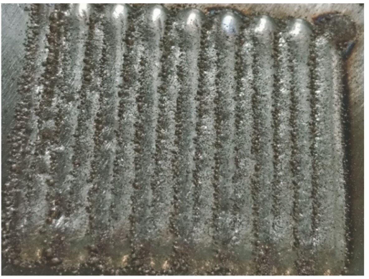
    Fig. 1. Surface of 316L coating when energy density is less than or equal to 19 J·mm-2
    Microstructures of 316L coating. (a) Cross-sectional morphology; (b) microstructure of cellular crystal; (b) microstructure of columnar crystal; (d) microstructure of overlapping zone
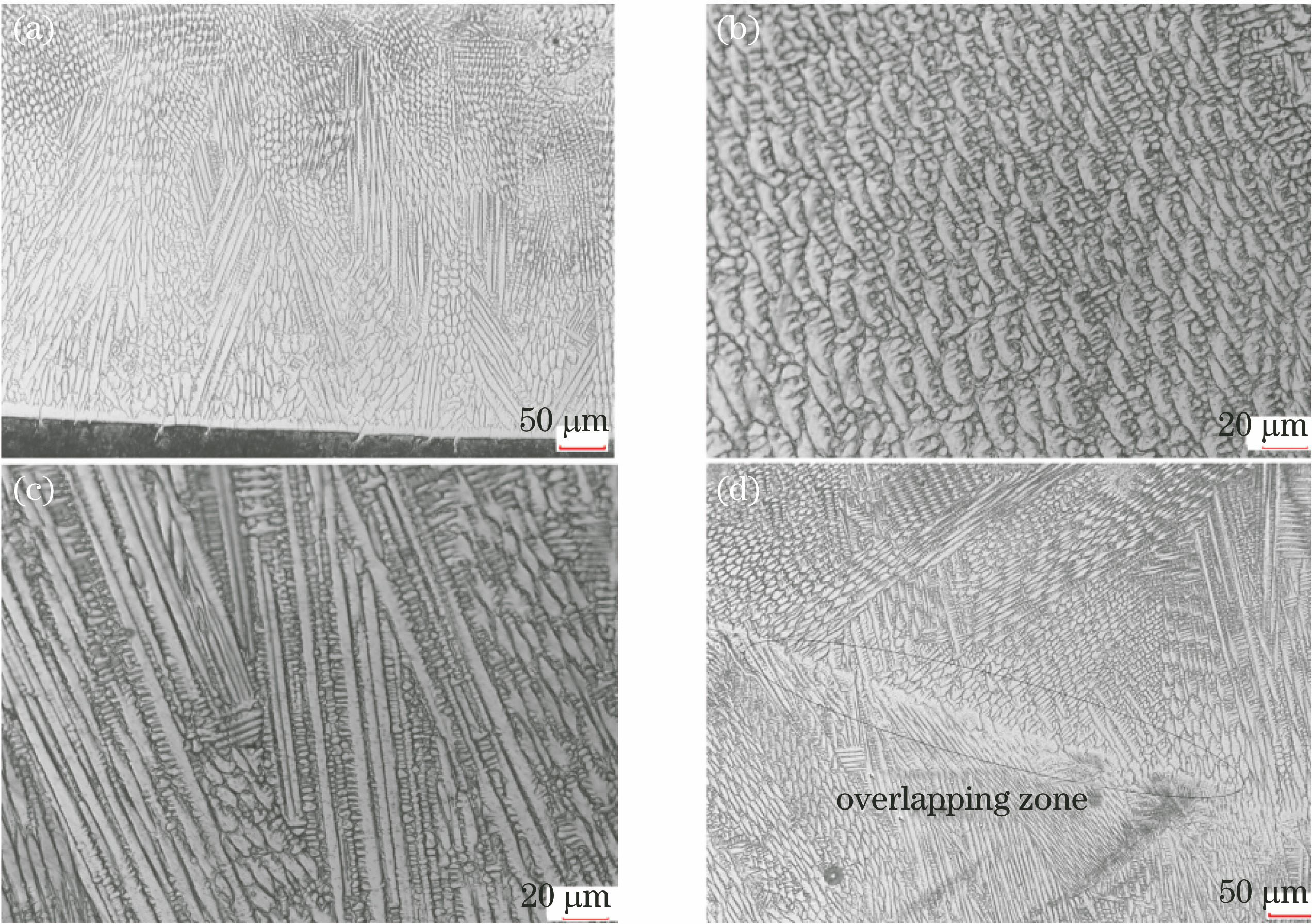
    Fig. 2. Microstructures of 316L coating. (a) Cross-sectional morphology; (b) microstructure of cellular crystal; (b) microstructure of columnar crystal; (d) microstructure of overlapping zone
    Relationship between hardness and energy density of 316L coating and schematic of microstructures. (a) Hardness of coating under different energy densities; (b)-(d) microstructures under energy densities of 19, 28, and 44 J·mm-2, respectively
    Fig. 3. Relationship between hardness and energy density of 316L coating and schematic of microstructures. (a) Hardness of coating under different energy densities; (b)-(d) microstructures under energy densities of 19, 28, and 44 J·mm-2, respectively
    SEM image of 316L coating under magnification of 3000
    Fig. 4. SEM image of 316L coating under magnification of 3000
    Polarization curves. (a) Substrate; (b) 316L coating
    Fig. 5. Polarization curves. (a) Substrate; (b) 316L coating
    Corrosion morphologies of substrate and 316L coating after suffering acid salt spray tests with different time. (a) Substrate after 16 h; (b) 316L coating after 16 h; (c) 316L coating after 96 h; (d) 316L coating after 350 h
    Fig. 6. Corrosion morphologies of substrate and 316L coating after suffering acid salt spray tests with different time. (a) Substrate after 16 h; (b) 316L coating after 16 h; (c) 316L coating after 96 h; (d) 316L coating after 350 h
    Micro corrosion morphologies of 316L coating after suffering acid salt spray for 350 h. (a) 100×; (b) 500×
    Fig. 7. Micro corrosion morphologies of 316L coating after suffering acid salt spray for 350 h. (a) 100×; (b) 500×
    FeCCrMoSiMnNi
    Bal.≤0.0816-182-31210-14
    Table 1. Elemental compositions of 316L metal powder (mass fraction, %)
    Power /WScanningspeed /(mm·s-1)Powderfeeding /(r·min-1)Energydensity /(J·mm-2)
    2200101.244.00
    2800202.028.00
    3400303.422.67
    3800402.419.00
    3800404.019.00
    3800503.015.20
    Table 2. Processing parameters of 316L metal powder
    PositionCrFeNiMo
    P117.2066.4213.233.16
    P218.9265.0313.043.01
    Table 3. Elemental contents of P1 and P2 points (mass fraction, %)
    Peng Liu, Zhikai Chen, Quanming Jin, Qinghai Zhu. Microstructure and Corrosion Behaviors of 316L Coating Fabricated by Laser Cladding[J]. Laser & Optoelectronics Progress, 2020, 57(3): 031402
    Download Citation