• Opto-Electronic Advances
  • Vol. 6, Issue 7, 230007 (2023)
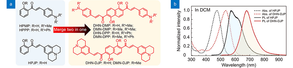
Chang-Cun Yan1,2,3,†, Zong-Lu Che2,†, Wan-Ying Yang2, Xue-Dong Wang2,*, and Liang-Sheng Liao1,2,*
Author Affiliations
  • 1Macao Institute of Materials Science and Engineering, Macau University of Science and Technology, Taipa, Macau SAR 999078, China
  • 2Institute of Functional Nano & Soft Materials, Jiangsu Key Laboratory for Carbon-Based Functional Materials & Devices, Joint International Research Laboratory of Carbon-Based Functional Materials and Devices, Soochow University, Suzhou 215123, China
  • 3Jiangsu Engineering Laboratory of Novel Functional Polymeric Materials, Jiangsu Key Laboratory of Advanced Negative Carbon Technologies, College of Chemistry, Chemical Engineering and Materials Science, Soochow University, Suzhou 215123, China
  • show less
    DOI: 10.29026/oea.2023.230007 Cite this Article
    Chang-Cun Yan, Zong-Lu Che, Wan-Ying Yang, Xue-Dong Wang, Liang-Sheng Liao. Deep-red and near-infrared organic lasers based on centrosymmetric molecules with excited-state intramolecular double proton transfer activity[J]. Opto-Electronic Advances, 2023, 6(7): 230007 Copy Citation Text show less
    (a) Chemical structures of the template and target compounds. (b) The normalized UV-vis absorption and PL spectra of HPJP and DHN-DJP in DCM solutions.
    Fig. 1. (a) Chemical structures of the template and target compounds. (b) The normalized UV-vis absorption and PL spectra of HPJP and DHN-DJP in DCM solutions.
    (a) Diagram of the ESDPT process in DHNs. (b) Calculated relative energies (kcal/mol) on S0 and S1 of DHN-DMP in vacuum.
    Fig. 2. (a) Diagram of the ESDPT process in DHNs. (b) Calculated relative energies (kcal/mol) on S0 and S1 of DHN-DMP in vacuum.
    (a, c, e) The normalized UV‒vis absorption and PL spectra of DMN- and DHN-doped PS films. (b, d, f) The decay plots and fitted curves of DHNs.
    Fig. 3. (a, c, e) The normalized UV‒vis absorption and PL spectra of DMN- and DHN-doped PS films. (b, d, f) The decay plots and fitted curves of DHNs.
    (a) Schematic diagram of a single DHN-doped microsphere. (b) PL micrograph of a single DHN-doped microsphere. Inset: scanning electron microscopy image of a single DHN-doped microsphere. (c) Partial magnifications of PL spectra of DHN-doped microspheres with different sizes. (d) The related curve of λ2/Δλ (λ: emission wavelength; Δλ: the space between the individual resonance peaks) at 700 nm versus D (D: diameter of selected microsphere). Inset: the simulated electric energy density in the cross-section of a microsphere with dimeter D = 10 µm. Red corresponds to the highest field density and blue is the lowest field density.
    Fig. 4. (a) Schematic diagram of a single DHN-doped microsphere. (b) PL micrograph of a single DHN-doped microsphere. Inset: scanning electron microscopy image of a single DHN-doped microsphere. (c) Partial magnifications of PL spectra of DHN-doped microspheres with different sizes. (d) The related curve of λ2λ (λ: emission wavelength; Δλ: the space between the individual resonance peaks) at 700 nm versus D (D: diameter of selected microsphere). Inset: the simulated electric energy density in the cross-section of a microsphere with dimeter D = 10 µm. Red corresponds to the highest field density and blue is the lowest field density.
    PL spectra of (a) DHN-DMP-, (d) DHN-DPP- and (g) DHN-DJP-doped PS microspheres under different pump densities. (b) Plots of lasing intensity as a function of pump density of a (b) DHN-DMP-, (e) DHN-DPP- and (h) DHN-DJP-doped PS microsphere. Insets: brightfield micrographs of the PS microspheres used in laser measurements. (c) 2D mappings of lasing intensity versus the number of pulses of a (c) DHN-DMP-, (f) DHN-DPP- and (i) DHN-DJP-doped PS microsphere, pumping density: 47.8 µJ/cm2.
    Fig. 5. PL spectra of (a) DHN-DMP-, (d) DHN-DPP- and (g) DHN-DJP-doped PS microspheres under different pump densities. (b) Plots of lasing intensity as a function of pump density of a (b) DHN-DMP-, (e) DHN-DPP- and (h) DHN-DJP-doped PS microsphere. Insets: brightfield micrographs of the PS microspheres used in laser measurements. (c) 2D mappings of lasing intensity versus the number of pulses of a (c) DHN-DMP-, (f) DHN-DPP- and (i) DHN-DJP-doped PS microsphere, pumping density: 47.8 µJ/cm2.
    Chang-Cun Yan, Zong-Lu Che, Wan-Ying Yang, Xue-Dong Wang, Liang-Sheng Liao. Deep-red and near-infrared organic lasers based on centrosymmetric molecules with excited-state intramolecular double proton transfer activity[J]. Opto-Electronic Advances, 2023, 6(7): 230007
    Download Citation