
Yingqi Liang, Guobin Mao, Junbiao Dai, Yingxin Ma. Biofunctionalized semiconductor quantum dots for virus detection[J]. Journal of Semiconductors, 2023, 44(2): 023101
- Journal of Semiconductors
- Vol. 44, Issue 2, 023101 (2023)
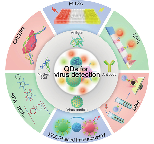
Fig. 1. (Color online) Summary of potential applications of semiconductor quantum dots for virus detection.
![(Color online) The different QDs based on types of ligands used for synthesis. (a) One-step preparation of DNA-QDs using Na2TeO3 as the tellurium source and N-acetyl-L-cysteine as the surface ligand[46]. (b) Amino group and tetramercaptan as the ligands for QDs synthesis using photoligation strategy[53]. (c) Imidazole and dithiol co-stabilized QDs to maintain strong interaction between dithiol and the metal ions and cause anti-oxidation of imidazole[37]. Modified with permission from (a) Ref. [46] Copyright 2014 American Chemical Society, (b) Ref. [53] Copyright 2013 American Chemical Society and (c) Ref. [37] Copyright 2015 American Chemical Society.](/richHtml/jos/2023/44/2/023101/jos_44_2_023101_f2.jpg)
Fig. 2. (Color online) The different QDs based on types of ligands used for synthesis. (a) One-step preparation of DNA-QDs using Na2TeO3 as the tellurium source and N-acetyl-L-cysteine as the surface ligand[46]. (b) Amino group and tetramercaptan as the ligands for QDs synthesis using photoligation strategy[53]. (c) Imidazole and dithiol co-stabilized QDs to maintain strong interaction between dithiol and the metal ions and cause anti-oxidation of imidazole[37]. Modified with permission from (a) Ref. [46] Copyright 2014 American Chemical Society, (b) Ref. [53] Copyright 2013 American Chemical Society and (c) Ref. [37] Copyright 2015 American Chemical Society.
Fig. 3. (Color online) Biofunctionalization of QDs. (a) The synthesis of hyaluronic acid functionalized QDs through electrostatic interaction[54]. (b) The preparation of azide-DNA functionalized QDs for GOx labeling through copper-free catalytic click reaction for the blood glucose detection[62]. (c) The design of two-dimensional QD molecular beacons through the coordination interaction of Cd2+ and dithiol[65]. Modified with permission from (a) Ref. [54] Copyright 2009 American Chemical Society, (b) Ref. [60] Copyright 2021 American Chemical Society and (c) Ref. [62] Copyright The Royal Society of Chemistry 2015.
Fig. 4. (Color online) Construction of the nucleic acid hybridization probe for virus detection. (a) QD-DNA complexes were prepared by electrostatic interaction between Dabcyl modified DNA and carbon QDs[71]. (b) π–π stacking interactions between CNTs and aromatic nucleotide in DNA functionalized CdTe QDs[18]. (c) The conjugation of the plasmonic Au NPs and CdSeS QDs through the covalent coupling to form the plasmonic Au NP-QDs nanohybrids for Zika virus detection[75]. (d) The preparation of QD-NBs and the detection of HIV-1 genomic RNAs detection in the living cell[66]. Modified with permission from (a) Ref. [68] 2022 Elsevier B.V. All rights reserved, (b) Ref. [69] 2012 Elsevier B.V. All rights reserved, (c) Ref. [72] 2017 Elsevier B.V. All rights reserved and (d) Ref. [63] Copyright 2019 American Chemical Society.
Fig. 5. (Color online) Distinct kinds of nucleic acid amplification method for virus detection. (a) The testing workflow of a rapid PCR lateral flow assay for the simultaneous detection of SARS- CoV-2 and influenza B virus. The nucleic acid was amplified by the rapid water bath RT-PCR, and the labeled amplicons were detected by using test strips[77]. (b) The one-step loop-mediated isothermal amplification (LAMP) assay with cysteamine-modified CdSeS/ZnS QDs for SARS-CoV-2 detection. LAMP produced the negatively charged polyphosphate, which can aggregate with the positively charged cysteamine modified CdSeS/ZnS QDs lead to fluorescence quenching of QDs[80]. (c) HCV RNA ultrasensitive determination based on CHA-induced the in-situ degradation of silver nanocomposite in GQD/Ag NCs to recover the fluorescence[82]. Modified with permission from (a) Ref. [74] Copyright The Royal Society of Chemistry 2022, (b) Ref. [77] Copyright 2022 American Chemical Society and (c) Ref. [79] 2022 Elsevier B.V. All rights reserved.
Fig. 6. (Color online) Signal enhancement methods of the probes for virus detection. (a) Detection of attomolar HIV-1 DNA using encapsulated QDs-encapsulated liposome complexes and single-particle detection techniques[83]. (b) The simultaneous detection of HIV-1 and HIV-2 via QD layer-by-layer assembled polystyrene microsphere by the interaction of SA and biotin[84]. (c) One-step simultaneous detection of EV71 infection-related miRNA based on DSN-assisted target amplification followed by the magnetic separation[88]. (d) The detection of African swine fever virus by combining CRISPR-Cas mediated nucleic acid probe cleavage and highly sensitive reporter of QDs[90]. (e) SARS-CoV-2 detection by combination of reverse-transcription recombinase-aided amplification assisted-CRISPR/Cas13a with QDs microspheres[91]. Modified with permission from (a) Ref. [80] Copyright 2013 American Chemical Society, (b) Ref. [81] Copyright 2015 John Wiley & Sons, Ltd, (c) Ref. [82] Copyright 2020 American Chemical Society, (d) Ref. [84] Copyright 2020 American Chemical Society and (e) Ref. [85] 2022 Elsevier B.V. All rights reserved.
Fig. 7. (Color online) Heterogeneous immunoassay. ELISA: (a) Fluorescence ELISA based on GOx-induced fluorescence quenching of H2O2-sensitvie CdTe QDs for the surface antigen detection of hepatitis B[94]; (b) Dual-modal immunoassay of rabies virus based on QDs and HRP-Ab co-modified silica nanospheres. The colorimetric signal was obtained by HRP-catalyzed TMA oxidation and the fluorescence signal was captured from loading QDs[95]; (c) Photoelectrochemical immunoassay of HIV-1 p24 antigen using DNA as ELISA label tag. Following the sandwich immunobinding, the DNA tags could be released and subsequent dipurinization of oligonucleotide strands could enable the oxidation of free nucleobases at a CdTe QDs modified ITO transducer[96]. MBIA: (d) Magnetic immunoassay of H1N1 virus based on immune-complex formation between QD-embedded silica nanoparticle and magnetic beads[97]; (e) The preparation of CdSeTeS QD/AuNPs nanocomposites and the close covalent attachment of AuNPs with CdSeTeS QDs effectively quenched the fluorescence signal which was recovered after NoV-LPs entrapment[98]; (f) Digital single virus immunoassay for the multiplex virus detection using fluorescent magnetic multifunctional nanospheres (FMNs) as both capture carriers and signal labels[100]. LFIA: (g) The fabrication of SiTQD probes and their application in ICA-based biosensor for the simultaneous detection of SARS-CoV-2 and FluA[102]; (h) SARS-CoV-2 spike protein detection by fabricating dual-functional SiO2@Au/QD labels. The SiO2@Au/QD labels were constructed by the layer-by-layer electrostatic interaction of SiO2@PEI, Au NPs, and QDs[103]. Modified with permission from (a) Ref. [88] 2018 Elsevier B.V. All rights reserved, (b) Ref. [89] Copyright 2020 American Chemical Society, (c) Ref. [90] Copyright 2015 American Chemical Society, (d) Ref. [91] 2020 Elsevier B.V. All rights reserved, (e) Ref. [92] 2018 Elsevier B.V. All rights reserved, (f) Ref. [94] Copyright 2019 American Chemical Society, (g) Ref. [96] 2021 Elsevier B.V. All rights reserved and (h) Ref. [97] 2021 Elsevier B.V. All rights reserved.
Fig. 8. (Color online) Homogeneous immunoassay. FRET: (a) EV71 detection caused by the interaction of QDs and Ru-Ab complex[104]; (b) EV71 and CVB3 simultaneous determination by the interaction of dual-color QDs-Ab complex and GO[105]; (c) Norovirus biosensor developed by combining LSPR from Au NPs to CdSeTeS QDs which blocked FRET from QDs to Au NPs[98]. QDSMs: (d) QDs-loaded spore-based monodisperse microparticles for immunoassay of parvovirus antibody via flow cytometry[106]. UCAD: (e) The workflow for the proximity-induced CRISPR-based SRAS-CoV-2 antibody detection. Proximity binding of the DNA probes to the SRAS-CoV-2 antibody can induce the dsDNA formation, primer extension and RPA, and CRISPR/Cas12a system to produce the fluorescence signal[108]. Modified with permission from (a) Ref. [98] Copyright 2011 American Chemical Society, (b) Ref. [99] Copyright 2012 American Chemical Society, (c) Ref. [92] 2015 Elsevier B.V. All rights reserved, (d) Ref. [100] Copyright 2022 The Author(s) Springer Nature and (e) Ref. [102] Copyright 2022 The Author(s) Springer Nature.

Set citation alerts for the article
Please enter your email address
© Copyright 2018-2021 | Chinese Laser Press. All Rights Reserved 沪ICP备15018463号-20