
Wei Luo, Tai Li, Yongde Li, Houjin Wang, Ye Yuan, Shangfeng Liu, Weiyun Wang, Qi Wang, Junjie Kang, Xinqiang Wang. Watts-level ultraviolet-C LED integrated light sources for efficient surface and air sterilization[J]. Journal of Semiconductors, 2022, 43(7): 072301
- Journal of Semiconductors
- Vol. 43, Issue 7, 072301 (2022)
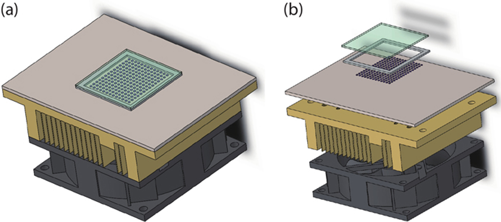
Fig. 1. (Color online) Schematic of UVC-LED ILS: (a) assembly diagram, (b) explosive diagram.
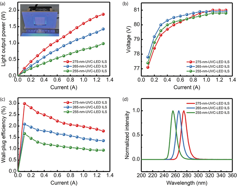
Fig. 2. (Color online) Characterization of three UVC-LED ILSs: (a) LOP vs. current, (b) voltage vs. current, (c) WPE vs. current, (d) EL spectra.
Fig. 3. (Color online) (a) Irradiance at different distances from the 275-nm-UVC-LED ILS under an injection current of 0.8 A. The inset shows that the reciprocal of irradiance is approximately proportional to the square of distance. (b) Irradiance distribution of the central axis of light-emitting area (x = 0 at the center of light-emitting area). (c) Temperature change with time after the 275-nm-UVC-LED ILS was turned on. The inset shows the locations for temperature measurements: p1 and p2 are the center and the edge of the quartz glass on light-emitting area, respectively, and p3 is located on AlN ceramic substrate and close to the light-emitting area. (d) Irradiance at a distance of 5 cm from the light-emitting surface of the 275-nm-UVC-LED ILS under different current injection.
Fig. 4. Micrographs of the Microorganism Colonies of (a, d) Escherichia coli, (b, e) Staphylococcus aureus, (c, f) Candida albicans before (a, b, c) and after (d, e, f) one-second sterilization by the 275-nm-UVC-LED ILS.
Fig. 5. (Color online) (a) Schematic diagram and (b) photo of the air sterilization module based on 275-nm-UVC-LED ILS. (c) Photo of UVC-LED array viewed from the inlet direction.
| ||||||||||||||||||||||||||||||||||||||||||||||
Table 0. Sterilization effects of the air sterilization module based on the 275-nm-UVC-LED ILS on Staphylococcus albus in the air of 20 m3 confined room.
|
Table 0. Sterilization effects of three UVC-LED ILSs with different light-emitting wavelengths on Staphylococcus aureus.
|
Table 0. Sterilization effects of the 275-nm-UVC-LED ILS on different microorganisms.

Set citation alerts for the article
Please enter your email address
© Copyright 2018-2021 | Chinese Laser Press. All Rights Reserved 沪ICP备15018463号-20