
Jianwei Chen, Hui Gong, Jing Yuan. Multispectral Imaging Technology and Its Applications in Biomedicine[J]. Laser & Optoelectronics Progress, 2021, 58(4): 0400001
- Laser & Optoelectronics Progress
- Vol. 58, Issue 4, 0400001 (2021)
![MSI systems based on different spectral separation elements[19]. (a) Rotating filter; (b) liquid crystal tunable filter; (c) planar compound eye imaging system; (d) multi-layer curved compound eye MSI system](/richHtml/lop/2021/58/4/0400001/img_1.jpg)
Fig. 1. MSI systems based on different spectral separation elements[19]. (a) Rotating filter; (b) liquid crystal tunable filter; (c) planar compound eye imaging system; (d) multi-layer curved compound eye MSI system
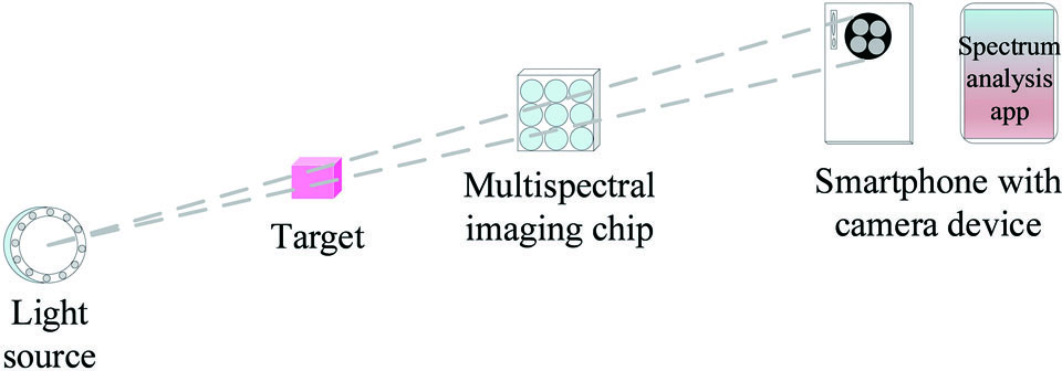
Fig. 2. Smartphone-based portable MSI system
Fig. 3. Multispectral microscopic imaging systems. (a) Multi-wavelength fluorescent microscopy; (b) fiber-based multispectral endomicroscopy; (c) multispectral optoacoustic tomography
Fig. 4. Multilayer composite metasurfaces used for multispectral achromatic imaging[37]. (a) Diagram of multilayer composite structure; (b) spectrum taken under white light illumination and distribution of focal spot of red light (650 nm); (c) foci of three characteristic peaks
Fig. 5. MSI technology used for skin tissue detection. (a) Multispectral imaging for skin slice via NIR MSI to distinguish healthy tissues from diseased tissues[51]; (b) MSOT for in vivo detection of skin melanin and blood oxygenation level[52]
Fig. 6. Assessment of breast tumor margins via MSOT[59]. (a) Ultrasound image of the excised specimen. Dashed area is tumor tissue; (b) multispectral optoacoustic image shows lipids and deoxyhemoglobin; (c) enlarged view shows that deoxyhemoglobin signal can be used to identify tumor margin
Fig. 7. Multispectral biometric feature recognition. (a) Extract and analyze multispectral characteristics of conjunctival vessels and iris textures[66]; (b) quaternion model for multispectral palmprint information recognition[74]; (c) hierarchical fusion of multi-spectral images and feature of face for face recognition[83]

Set citation alerts for the article
Please enter your email address
© Copyright 2018-2021 | Chinese Laser Press. All Rights Reserved 沪ICP备15018463号-20