Journals >Chinese Journal of Lasers
Contents


2020
Volume: 47 Issue 2
43 Article(s)
Export citation format
[in Chinese]

Introduction for Special Issue on Sixty-Year Crossing and Integration for Laser and Biomedicine
[in Chinese], [in Chinese], and [in Chinese]
Chinese Journal of Lasers
- Publication Date: Jan. 01, 1900
- Vol. 47, Issue 2, 207000 (2020)

Biomedical Photonics and Laser Medicine
Mueller Matrix Polarimetry: A Label-Free, Quantitative Optical Method for Clinical Diagnosis
Shen Yuanxing, Yao Yue, He Honghui, Liu Shaoxiong, and Ma Hui
Of late, with the emergence of new optical devices and technological advances in data processing, polarization techniques are being increasingly used in biomedicine. Mueller matrix calculus is suitable for describing the polarization properties of biomedical specimens because of its mathematical completeness and compatOf late, with the emergence of new optical devices and technological advances in data processing, polarization techniques are being increasingly used in biomedicine. Mueller matrix calculus is suitable for describing the polarization properties of biomedical specimens because of its mathematical completeness and compatibility with common optical equipment. Compared with traditional non-polarization optical methods, Mueller matrix polarimetry is sensitive to the scattering induced by subwavelength structures and can provide more information about anisotropic optical properties, including the birefringence and diattenuation of a sample. In this review, we introduce Mueller matrix calculus and related technologies that have great application potential in biomedical studies, including the Mueller matrix decomposition and transformation methods, transmission Mueller matrix microscopes, backscattering Mueller matrix imaging equipment, Mueller matrix endoscopes, and polarization staining techniques. Further, we summarize the improvements in clinical diagnosis made using Mueller matrix polarimetry, such as detection of liver cancer, gastrointestinal cancer, and breast ductal carcinoma tissues. As a label-free, noninvasive, quantitative, and rapid imaging method, Mueller matrix polarimetry has broad application prospects in biomedical studies and clinical diagnosis..
Chinese Journal of Lasers
- Publication Date: Feb. 01, 2020
- Vol. 47, Issue 2, 207001 (2020)
Accurate Characterization of Spatial Orientations of Fiber-Like Structures in Biological Tissues and Its Applications
Liu Zhiyi, Meng Jia, Qiu Jianrong, Han Tao, Wang Di, Zhuo Shuangmu, and Ding Zhihua
Fiber-like structure is one of the basic structures found in biological tissues. The spatial orientations of fiber-like structures change with the initiation and progression of some diseases. In this study, we present a brief overview of quantitative orientation analysis methods for fiber-like structures within biologiFiber-like structure is one of the basic structures found in biological tissues. The spatial orientations of fiber-like structures change with the initiation and progression of some diseases. In this study, we present a brief overview of quantitative orientation analysis methods for fiber-like structures within biological tissues and main applications of these methods. We especially focus on the research progress of spatial orientation information in important disease models, including wound healing, osteoarthritis, breast cancer, peritoneal metastasis, and brain injury. Additionally, we explore the relations between tissue structure and function via specific engineered tissues. A highly sensitive and highly accurate description of the fiber-like structures within biological tissues serves as a novel method for studying disease initiation and progression, shows potential for early disease diagnosis, and improves our understanding of the mechanisms underlying some disorders. Finally, future potential applications of the orientation analysis methods are explored..
Chinese Journal of Lasers
- Publication Date: Feb. 01, 2020
- Vol. 47, Issue 2, 207002 (2020)
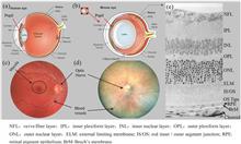
Review of Advances in Ophthalmic Optical Imaging Technologies from Several Mouse Retinal Imaging Methods
Zhang Pengfei, Zhang Tingwei, Song Weiye, Lu Yiming, and Jian Yifan
Animal studies play very important roles in the basic science research. Recently, several optical imaging methods, which are widely adopted in human retinal imaging, have been successfully applied into animal retina researches. By providing high-resolution cellular details about the retina without the need for histologAnimal studies play very important roles in the basic science research. Recently, several optical imaging methods, which are widely adopted in human retinal imaging, have been successfully applied into animal retina researches. By providing high-resolution cellular details about the retina without the need for histological section, these optical imaging methods provide powerful tools for researchers to study using the animal retina. Correspondingly, several new technologies have been developed for animal retina researches, which are also applicable for human retinal imaging, or provide new insights in understanding the human retina function mechanism. Based on their own research experiences in in vitro optical imaging methods for mouse retinas, the authors review several latest break-through technology developments in animal and human retinal imaging with high resolution and with a focus on demonstration of the “can-do” ability of the current technologies, hoping it will provide new insights to promote the advancements for ophthalmic imaging in both species..
Chinese Journal of Lasers
- Publication Date: Feb. 01, 2020
- Vol. 47, Issue 2, 207003 (2020)
Optical Coherence Microscopy and Its Application
Han Tao, Qiu Jianrong, Wang Di, Meng Jia, Liu Zhiyi, and Ding Zhihua
Optical coherence microscopy (OCM) utilizes a coherent detection method for optical microscopy. Having advantages, such as high axial resolution, high signal-to-noise ratio, and label-free imaging in optical coherence tomography (OCT), the OCM can achieve a micron-scale spatial resolution by utilizing a high-power objeOptical coherence microscopy (OCM) utilizes a coherent detection method for optical microscopy. Having advantages, such as high axial resolution, high signal-to-noise ratio, and label-free imaging in optical coherence tomography (OCT), the OCM can achieve a micron-scale spatial resolution by utilizing a high-power objective for obtaining high lateral resolution. Initially, the basic principle and implementation scheme of the OCM technology are introduced; subsequently, the principles and research progress of the OCM technology around the world are summarized. This study also examines some advanced OCM technologies considering the problems that how to realize ultra-high-resolution imaging and that the depth of focus limits the imaging depth. The OCM technology has broad application prospect in biomedicine, material detection, and other fields..
Chinese Journal of Lasers
- Publication Date: Feb. 01, 2020
- Vol. 47, Issue 2, 207004 (2020)

Coherent Raman Scattering Microscopy Technique and Its Biomedical Applications
Li Zilin, Li Shaowei, Zhang Silu, Shen Binglin, Qu Junle, and Liu Liwei
As a new imaging technique, coherent Raman scattering (CRS) microscopy has been widely used in chemical structure and composition analysis because of its advantages of label-free, high specificity, and on-invasion. In recent years, the mutual cross-over and integrated development of photonics, biomedicine, and microscoAs a new imaging technique, coherent Raman scattering (CRS) microscopy has been widely used in chemical structure and composition analysis because of its advantages of label-free, high specificity, and on-invasion. In recent years, the mutual cross-over and integrated development of photonics, biomedicine, and microscopic imaging technology has greatly promoted the application of CRS microscopy in biomedicine. This paper briefly introduces the basic principle of CRS microscopy and its classification, then explains the most widely used implementation of CRS, and summarizes the recent applications of CRS microscopic imaging in biomedicine, including detection, lipid analysis, and protein conformation change. Finally, the future development of CRS is discussed..
Chinese Journal of Lasers
- Publication Date: Feb. 01, 2020
- Vol. 47, Issue 2, 207005 (2020)
Advances in Optical Imaging for Monitoring Photodynamic Therapy Dosimetry
Li Wenbo, Shen Yi, and Li Buhong
Photodynamic therapy (PDT) has been widely used as a precise targeted therapeutic modality in the clinical treatments of malignant tumor and benign diseases. The utilization of advanced optical imaging techniques in the real-time quantification of PDT dosimetric parameters is essential in predicting PDT efficiency and Photodynamic therapy (PDT) has been widely used as a precise targeted therapeutic modality in the clinical treatments of malignant tumor and benign diseases. The utilization of advanced optical imaging techniques in the real-time quantification of PDT dosimetric parameters is essential in predicting PDT efficiency and providing personalized and precise treatment. In this study, four important parameters in PDT dosimetry were introduced (i.e., photosensitizer, ground-state oxygen, singlet oxygen, and vascular response). Furthermore, the advanced optical imaging techniques currently developed for monitoring the aforementioned dosimetric parameters are summarized, and the advantages and limitations of each optical imaging technique is comparatively analyzed. Finally, challenges in clinical translation of optical imaging techniques to the clinical application of PDT are briefly discussed..
Chinese Journal of Lasers
- Publication Date: Feb. 01, 2020
- Vol. 47, Issue 2, 207006 (2020)
Review of Tissue Optical Clearing Methods for Imaging Whole Organs
Yu Tingting, and Zhu Dan
The technological development in modern optical imaging and fluorescent labeling has provided important tools for obtaining high-resolution three-dimensional (3D) structural information about biological tissues. However, the opaque nature of most biological tissues limits the light penetration depth, which limits theirThe technological development in modern optical imaging and fluorescent labeling has provided important tools for obtaining high-resolution three-dimensional (3D) structural information about biological tissues. However, the opaque nature of most biological tissues limits the light penetration depth, which limits their applications for large tissue specimens or organs. In recent years, optical clearing methods that employ various physical and chemical means have been proposed for reducing light attenuation and improving the imaging depth, thereby providing novel perspectives for 3D imaging of large samples or whole organs. This paper reviews the tissue optical clearing methods for imaging whole organs from three aspects: in vitro optical clearing methods for tissues, whole-mount labeling methods, and 3D imaging techniques..
Chinese Journal of Lasers
- Publication Date: Feb. 01, 2020
- Vol. 47, Issue 2, 207007 (2020)
Application of Second Harmonic Generation in Biomedical Imaging
Zhang Ziyi, Wang Mingxue, Liu Zhihe, Fang Xiaofeng, and Wu Changfeng
Second harmonic generation (SHG) is a novel optical imaging technology that has been recently developed. SHG has attracted considerable attention as a new tool for the biological structure and durable tracking. The SHG technology eliminates many disadvantages associated with the classical fluorescent probes. It is an iSecond harmonic generation (SHG) is a novel optical imaging technology that has been recently developed. SHG has attracted considerable attention as a new tool for the biological structure and durable tracking. The SHG technology eliminates many disadvantages associated with the classical fluorescent probes. It is an ideal in vivo imaging method and exhibits good biomedical application prospects. In this study, we introduce the principle of SHG and its imaging device, classify the SHG media, review the application of SHG in biomedical imaging, and prospect future opportunities and challenges..
Chinese Journal of Lasers
- Publication Date: Feb. 01, 2020
- Vol. 47, Issue 2, 207008 (2020)
Quantitative Fluorescence Resonance Energy Transfer Measurement Based on Spectral Unmixing
Yin Ao, and Chen Tongsheng
Owing to its inherent ability to overcome the spectral crosstalk, high sensitivity, and non-destructivity characteristic, fluorescence resonance energy transfer (FRET) quantitative measurement (spFRET) method based on spectral unmixing has been generally regarded as the most promising approach of live-cell FRET measureOwing to its inherent ability to overcome the spectral crosstalk, high sensitivity, and non-destructivity characteristic, fluorescence resonance energy transfer (FRET) quantitative measurement (spFRET) method based on spectral unmixing has been generally regarded as the most promising approach of live-cell FRET measurement. This paper first briefly introduced the quantitative FRET measurement method and the related research advances on FRET technology. Second, the principle, development process, and robustness of spFRET based on linear separation of emission spectra (Em-unmixing) and linear separation of excitation emission spectra (ExEm-unmixing), respectively, were introduced. Finally, the potential advantages of the two spFRET technologies in live-cell FRET applications was also provided and discussed..
Chinese Journal of Lasers
- Publication Date: Feb. 01, 2020
- Vol. 47, Issue 2, 207009 (2020)
Optogenetics Based on Light-Gated Protein-Protein Interactions and Its Applications
Huang Peiyuan, Song Yutong, Zhang Ning, Zhao Zhihao, and Duan Liting
Optogenetics is an emerging technique that exploits light to control cells by combining optics and genetics techniques. In optogenetic systems, cells are genetically modified to express photosensitive proteins and consequently become responsive to light pulses. Optogenetics has revolutionized neuroscience research by fOptogenetics is an emerging technique that exploits light to control cells by combining optics and genetics techniques. In optogenetic systems, cells are genetically modified to express photosensitive proteins and consequently become responsive to light pulses. Optogenetics has revolutionized neuroscience research by facilitating selective and rapid control of targeted neurons expressing light-gated ion channels. In addition to light-gated ion channels, photosensitive proteins based on light-gated protein-protein interactions are widely used in optogenetic research. In this review, we discuss these common photosensitive proteins and summarize optogenetic applications in optical control of gene expression, phase separation, biosynthesis, and organelle distribution based on light-gated protein-protein interactions..
Chinese Journal of Lasers
- Publication Date: Feb. 01, 2020
- Vol. 47, Issue 2, 207010 (2020)
Optical Nanobiosensors with Different Structures and Their Applications in Tumor Screening
Jiang Tianshu, Zhang Ruotong, Dong Changzi, Jin Weiqiu, Jia Minglong, and Zhang Zhenxi
Tumor is an important problem that modern medicine needs to overcome. Early screening and treatment of tumor require immediate attention in clinical practice. Therefore, this paper describes the basic principles and detection characteristics of several common tumor marker nanobiosensors based on nanoparticle, nanowire,Tumor is an important problem that modern medicine needs to overcome. Early screening and treatment of tumor require immediate attention in clinical practice. Therefore, this paper describes the basic principles and detection characteristics of several common tumor marker nanobiosensors based on nanoparticle, nanowire, nanotube, and nanoarray material. Core-shell nanoparticles have abundant modification function. Nanowires are often made into field-effect tube to detect tumor markers. Based on the scale effect, nanotubes are primarily used in transport of carriers and detection platform. Metal and metal-oxide nanoarrays can detect cancer cells using the principle of electrochemical impedance spectroscopy. In addition to the advantages and application characteristics determined by different structures, nanobiosensors have the overall advantages of rapid and convenient detection of tumor cells and low detection limit compared with traditional detection methods. Therefore, nanobiosensors offer great potential in medical detection and tumor research..
Chinese Journal of Lasers
- Publication Date: Feb. 01, 2020
- Vol. 47, Issue 2, 207011 (2020)
Recent Advances in Nanophotosensitizers for Antibacterial Photodynamic Therapy
Zhang Chang, Ren En, Pang Xin, Li Lei, and Liu Gang
Antibacterial photodynamic therapy (APDT), which is a non-invasive treatment method, is based on the interaction between near-infrared light and a nontoxic photosensitizer concentrated at the lesion site to generate reactive oxygen species (ROS). These species are highly cytotoxic in virtually all bacteria. With the deAntibacterial photodynamic therapy (APDT), which is a non-invasive treatment method, is based on the interaction between near-infrared light and a nontoxic photosensitizer concentrated at the lesion site to generate reactive oxygen species (ROS). These species are highly cytotoxic in virtually all bacteria. With the development of biomaterials and nanotechnology, advances in nano-biotechnology have resulted in the optimization of biocompatibility and biosafety of small-molecule photosensitizers. The targeting ability has improved and the quantum yield under illumination has significantly increased. Nanotechnology exhibits excellent clinical application prospects with respect to antimicrobial therapy. In this review, recent applications and developments of APDT are summarized by combining various modification strategies and mechanisms for nanophotosensitizers..
Chinese Journal of Lasers
- Publication Date: Feb. 01, 2020
- Vol. 47, Issue 2, 207012 (2020)
Probes for Endoscopic Optical Coherence Tomography: Minimized Design and Depth of Focus Extension
Qiu Jianrong, Han Tao, Wang Di, Meng Jia, Liu Zhiyi, and Ding Zhihua
Minimized probe is a common requirement in endoscopic optical coherence tomography (OCT). We introduce the development of mainstream designs based on ball lens, fiber lens, graded index fiber, free form lens and free-lens, summarize their advantages and disadvantages, and put forward some suggestions for miniaturizatioMinimized probe is a common requirement in endoscopic optical coherence tomography (OCT). We introduce the development of mainstream designs based on ball lens, fiber lens, graded index fiber, free form lens and free-lens, summarize their advantages and disadvantages, and put forward some suggestions for miniaturization of the probe. The development of probe with extended depth of focus (DOF) poses significance on imaging subcellular structure of human internal organs. We review several important techniques of DOF extension suitable for miniature probes, among which the probe based on mode interference is believed to have great potential because of its easy fabrication, compact structure, high light transmission efficiency, optimized working distance and DOF, and uniformity of axial light intensity..
Chinese Journal of Lasers
- Publication Date: Feb. 01, 2020
- Vol. 47, Issue 2, 207013 (2020)
Progress in Biomedical Imaging Based on Terahertz Quantum Cascade Lasers
Fu Zhanglong, Li Ruizhi, Li Hongyi, Shao Dixiang, and Cao Juncheng
Terahertz (THz) waves are ideal for biomedical imaging because of its natural non-ionization properties, sensitivity to moisture, and penetration depth in biomedical tissues. The THz quantum cascade laser (QCL) has advantages of high power, good-quality spot, fast modulation rate, and tiny size. Compared with traditionTerahertz (THz) waves are ideal for biomedical imaging because of its natural non-ionization properties, sensitivity to moisture, and penetration depth in biomedical tissues. The THz quantum cascade laser (QCL) has advantages of high power, good-quality spot, fast modulation rate, and tiny size. Compared with traditional biomedical imaging systems, the biomedical imaging systems based on THz QCL have a superior signal-to-noise ratio, higher imaging resolution, faster imaging speed, and more compact structure. This study reviewed the progress on the research of biomedical imaging systems based on THz QCL. Furthermore, this study also summarized the advantages of THz bioimaging, THz QCL biomedical imaging, biomedical imaging systems, and biomedical imaging goals. Moreover, the direction of future developments is viewed..
Chinese Journal of Lasers
- Publication Date: Feb. 01, 2020
- Vol. 47, Issue 2, 207014 (2020)
Advances in Functional Optical Coherence Tomography and Neuroimaging of Stroke
Yang Shanshan, Yao Lin, Liu Kaiyuan, and Li Peng
Functional imaging techniques have been continuously developed based on optical coherence tomography (OCT). OCT angiography (OCTA) technique employs the relative motion of red blood cells and surrounding tissues as the endogenous label for blood flow. By analyzing the dynamic optical scattering characteristics of spatiFunctional imaging techniques have been continuously developed based on optical coherence tomography (OCT). OCT angiography (OCTA) technique employs the relative motion of red blood cells and surrounding tissues as the endogenous label for blood flow. By analyzing the dynamic optical scattering characteristics of spatially scattered signals in OCT, the blood flow motion information is extracted. Thus, OCTA enables an in vivo, label-free, and 3D high-resolution blood flow imaging by distinguishing dynamic blood flow areas and surrounding static tissues in three-dimensional space. The optical attenuation coefficient (OAC) algorithm evaluates the degree of tissue damage and accurately reveals tissue activity by analyzing the attenuation characteristics of spatially scattered signals in OCT with depth. OCTA technology and OAC algorithm enable in vivo, label-free, 3D high-resolution, and long-term monitoring of stroke progression in real time, including real-time assessment of ischemia and blood flow reperfusion, and tissue damage and its degree of recovery. A systematic review is made on the development of OCTA technology and OAC algorithm, and the progress of related stroke research is further introduced. The above-mentioned OCT technology has important application value in the field of biomedicine..
Chinese Journal of Lasers
- Publication Date: Feb. 01, 2020
- Vol. 47, Issue 2, 207015 (2020)
Biomedical Photoacoustic Microscopy: Advances in Technology and Applications
Long Xiaoyun, and Tian Chao
Combing the advantages of optical and ultrasound imaging, photoacoustic tomography is an emerging, fast growing biomedical imaging modality, possessing high resolution, superb contrast and deep tissue penetration. Photoacoustic microscopy (PAM) is an important embodiment of photoacoustic tomography. It has been extensiCombing the advantages of optical and ultrasound imaging, photoacoustic tomography is an emerging, fast growing biomedical imaging modality, possessing high resolution, superb contrast and deep tissue penetration. Photoacoustic microscopy (PAM) is an important embodiment of photoacoustic tomography. It has been extensively studied for preclinical and clinical applications by taking advantage of its ability to provide anatomical and functional information of live bodies noninvasively. To make people better understand the emerging technique, this review reviews the development status, latest technology, and application advances of PAM. We first introduce the working principle and typical system configurations and then discuss major advances in spatial resolution, imaging depth, scanning methods, signal detection methods, and multimodality imaging. Subsequently, we review the current status of biomedical applications of PAM. Finally, technical challenges of PAM in the translation to clinical settings are discussed..
Chinese Journal of Lasers
- Publication Date: Feb. 01, 2020
- Vol. 47, Issue 2, 207016 (2020)
Progress in Research on Rare-Earth Upconversion Luminescent Nanomaterials and Bio-Sensing
Xie Yingling, Shen Bo, Zhou Bingshuai, Liu Min, Fei Hongtian, Sun Jiao, and Dong Biao
Rare earth doped upconversion nanoluminescent materials (UCNP) can convert low-frequency photons into high-frequency photons, usually near-infrared light excitation and visible light emission. This unique optical property makes it promising for biological applications. In recent years, UCNPs have made significant progrRare earth doped upconversion nanoluminescent materials (UCNP) can convert low-frequency photons into high-frequency photons, usually near-infrared light excitation and visible light emission. This unique optical property makes it promising for biological applications. In recent years, UCNPs have made significant progress in the imaging and sensing fields. This paper reviews the important progress in synthesis, surface modification, and bioassay using UCNPs, covering important advances in biological detection, and including upconversion fluorescence-based temperature, ions, small molecules, and important proteins and nucleic acids..
Chinese Journal of Lasers
- Publication Date: Feb. 01, 2020
- Vol. 47, Issue 2, 207017 (2020)
Rapid Histological Imaging Using Stimulated Raman Scattering Microscopy
Zhang Bohan, Guo Li, Yao Lie, Zou Xiang, and Ji Minbiao
Rapid histological imaging of pathological tissues with sufficient diagnostic information has great potential to aid doctors in intraoperative decision making. Stimulated Raman scattering (SRS) microscopy is an emerging label-free imaging modality capable of obtaining histological images of tissues without the need forRapid histological imaging of pathological tissues with sufficient diagnostic information has great potential to aid doctors in intraoperative decision making. Stimulated Raman scattering (SRS) microscopy is an emerging label-free imaging modality capable of obtaining histological images of tissues without the need for time-consuming tissue processing, such as fixing, sectioning, and staining. An increasing number of studies have demonstrated SRS microscopy as a “virtual histology” tool for rapid diagnosis of various diseases. In this review, we focus on the basic principles and current developments of SRS microscopy as well as its applications for rapid tissue histology..
Chinese Journal of Lasers
- Publication Date: Feb. 01, 2020
- Vol. 47, Issue 2, 207018 (2020)
Research Progress of Miniaturized Photoacoustic Imaging Technology in Biomedical Field
Liu Qiang, Jin Tian, Chen Qian, and Xi Lei
Photoacoustic imaging (PAI) has been widely used in the fields of cardiovascular and cerebrovascular research, cancer diagnostic, brain science, and early diagnosis to diseases owing to its distinguished characteristics, such as noninvasion, high resolution, and high contrast. With the recent progress in opto-electro-mPhotoacoustic imaging (PAI) has been widely used in the fields of cardiovascular and cerebrovascular research, cancer diagnostic, brain science, and early diagnosis to diseases owing to its distinguished characteristics, such as noninvasion, high resolution, and high contrast. With the recent progress in opto-electro-mechanical technology, the miniaturized PAI technology has developed rapidly. This study focusses on the miniaturization of PAI system and provides a review for the development of PAI from the viewpoint of hand-held, wearable, portable, and endoscopic PAI devices..
Chinese Journal of Lasers
- Publication Date: Feb. 01, 2020
- Vol. 47, Issue 2, 207019 (2020)
Applications of Holographic Optical Tweezers in Biological Research
Liang Yansheng, Yao Baoli, and Lei Ming
As noninvasive tools of high-resolution micromanipulation and force measurement, optical tweezers have been widely applied to researches in life science. Holographic optical tweezers show higher flexibility than the conventional single-trap optical tweezers in producing arbitrarily patterned trap arrays with the help oAs noninvasive tools of high-resolution micromanipulation and force measurement, optical tweezers have been widely applied to researches in life science. Holographic optical tweezers show higher flexibility than the conventional single-trap optical tweezers in producing arbitrarily patterned trap arrays with the help of the spatial light modulator, which has significant potential in application to the biomedical research. In this paper, we review the basic principle of holographic optical tweezers, hologram algorithm, and the progress in the applications of holographic optical tweezers in biological research. We expect that this review will provide a helpful reference to the community that will apply holographic optical tweezers to the biological research..
Chinese Journal of Lasers
- Publication Date: Feb. 01, 2020
- Vol. 47, Issue 2, 207020 (2020)
Cell Optoporation Characterization Method for Gold-Nanoparticle Mediation
Gu Qing, Wang Jiazhuang, Du Xiaofan, Wang Jing, Zhang Zhenxi, and Yao Cuiping
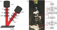
A new characterization method, namely membrane voltage measurement method, is proposed. Because the recovery time of membrane potential is dependent on the size of optoporation, establishing the relationship between the size of cytomembrane optoporation and the laser energy threshold can provide theoretical support forA new characterization method, namely membrane voltage measurement method, is proposed. Because the recovery time of membrane potential is dependent on the size of optoporation, establishing the relationship between the size of cytomembrane optoporation and the laser energy threshold can provide theoretical support for importing different exogenous substances into cells. In this study, gastric cancer cells are cultured with gold nanoparticles. On the premise that cells are not affected by the toxicity of gold nanoparticles, gastric cancer cells incubated with gold nanoparticles are irradiated using a nanosecond-pulsed laser with different energies. Propidium iodide and calcein-AM are used for staining verification of perforated cells. The results indicate that optoporation of cell membrane can be successfully achieved with 400 nanoparticles (diameter of 100 nm) per cell and 20 mJ/cm 2 energy density by 532 nm pulsed laser, without obvious dead cells. During the optoporation, the membrane potential is measured using optical mapping technique. It is found that the membrane potential firstly increases and then restores with 50 mV maximum increment and 250 s recovery time. These results confirm that the cell membrane damage can be recovered by optoporation and characteristiced by membrane potential change..
Chinese Journal of Lasers
- Publication Date: Feb. 01, 2020
- Vol. 47, Issue 2, 207021 (2020)
Low Photon Count Fluorescence Lifetime Analysis Based on Alternating Descent Conditional Gradient Method
Zhang Xiao, Lin Danying, Niu Jingjing, Liu Xiongbo, Zhang Jiao, Yu Bin, Zhang Wei, and Qu Junle
The development of fluorescence lifetime analysis method suitable for low photon count is of great significance for the evelopment and application of fast fluorescence lifetime imaging microscopy (FLIM). In this paper, we consider the fluorescence lifetime analysis, inspired by the compression sensing algorithm in highThe development of fluorescence lifetime analysis method suitable for low photon count is of great significance for the evelopment and application of fast fluorescence lifetime imaging microscopy (FLIM). In this paper, we consider the fluorescence lifetime analysis, inspired by the compression sensing algorithm in high-density single molecule localization microscopy, as a sparse inverse problem, and we propose an alternating descent conditional gradient (ADCG) based method for fluorescence lifetime analysis. Through the analysis of simulation data and experimental data, we demonstrate that the ADCG-FLIM algorithm can be appropriately implemented to analyze fluorescence lifetime even in the case of low photon count, thereby benefiting the development and application of live cell fast FLIM..
Chinese Journal of Lasers
- Publication Date: Feb. 01, 2020
- Vol. 47, Issue 2, 207022 (2020)
Effect of 0.1 THz Radiation on Excitability of Hippocampal Neurons in Sprague Dawley Rats
Zhang Xinxin, He Mingxia, Zhao Jinwu, Chen Xieyu, Liu Liyuan, Lu Xiaoyun, Tian Tian, Chen Mengqiu, and Wang Pu
In this work, the effect of terahertz (THz) radiation on the excitability of hippocampal neurons is studied by changing the neuronal membrane potential and using hippocampal neurons in Sprague Dawley (SD) rats irradiated by a THz source with frequency of 0.1 THz and power density of 2.65 mW/cm 2 for 5, 15, and 25 min, In this work, the effect of terahertz (THz) radiation on the excitability of hippocampal neurons is studied by changing the neuronal membrane potential and using hippocampal neurons in Sprague Dawley (SD) rats irradiated by a THz source with frequency of 0.1 THz and power density of 2.65 mW/cm 2 for 5, 15, and 25 min, respectively. The results show that THz irradiation for 15 and 25 min causes a significant depolarization of the hippocampal neurons, thereby increasing neuron excitability. To explore the mechanism behind this THz radiation-induced excitability of neurons, the intracellular concentrations of Ca 2+, Na +, and K + are determined. The results show that Ca 2+ and Na + concentrations in the hippocampal neurons increase and K + concentration decreases after irradiation by the THz source. Our study shows that the irradiation with frequency of 0.1 THz and power density of 2.65 mW/cm 2 can promote neuronal excitation by regulating the concentration of charged ions in the hippocampal neurons. This finding may provide the preliminary experimental basis for the application of THz radiation technology to the biomedical field..
Chinese Journal of Lasers
- Publication Date: Feb. 01, 2020
- Vol. 47, Issue 2, 207023 (2020)
Compressed Sensing STORM Super-Resolution Image Reconstruction Based on Noise Correction-Principal Component Analysis Preprocessing Algorithm
Pan Wenhui, Chen Bingling, Zhang Jianguo, Gu Zhenyu, Xiong Jia, Zhang Dan, Yang Zhigang, and Qu Junle
The low temporal resolution of stochastic optical reconstruction microscopy (STORM) limits its ability to observe dynamic events in live cells. Further, the post-processing analysis and reconstruction algorithms have an important effect on super-resolution images. In this study, we report a new noise-correction principThe low temporal resolution of stochastic optical reconstruction microscopy (STORM) limits its ability to observe dynamic events in live cells. Further, the post-processing analysis and reconstruction algorithms have an important effect on super-resolution images. In this study, we report a new noise-correction principal component analysis method for single-molecule localization microscopy against fluorescent spot overlapping and excessive background noise in a single frame of images owing to high-density labeling and high camera-sampling frequency. The proposed method can improve the positioning accuracy of existing localization methods by pre-processing the raw images acquired by the single molecule localization microscopy before reconstruction. In addition, this method can accurately distinguish the overlapping molecules. Therefore, it is suitable for samples exhibiting a high fluorophore density. Thus, the proposed method improves the temporal resolution of super-resolution imaging, providing a powerful technical support for the STORM imaging of live cells..
Chinese Journal of Lasers
- Publication Date: Feb. 01, 2020
- Vol. 47, Issue 2, 207024 (2020)
Identification of Human Coronary Atherosclerotic Plaques using Spectrum- and Time-Resolved Multiphoton Microscopy
Zhang Rongli, Li Hui, Wu Yueheng, Yu Jia, Liu Shangmin, Zheng Wei, and Lin Zhanyi
This study evaluated 8 unstained human coronary tissues ex vivo using spectrum- and time-resolved multiphoton microscopy. First, according to the spectra, the elastin fibers and collagen fibers in the coronary arterial intima can be separated clearly. Second, the ratios of the two types of fiber signals were calculatedThis study evaluated 8 unstained human coronary tissues ex vivo using spectrum- and time-resolved multiphoton microscopy. First, according to the spectra, the elastin fibers and collagen fibers in the coronary arterial intima can be separated clearly. Second, the ratios of the two types of fiber signals were calculated to assess changes in the relative content of the collagen and elastin fibers in the coronary arterial wall caused by an atherosclerotic lesion. Third, we assessed biochemical variations of the elastin fibers in the coronary atherosclerotic tissues by measuring fluorescence lifetime and found that the coronary atherosclerotic plaque has a lower mean fluorescence lifetime than a normal tissue. This study demonstrates that spectrum- and time-resolved multiphoton microscopy can effectively identify coronary atherosclerotic plaques, thus indicating its potential as a novel research tool for studying coronary arteriosclerotic lesions in the future..
Chinese Journal of Lasers
- Publication Date: Feb. 01, 2020
- Vol. 47, Issue 2, 207025 (2020)
Ultrasensitive Quantitative Detection of Alpha-Fetoprotein Based on SERS Spectroscopy
Wang Tingyin, Wang Yunyi, Lin Xueliang, Xu Yunchao, Lin Huijing, Liu Xiaokun, and Feng Shangyuan
In this paper, the ultrasensitive detection of an alpha-fetoprotein (AFP) is performed using a combination of two-dimensional (2D) surface-enhanced Raman scattering (SERS) and an AFP aptamer. We construct SERS "hot spots" through complementary base-pairing between substrates modified with AFP aptamer and silvIn this paper, the ultrasensitive detection of an alpha-fetoprotein (AFP) is performed using a combination of two-dimensional (2D) surface-enhanced Raman scattering (SERS) and an AFP aptamer. We construct SERS "hot spots" through complementary base-pairing between substrates modified with AFP aptamer and silver nanoparticles modified with aptamer complementary sequence. The complementary adaptor sequence is also modified using Raman signal labeled molecule ROX, but the addition of the AFP damages the structure of nano-gap "hot spots", resulting in a decrease in intensity of the SERS signal. The ultrasensitive quantitative detection of AFP is realized using the changed working curve of the SERS signal of ROX. The detection limit of this method is 145 fg/mL, which is one order of magnitude higher than that of the traditional clinical detection method. The designed AFP SERS probe also exhibits good specificity and anti-interference ability. These results show that this new approach may provide a rapid and effective method for the accurate detection of AFP..
Chinese Journal of Lasers
- Publication Date: Feb. 01, 2020
- Vol. 47, Issue 2, 207026 (2020)
Cherenkov-Excited Luminescence Scanned Tomography Reconstruction Based on Approximate Message Passing
Feng Jinchao, Chang Di, Li Zhe, Sun Zhonghua, and Jia Kebin
As a new molecular imaging technology, Cherenkov-excited luminescence scanned imaging (CELSI) has merits of high spatial resolution and large imaging depth, therefore showing a potential for monitoring the physiological changes of tumors during radiotherapy. In our previous work, we developed a tomographic technique foAs a new molecular imaging technology, Cherenkov-excited luminescence scanned imaging (CELSI) has merits of high spatial resolution and large imaging depth, therefore showing a potential for monitoring the physiological changes of tumors during radiotherapy. In our previous work, we developed a tomographic technique for CELSI based on Tikhonov method, which is problematic to reconstruct accurate fluorescent targets with position depth larger than 3 cm or with low contrast. To overcome this problem, we develop a sparse reconstruction method for tomographic CELSI based on approximate message passing. To demonstrate the merits of the proposed algorithm, we compare it with traditional Tikhonov regularization and three sparse based reconstruction algorithms. Our results show that the proposed method can achieve best performance in terms of mean-square error and contrast noise ratio..
Chinese Journal of Lasers
- Publication Date: Feb. 01, 2020
- Vol. 47, Issue 2, 207027 (2020)
Identifying Single Cell Types via Whispering Gallery Mode Optical Microcavities
Wang Yaping, Wang Xiuhong, and Wang Pu
Whispering gallery mode is a type of optical mode where photons move in a quasi-two-dimensional plane, and the total reflection occurs at the boundary of the microcavity without reflecting out of the cavity. This mode has a high Q value and small mode volume, and it is extremely sensitive to changes in the surrounding Whispering gallery mode is a type of optical mode where photons move in a quasi-two-dimensional plane, and the total reflection occurs at the boundary of the microcavity without reflecting out of the cavity. This mode has a high Q value and small mode volume, and it is extremely sensitive to changes in the surrounding environment. A broadband fluorescence can be transformed into narrow-spectrum laser output by using the whispering gallery mode. In this paper, polystyrene microspheres doped with the dragon green fluorescent dye are used as whispering gallery mode optical microcavity. Through the phagocytosis of cells, the fluorescent microspheres reach inside cells and then are pumped by nanosecond pulsed laser to achieve the output of whispering gallery mode laser in cells. In comparison with the laser output in the pure-water environment, a redshift of the intracellular fluorescent microsphere whispering gallery mode resonance emission can be observed, and the redshift is related to cell type; therefore, it can be used for unlabeled identification of cell type..
Chinese Journal of Lasers
- Publication Date: Feb. 01, 2020
- Vol. 47, Issue 2, 207028 (2020)
Label-Free Imaging of β-Amyloid Plaques and Photodynamic Degradation
Huang Yanxia, Xu Hao, Luan Ping, Ohulchanskyy Tymish Y, and Qu Junle
Formation of β-Amyloid (Aβ) plaques is one of the most significant pathological features of Alzheimer’s disease (AD). Detection and degradation of Aβ plaques are crucial in AD treatment. When Aβ monomers aggregate to form plaques, they produce strong autofluorescence. In this study, we investigate label-free imaging ofFormation of β-Amyloid (Aβ) plaques is one of the most significant pathological features of Alzheimer’s disease (AD). Detection and degradation of Aβ plaques are crucial in AD treatment. When Aβ monomers aggregate to form plaques, they produce strong autofluorescence. In this study, we investigate label-free imaging of Aβ plaques by using the nonlinear optical imaging method and study their degradation via photodynamic effect of the photosensitizer. Moreover, we study the relationship between photosensitizers with different concentrations and degradation effect of Aβ plaques. Corresponding liposomes are prepared and they are applied to the degradation of Aβ plaques. The potential applications of label-free optical imaging and photodynamic therapy in AD research are discussed, and a new way for optimizing AD diagnosis and treatment is provided..
Chinese Journal of Lasers
- Publication Date: Feb. 01, 2020
- Vol. 47, Issue 2, 207029 (2020)
A Method of Backscattering Micro-Spectrum Classification Based on Principal Component Analysis and Fuzzy Cluster Analysis
Wang Cheng, Jiao Tong, Lu Yufei, Xu Kang, Li Sen, Liu Jing, and Zhang Dawei
Rapid detection of foodborne pathogens is one of the most effective ways to overcome food safety problems. To realize a rapid, efficient and label-free detection and classification of foodborne pathogens, this study aims to improve the performance of existing optical fiber confocal backscattering spectrum system. ThrouRapid detection of foodborne pathogens is one of the most effective ways to overcome food safety problems. To realize a rapid, efficient and label-free detection and classification of foodborne pathogens, this study aims to improve the performance of existing optical fiber confocal backscattering spectrum system. Through this process, the light field diameter is reduced to fit small biological samples, and single spectrum level detection can be achieved. Furthermore, the backscattering micro-spectrum of three categories of common foodborne pathogens (Salmonella enteritidis, Escherichia coli, and Salmonella typhimurium) with similar morphology is measured without labels. A multivariate analysis model is established by combining principal component analysis (PCA) and fuzzy cluster analysis (FCA) at the characteristic wavelength range of 500--800 nm. Results show that the top five principal components contain 80.41% characteristic spectral information. The scores of the top five principal components are taken as the variables for the FCA. The accuracy of 100%, according to the degree matrix of membership, is achieved for the clustering results of three kinds of bacteria. Also, results show that optical fiber confocal backscattering spectroscopy, combined with PCA and FCA, can be used to analyze and classify a single spectrum rapidly, efficiently, and without labels..
Chinese Journal of Lasers
- Publication Date: Feb. 01, 2020
- Vol. 47, Issue 2, 207030 (2020)
Method for Generating Parallelized Fluorescence Depletion Patterns Based on Optical Wedges
Zhang Shuochen, and Feng Jihong
Herein, a novel method for generating parallelized fluorescence depletion patterns based on optical wedges is proposed. Optical wedges and matching reflectors are used to control the stimulated emission depletion (STED) beam inclination angle of the tube lens object space to make full use of the numerical aperture of tHerein, a novel method for generating parallelized fluorescence depletion patterns based on optical wedges is proposed. Optical wedges and matching reflectors are used to control the stimulated emission depletion (STED) beam inclination angle of the tube lens object space to make full use of the numerical aperture of the microscope objective to produce parallelized fluorescence depletion patterns with small periodicity. Simulation results show that a square lattice-like parallelized fluorescence depletion pattern with periodicity as small as 282.0 nm×283.6 nm is generated when the wavelength of the STED beam is 760 nm and numerical aperture of the microscope objective is 1.4, thereby achieving relatively high imaging resolution..
Chinese Journal of Lasers
- Publication Date: Feb. 01, 2020
- Vol. 47, Issue 2, 207031 (2020)
Tumor-Specific Imaging of Small Animals Based on Multi-Angle Optoacoustic Mesoscopy Imaging Method
Lu Tong, Gao Feng, Song Shaoze, Chen Tingting, Miao Shichao, and Li Jiao
In this study, we construct a high-sensitivity optoacoustic mesoscopy (OPAM) experimental system with a multi-angle scanning mode that can simultaneously perform high-sensitivity measurements and achieve multi-angle information acquisition. This system is employed to acquire the structural and functional information reIn this study, we construct a high-sensitivity optoacoustic mesoscopy (OPAM) experimental system with a multi-angle scanning mode that can simultaneously perform high-sensitivity measurements and achieve multi-angle information acquisition. This system is employed to acquire the structural and functional information related to small-animal tumor models. The spatial resolution of this OPAM experimental system is verified via a phantom experiment that satisfies the OPAM imaging requirements. Further, by applying the OPAM experimental system on two types of small-animal tumor models, their structural images and obvious type characteristics are presented. Subsequently, the blood oxygen saturation values of the tumors are obtained using the OPAM experimental system under the dual-wavelength measurement condition. The experimental results denote that the OPAM experimental system can provide valuable reference and guidance for oncology research and that the proposed system exhibits considerable application prospects in the field of biomedical research..
Chinese Journal of Lasers
- Publication Date: Feb. 01, 2020
- Vol. 47, Issue 2, 207032 (2020)
Antimonene Nanoflakes as a Photoacoustic Imaging Contrast Agent for Tumor in vivo Imaging
Yu Jingwen, Wang Xiuhong, Feng Jinchao, Zhang Na, and Wang Pu
Photoacoustic imaging, a novel biomedical imaging technique that combines the advantages of optical imaging and acoustic imaging, offers high-resolution biological tissue imaging to facilitate the observation of deeper imaging sites. In other words, it breaks the “soft limit” of conventional optical bioimaging techniquPhotoacoustic imaging, a novel biomedical imaging technique that combines the advantages of optical imaging and acoustic imaging, offers high-resolution biological tissue imaging to facilitate the observation of deeper imaging sites. In other words, it breaks the “soft limit” of conventional optical bioimaging techniques. However, many diseases, especially in the early stage, present no obvious photoacoustic contrast; therefore, it is crucial to identify effective exogenous photoacoustic contrast agents. Here we introduce a novel two-dimensional material, antimonene nanoflakes (AMNFs), which demonstrates great optical absorption from 300 nm to 900 nm as well as excellent photothermal conversion efficiency and photoacoustic performance. This material is expected to be useful as a contrast agent, helping to achieve excellent photoacoustic imaging of ultra-small tumors in vivo..
Chinese Journal of Lasers
- Publication Date: Feb. 01, 2020
- Vol. 47, Issue 2, 207033 (2020)
Assessment of Bacterial Inflammation Based on Optical Coherence Tomography Angiography
Liu Yubin, Chen Zhiyi, and Yuan Zhen
Optical coherence tomography angiography (OCTA) is an important tool for investigating microvascular networks and microcirculation in living tissue. In this study, OCTA was employed for noninvasive in vivo monitoring and assessment of inflammation induced by bacteria in a mouse ear model. Imaging results demonstrated tOptical coherence tomography angiography (OCTA) is an important tool for investigating microvascular networks and microcirculation in living tissue. In this study, OCTA was employed for noninvasive in vivo monitoring and assessment of inflammation induced by bacteria in a mouse ear model. Imaging results demonstrated that OCTA can monitor changes in microvascular density and morphology of blood vessels caused by immunovascular responses during the inflammatory process with a high degree of resolution and sensitivity. Distinctly enhanced OCT signals from the mouse ear were observed following bacterial infection owing to an influx of red blood cells caused by the bacteria. A highly dense microvascular network noted in the palms of healthy subjects by OCTA, demonstrates the feasibility of OCTA for the clinical evaluation of inflammation. This method can improve the understanding of the pathological mechanisms of inflammation and can be useful in the clinical evaluation of inflammation..
Chinese Journal of Lasers
- Publication Date: Feb. 01, 2020
- Vol. 47, Issue 2, 207034 (2020)
Photoacoustic Microscopy Based on Highly Sensitive Ultrasound Transducer
Yang Chen, Jiao Yang, Zhu Xinle, Jian Xiaohua, George Sergiadis, and Cui Yaoyao
In photoacoustic microscopy (PAM), detection sensitivity is an important factor for determining imaging quality and depth, which can affect photoacoustic imaging applications in biomedical engineering. To improve the detection sensitivity and signal-to-noise ratio (SNR), a miniature amplifier was integrated close to anIn photoacoustic microscopy (PAM), detection sensitivity is an important factor for determining imaging quality and depth, which can affect photoacoustic imaging applications in biomedical engineering. To improve the detection sensitivity and signal-to-noise ratio (SNR), a miniature amplifier was integrated close to an ultrasound transducer for front-end signal amplification and output impedance matching. An optical resolution PAM was developed based on the fabricated prototype transducer to perform 3D imaging of phantoms and small vessels of the mouse ear. Experimental results show that the SNR is improved by over 10 dB. The proposed PAM has the potential to be used for monitoring and quantitatively analyzing weak physiological changes..
Chinese Journal of Lasers
- Publication Date: Feb. 01, 2020
- Vol. 47, Issue 2, 207035 (2020)
Orthotopic Coaxial Projective Imaging for Neurosurgical Navigation
Wu Bingxuan, Liu Peng, Li Xingyi, Shao Pengfei, and Xu Xiaorong
In this study, we report a novel stereotactic neurosurgical navigation system based on orthotopic coaxial projective imaging (CPI). The proposed system facilitates automatic registration of the position of the patient, real-time projection of the preoperatively reconstructed surgical target and the planned approach in In this study, we report a novel stereotactic neurosurgical navigation system based on orthotopic coaxial projective imaging (CPI). The proposed system facilitates automatic registration of the position of the patient, real-time projection of the preoperatively reconstructed surgical target and the planned approach in the surgical field, and precise intraoperative guidance with respect to the target and the approach. The average reprojection error of the system is 0.4 mm at its working distance via a coaxial calibration test, indicating a high photographic-projection coaxiality. The average projection error of the system in a stereotactic projection test is 1.2 mm at different projection angles, indicating a high stereotactic navigation accuracy. In a neurosurgery simulated by using a phantom model, the average projection error of the system is 0.3--3.1 mm in different operational conditions, indicating the technical feasibility of the system for neurosurgical navigation. Unlike the conventional stereotactic surgical navigation systems that require a display screen, the CPI system avoids frequent viewpoint changes between the screen and the surgical field, eliminates the possible operating errors, and improves the surgical efficiency. The proposed system may be applied to surgically treating many neurological diseases, including brain tumors and brain hematomas..
Chinese Journal of Lasers
- Publication Date: Feb. 01, 2020
- Vol. 47, Issue 2, 207036 (2020)
Positioning Method Based on Microphotodiodes and Ultrasound Transducers for Epilepsy Surgery
Shen Zhitian, Shao Weiwei, Jiao Yang, Xu Jie, Ma Hongtao, and Cui Yaoyao
Epilepsy surgery often depends on the accurate positioning of lesions; however, current positioning methods have some limitations. The electrocorticogram (ECoG) in clinical inspection has a high temporal resolution, but its spatial resolution is below the accepted standard. Based on the neurovascular coupling mechanismEpilepsy surgery often depends on the accurate positioning of lesions; however, current positioning methods have some limitations. The electrocorticogram (ECoG) in clinical inspection has a high temporal resolution, but its spatial resolution is below the accepted standard. Based on the neurovascular coupling mechanism in the human brain, we propose a new epileptic foci detection method by detecting cerebral microvascular blood flow with microphotodiodes and ultrasound transducers. The results of experiments conducted on animals show that the light signal on the surface of the cortex has good correspondence with the ECoG signal. The spectra of the ultrasound signal also matches that of the electrical signal within a depth of 1 mm below the cortex. Therefore, we believe that this method can improve the spatial resolution of epileptic lesion detection, and it can potentially be used for depth inspection..
Chinese Journal of Lasers
- Publication Date: Feb. 01, 2020
- Vol. 47, Issue 2, 207037 (2020)
Light Focusing Through Scattering Medium Based on Binary Transmission Matrix
Si Ke, Tang Liming, Du Jichao, Wu Chenxue, Xu Xiaobin, Hu Lejia, Chen Jiajia, and Gong Wei
In this paper, a speckle recovery algorithm for light through scattering media that can achieve focusing at any position in a large field of view is proposed. The transmission matrix of the scattering medium is measured and binarized by simulating the optical path, and the digital micro-mirror device is used to modulatIn this paper, a speckle recovery algorithm for light through scattering media that can achieve focusing at any position in a large field of view is proposed. The transmission matrix of the scattering medium is measured and binarized by simulating the optical path, and the digital micro-mirror device is used to modulate the binary amplitude of the incident light to achieve single-point or multi-point focusing through the scattering medium. Due to the independence of different focus positions, the algorithm can achieve large field-of-view focusing at any position. The simulation results show that the intensity enhancement factor of the focus position increases with the increase in the sampling number. Compared with the traditional three-step phase shift method, an enhancement ratio of 55% can be obtained by the proposed algorithm when the number of sampling is reduced by 1/3, which is 12% higher than that by the three-step phase shift method. The proposed algorithm exhibits great significance for realizing large field-of-view scanning and focusing through the scattering medium, making it applicable in the field of biomedical imaging..
Chinese Journal of Lasers
- Publication Date: Feb. 01, 2020
- Vol. 47, Issue 2, 207038 (2020)
Characterization of Cell Distribution Based on Optical Coherence Tomography Scattering
Shen Renqiang, Wang Ling, Xu Ming''en, and Peng Shichang
Cell distribution can be characterized quantitatively using a depth-resolved scattering coefficient calculation method based on optical coherence tomography (OCT). This method uses a single scattering model to obtain a depth-resolved scattering coefficient distribution map, and the law of cell distribution is verified Cell distribution can be characterized quantitatively using a depth-resolved scattering coefficient calculation method based on optical coherence tomography (OCT). This method uses a single scattering model to obtain a depth-resolved scattering coefficient distribution map, and the law of cell distribution is verified using the histogram statistics of the scattering coefficient. A multilayer scattering medium model is used to verify the effectiveness of the proposed method. The experiments reveal that the scattering coefficient distribution maps can resolve the cell distribution depth and provide a more obvious contrast between the cell and background compared with an intensity map. There are no significant differences in the distributions of the OCT intensity signal histograms of various cell suspensions. The histogram of the scattering coefficient contains the characteristic cell concentration peak, and the degree of deviation between the characteristic peak and background scattering characteristic peak is linear with the cell concentration. The cell distribution in the bio-3D printed hydrogel scaffold detected by the OCT scattering coefficient distribution map corresponds well with the H&E staining results, and the corresponding scattering coefficient histogram distribution exhibits peaks characteristic of the cell concentration..
Chinese Journal of Lasers
- Publication Date: Feb. 01, 2020
- Vol. 47, Issue 2, 207039 (2020)
Transmission of a Laser Emitted from an Interpolated Optical Fiber in Tissue Based on Monte Carlo Method
Ding Leming, Dai Lijuan, Zhang Lei, and Qian Zhiyu
To understand characteristic of the shapes of most tumors and the laser transmission mode as an internal light source during laser interstitial therapy, a double-layered media model in the shape of sphere with an interpolated optical fiber was established. Then, the modes of the photon transmitting in the tissue, at thTo understand characteristic of the shapes of most tumors and the laser transmission mode as an internal light source during laser interstitial therapy, a double-layered media model in the shape of sphere with an interpolated optical fiber was established. Then, the modes of the photon transmitting in the tissue, at the boundary of the sphere, and on the surface of the inserted optical fiber were set, assuming the photons were launched at the center of the model. Finally, the photon migration in the model was simulated based on the Monte Carlo (MC) method in the Visual Studio (VS) program. The simulation results show that the optical fiber primarily affects the movements of photons that are moving near the surface from which they were launched. The energy absorption by the tissue under the photon-launching surface is greater than that in the above the surface. The absorption of the inner boundary increases with decreasing inner sphere radius. Compared with the traditional MC method, this model has more similarities with a real laser interstitial thermotherapy situation, which is of considerable practical significance for predicting the thermal damage range of laser interstitial thermotherapy..
Chinese Journal of Lasers
- Publication Date: Feb. 01, 2020
- Vol. 47, Issue 2, 207040 (2020)
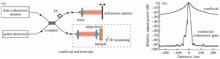
Intraocular Laser Surgery Combined Calibration Method Based on Bimodal Image Information
Yang Fan, Chen Cong, Shen Chaoyi, and Fan Licheng
In this work, a swept-source optical coherence tomography (SS-OCT) system was constructed using a dual fiber ring structure to obtain the depth spatial information of a planned surgical site in laser ophthalmic surgery. In the proposed system, a dichroic mirror was added to the sample arm to integrate it with a CMOS caIn this work, a swept-source optical coherence tomography (SS-OCT) system was constructed using a dual fiber ring structure to obtain the depth spatial information of a planned surgical site in laser ophthalmic surgery. In the proposed system, a dichroic mirror was added to the sample arm to integrate it with a CMOS camera system. After matching the geometric relationship between the coordinates of the OCT system and camera system, several scanning modes, including multi-angle linear scanning and annular scanning, were designed, achieving multimode scanning imaging at any given position of camera video images. The longitudinal resolution of the imaging system was 13.68 μm, while the lateral resolution was 29.8 μm. The imaging depth in the air was 17.25 mm. The measured maximum lateral deviation was 275 μm, and the maximum longitudinal deviation was 70 μm; thus, the requirements of the operation location were met. By using the proposed system, rapid depth information acquisition of the designated parts of the eye is possible. Therefore, the system is suitable for application in ophthalmic disease diagnosis and cataract surgery navigation..
Chinese Journal of Lasers
- Publication Date: Feb. 01, 2020
- Vol. 47, Issue 2, 207041 (2020)
Measurement of Heart Rate Based on Ballistocardiography
Kong Lingqin, Wu Yuheng, Pang Zongguang, Zhao Yuejin, Dong Liquan, Liu Ming, Hui Mei, Wang Weijie, Guo Ying, and Wang Xiatian
Non-contact ballistocardiography (BCG) is used to measure heart rate (HR) by measuring the periodic pressure of blood on the walls of blood vessels during circulation. This pressure causes the periodic weak mechanical movement of various parts of the body, including the head, which is very weak, and the BCG signal extrNon-contact ballistocardiography (BCG) is used to measure heart rate (HR) by measuring the periodic pressure of blood on the walls of blood vessels during circulation. This pressure causes the periodic weak mechanical movement of various parts of the body, including the head, which is very weak, and the BCG signal extracted from the body movement has a low signal-to-noise ratio, which limits the measurement accuracy of the heart rate. An optical lever is used to amplify the head motion, and a non-contact high precision heart rate detection algorithm named optical lever amplified BCG algorithm (OLA-BCG) is proposed. In the proposed method, a laser is used as the active light source. A plane mirror attached to the head is used to amplify the head motion. At the same time, the weighted centroid tracking algorithm is used to extract the motion trajectory of the head, and the interference noise is filtered out by independent component analysis to obtain BCG signal. Finally, the extracted BCG signal is analyzed and the heart rate is calculated. Experimental results show that the proposed OLA-BCG can effectively improve the signal-to-noise ratio and measurement accuracy..
Chinese Journal of Lasers
- Publication Date: Feb. 01, 2020
- Vol. 47, Issue 2, 207042 (2020)

© Copyright 2018-2021 | Chinese Laser Press. All Rights Reserved 沪ICP备15018463号-20