Abstract
A light purplish red sapphire is heat treated in an airtight crucible. The sample changes little in color after receiving heat treatment at 1100°C, but turns to light blue and blue after being treated at 1200°C and 1300°C, respectively. Before heating, the UV-VIS absorption spectra of the sample are dominated by the 551 nm broad absorption band contributed by the -electron transition of . After heating, the UV-VIS absorption spectra are dominated by the 563 nm broad absorption band contributed by the intervalence charge transfer of . The x ray photoelectron spectroscopy test reveals that the and ion contents increase with increasing temperature. The sapphire changing from light purplish red to blue in the heating process is owing to the fact that the and contents grow and the intervalence charge transfer of selectively absorbs UV-VIS light.Ruby and sapphire, both coming from the corundum family (), are precious stones. Corundum in its pure state is colorless but takes on some corresponding colors when it contains some amounts of Cr, Fe, Ti, or other transition metals[1]. Chromium lends a red tint to corundum while iron and titanium make corundum look blue. The color of corundum depends not only on the kinds of transition metals it contains but it is also closely related to their chemical valence state. For instance, the Geuda corundum mined from Sri Lanka looks milky white when not heat treated but presents an appealing blue tint after receiving treatment at a high temperature. It owes its color transition to the valence state change of the iron element in it[2]. A lot of research reports have been written on ruby and sapphire heat treatment[3–7]. The purpose of our experiment is to improve the color of low-quality sapphire produced in Burma by low-temperature heat treatment. In the course of the experiment, it is found that the color of sapphire experienced interesting changes. We used absorption spectroscopy and x ray fluorescence spectrometer tested samples before and after heat treatment, and analyzed the changes in chemical valence states of iron and titanium, as well as the interaction between the two. Changes in the color of the samples during the experiment and the principle of the phenomenon are highly consistent with the research results by Bristow et al.[8]; that is, the color of sapphire is contributed by the intervalence charge transfer (IVCT) of ions to .
The raw sample sapphire studied by this paper was light purplish red and semitransparent, having quite a few micro cracks. The raw stone was processed into a slice about 1.3 mm thick and large. The slice was soaked in diluted muriatic acid for 30 min before being rinsed clean using clear water. It was placed into an aluminum crucible, which was then made airtight and put into an electric furnace for heating to 1100°C, 1200°C, and 1300°C, respectively. The heating rate was 4°C/min and the stone was heated to the programmed temperature from room temperature, maintained at that temperature for 2 h, then allowed to cool back to room temperature inside the furnace.
An XRF-1800 model x ray fluorescence spectrometer was employed for chemical composition analysis. The condition in which the analysis was carried was as follows: a voltage of 40 kV and a tube current of 140 mA, a 4 kW thin window of x ray tube, a 75 μm beryllium window, an Rh target, a maximum scan rate of 300°/min, a 250 μm micro-zone analysis imaging, and an element measurement range of . The results are given as content of oxides. X ray photoelectron spectroscopy (XPS) is a versatile surface analysis technique that can be used for compositional and chemical state analysis. We test the content of Fe and Ti in different chemical valence states of the sample before and after thermal treatment by using an x ray photoelectron spectrometer. XPS testing was done using an ESCALAB 250Xi model x ray photoelectron spectrometer manufactured by Thermo Fisher, an American company. The major technical indices of this instrument include an analysis room vacuum pressure better than and a monochromatization XPS optimum energy resolution better than 0.45 eV, in the case of Ag 3d5/2 peak. Corrections for surface charging were applied using the C 1s signal (284.7 eV) of surface impurities, as a reference. All spectra were corrected by subtracting a Shirley-type background. The curve fitting was carried out using the ratio . Iron and titanium ions acting as the impurity ions exist in the crystal lattice of corundum after substituting Al ions. Their binding energy is different from that in the pure oxide state, and both kinds and the valence state of its surrounding cations will affect their binding energy values. The changes in the absorption spectra of samples in the visible light range are analyzed with a UV-VIS-NIR spectrometer before and after heat treatment. Absorption spectrum testing was performed using a CARY500 model UV-VIS-NIR spectrometer produced by Varian, an American manufacturer. This instrument offers a wavelength accuracy of and its resolution is superior to 0.05 nm.
Sign up for Chinese Optics Letters TOC. Get the latest issue of Chinese Optics Letters delivered right to you!Sign up now
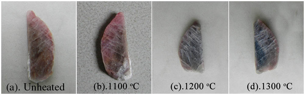
The sample slice looked light purplish red before heating and changed little after treating at 1100°C, but turned to light blue if treated at 1200°C. Heating at 1300°C made it look dark blue, as shown in Fig. 1.

Figure 1.Sample and its postheating color changes. (a) Photo of the unheated sample, photos of the sample heated at (b) 1100°C, (c) 1200°C, and (d) 1300°C.
Chemical composition testing was performed on the sample using XRF, the analysis results being given in Table 1. Due to their different formed environment and time, iron and titanium elements are not evenly distributed in the sample. In addition, it is the surface of the sample that XRF directly tested. Therefore, XRF’s test results represent the kind and content ratio of the elements in the surface area of samples. The sample contained chromium, iron, titanium as well as silicon, calcium, and gallium. It is known that the colors of the ruby and sapphire were largely contributed by transition metals like chromium, iron, and titanium[1]. The total iron is given as since and cannot be differentiated using this method. This is the same case with titanium.
A UV-VIS spectrometer was employed in the sample absorption spectra test before and after heating. Figure 2 summarizes the test results. The absorption spectra in Fig. 2 have been shifted vertically.
| Al2O3 | Cr2O3 | Fe2O3 | TiO2 | SiO2 | CaO | GaO |
|---|
| 96.0521 | 0.8090 | 1.9665 | 0.1302 | 0.3348 | 0.2788 | 0.0415 |
Table 1. XRF Testing Results of the Sample (wt.%)
Figure 2.Comparison of different UV-VIS absorption spectra of corundum samples before and after heat treatment. (a) UV-VIS absorption spectra of the unheated sample, UV-VIS absorption spectra of the sample heated at (b) 1100°C, (c) 1200°C, and (d) 1300°C.
Before heating, the sample had a wide absorption band in the 495–620 nm range centering on 551 nm, which was contributed primarily by the -electron transition of [1,9]. This absorption band produced varying absorption mainly in the green, yellow, and red color regions, hence making the sample light purplish red. Theoretically speaking, the chromium in the sample should have given off a strong fluorescent emission at 693 nm; the testing results demonstrated, however, that such a fluorescent emission peak was rather faint, possibly because of the presence of a relatively high iron content. After heat treatment, the center wavelength of the wide absorption band in the 495–620 nm range shifted. As the heat treatment temperature rose, the center wavelength of the wide absorption band shifted progressively toward a longer wavelength. Subsequent to heat treatment at 1300°C, the wide absorption band center was at 563 nm and the sample turned blue.
As shown in Fig. 3, after fitting the absorption spectra through a 1300°C heat treatment of the sample, we found the wide absorption band centered around 563 nm that is caused by an overlap from the and absorption spectra. -electron transition of caused the wide absorption band centered at 548 nm, and the IVCT of ions pair caused the wide absorption band centered at 548 nm[10,11]. Iron and titanium have been confirmed to be present on adjacent lattice sites in sapphire[12]. It was suggested by Eigenmann and Günthard that these could be in either valence state (II–IV or III–III pairs)[13]. Bristow et al.[8] confirmed the sapphire blue is contributed by IVCT between Ti4+ and Fe2+ when situated in neighboring lattice sites. Our analyses of the experimental phenomena, the absorption spectrum, and the XPS spectra well proved the sapphire blue genetic theory by Bristow et al.[8].
Figure 3.Observed and fitted absorption spectrum of the sapphire sample after it is heated at 1300°C (spectrum range from 490 to 640 nm).
Figure 4.Observed and fitted x ray photoelectron spectrometer spectrum of Fe 2p from the surface of the sapphire sample: (a) unheated sample, sample heated at (b) 1100°C, (c) 1200°C, and (d) 1300°C.
Figure 5.Observed and fitted x ray photoelectron spectrometer spectrum of Ti 2p from the surface of the sapphire sample: (a) unheated sample, sample heated at (b) 1100°C, (c) 1200°C, and (d) 1300°C.
The transition metals, iron and titanium to be more specific, in the sample were analyzed using XPS for their valence state and content percentage both before and after heating, the results being given in Table 2 and Figs. 4 and 5.
| | Binding energies (eV) | Percentage ( % ) |
|---|
| −Unheat | −1100°C | −1200°C | −1300°C |
|---|
| Fe2+ | 711.7 | 39.62 | 49.80 | 58.90 | 65.35 |
| Fe3+ | 713.6 | 60.38 | 50.20 | 41.10 | 34.65 |
| Ti3+ | 455.3 | 51.28 | 43.74 | 42.40 | 8.33 |
| Ti4+ | 458.8 | 48.72 | 56.26 | 57.60 | 91.67 |
Table 2. X-ray Photoelectron Spectrometer Analysis Results of Fe 2p and Ti 2p Spectrum at Different Temperatures
As shown in Fig. 4, Before the heat treatment, the sample contained more , comprising 60.38% of the total iron, and less , which accounted for 39.62% of the total iron[14–17]. The content dropped and the content rose with a rising heat treatment temperature. Following the heat treatment at 1300°C, the content of the dropped to 34.65% whereas that of rose to 65.35%. As shown in Fig. 5, Before the heat treatment, the and contents were comparative[16,18,19], with constituting 51.28% of the total titanium and 48.72%. The content dropped and the content rose with a rising heat treatment temperature. After a heat treatment at 1300°C, the content of dropped to 8.33%, whereas that of rose to 91.67%. The ion pairs, which absorb UV-VIS light selectively, increased in quantity after the heat treatment, thereby making the heated sample blue.
The heat treatment process reported by this Letter was completed in an airtight crucible. The crucible had not been vacuum pumped before heating and some oxygen remained in it as a result. From the test results, it may be inferred that the transition metals in the sample have undergone an oxidation-reduction reaction, with a portion of such a reaction taking place between iron and titanium in a manner illustrated by Formula-1. Ter lost one electron and was thus oxidized, whereas received one, thus reduced.
Given the XPS test results, it can be deduced that an oxidation-reduction reaction occurred between and , resulting in higher and contents. A broad absorption band centering on 563 nm in the UV-VIS absorption spectra was caused by the IVCT between the ion pair. The sample turned blue from light, and purplish red through the heat treatment, because the content of the ion pairs, which absorb the visible light selectively, became higher in the sample.
Major predecessors’ research on sapphire heat treatment is to control the heat treatment atmosphere, including the reducing atmosphere and the oxidizing atmosphere, with little consideration that the redox reaction between different transition metal elements occurres in internal sapphire. The test of this Letters is processed in a nonvacuum and airtight corundum crucible, and belongs to the weak oxidizing atmosphere. According to the UV-VIS absorption spectra and XPS analysis, the color transform of sapphire samples is attributed to a redox reaction between iron and titanium. The phenomenon and principle analyses are seldom mentioned in previous literature.
References
[1] D. S. Mcclure. J. Chem. Phys., 36, 2757(1962).
[2] K. Nassau. Gems Gemol., 17, 121(1981).
[3] J. S. D. Abraham. Gems Gemol., 18, 79(1982).
[4] P. Winotai, S. Saiseng, T. Sudyoadsuk. Int. J. Mod. Phys. Lett. B, 15, 873(2001).
[5] T. Kittiauchawal, P. Limsuwan, S. Eitssayeam, T. Tunkasiri. Int. J. Mod. Phys. B, 22, 4730(2008).
[6] S. Achiwawanich, N. Brack, B. D. James, J. Liesegang. Appl. Surf. Sci., 252, 8646(2006).
[7] S. W. Novak, C. W. Magee, T. Moses, W. Wuyi. Appl. Surf. Sci., 231, 917(2004).
[8] J. K. Bristow, S. C. Parker, C. R. A. Catlow, S. M. Woodley, A. Walsh. Chem. Commun., 49, 5259(2013).
[9] L. E. Cartier. J. Gemmol., 31, 171(2009).
[10] J. M. García-Lastra, M. T. Barriuso, J. A. Aramburu, M. Moreno. Phy. Rev. B., 72, 113104(2005).
[11] J. Ferguson, P. E. Fielding. Chem. Phys. Lett., 10, 262(1971).
[12] R. G. Burns, V. M. Burns. J. Adv. Ceram., 10, 46(1984).
[13] K. Eigenmann, H. H. Günthard. Chem. Phys. Lett., 13, 58(1972).
[14] T. Yamashita, P. Hayes. Appl. Surf. Sci., 254, 2441(2008).
[15] T. Fujii, F. M. F. D. Groot, G. A. Sawatzky, F. C. Voogt, T. Hibma, K. Okada. Phys. Rev. B, 59, 3195(1999).
[16] B. A. V. Hassel, A. J. Burggraaf. Appl. Phys. A, 52, 410(1991).
[17] H. L. Saadon. Chin. Opt. Lett., 13, 071901(2015).
[18] X. Sun, J. Wei, W. Wang, H. Lu. Chin. Opt. Lett., 13, 071401(2015).
[19] D. Wang, M. Chen, X. Liu, X. Gao. Chin. Opt. Lett., 13, 081404(2015).