• Opto-Electronic Advances
  • Vol. 4, Issue 11, 210048-1 (2021)
Moram Sree Satya Bharati and Venugopal Rao Soma*
Author Affiliations
  • Advanced Centre of Research in High Energy Materials (ACRHEM), University of Hyderabad, Telangana 500046, India
  • show less
    DOI: 10.29026/oea.2021.210048 Cite this Article
    Moram Sree Satya Bharati, Venugopal Rao Soma. Flexible SERS substrates for hazardous materials detection: recent advances[J]. Opto-Electronic Advances, 2021, 4(11): 210048-1 Copy Citation Text show less
    A schematic depicting the various parameters influencing the SERS signal.
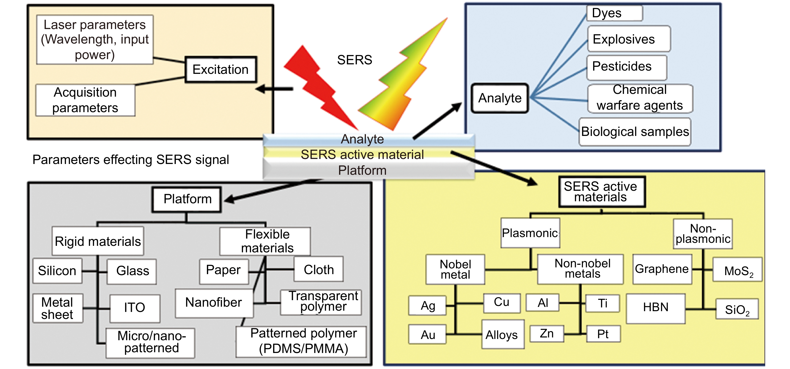
    Fig. 1. A schematic depicting the various parameters influencing the SERS signal.
    The ideal requirements of SERS substrates are summarized in this schematic.
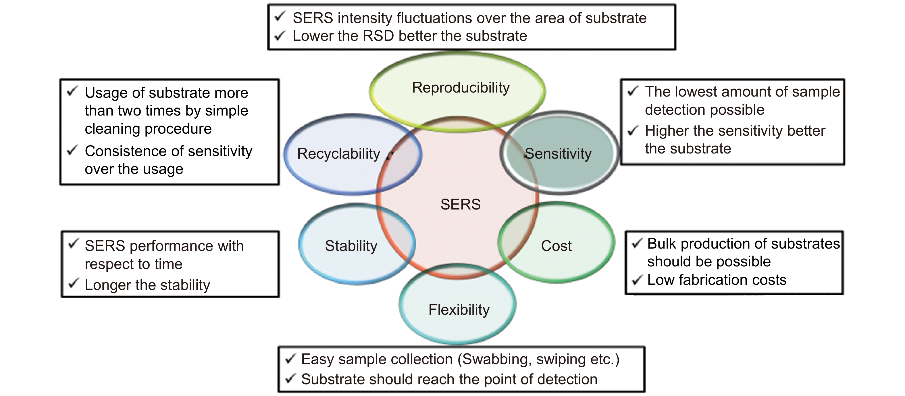
    Fig. 2. The ideal requirements of SERS substrates are summarized in this schematic.
    Year wise publications on flexible SERS substrates obtained through a search in SCOPUS.
    Fig. 3. Year wise publications on flexible SERS substrates obtained through a search in SCOPUS.
    Explosive trace detection using flexible SERS substrates detection of TNT, RDX, and PETN using self-assembly triangular nanoprisms on adhesive tape. Figure reproduced with permission from ref.58, Royal Society of Chemistry.
    Fig. 4. Explosive trace detection using flexible SERS substrates detection of TNT, RDX, and PETN using self-assembly triangular nanoprisms on adhesive tape. Figure reproduced with permission from ref.58, Royal Society of Chemistry.
    Various fabrication techniques used for paper-based SERS substrates. (a) Vapor deposition. (b) Inkjet printing. (c) Dipping. (d) Pen-on-paper. (e) Drop-casting on hydrophilic wells. (f) Self assembling. (g) In-situ reduction. Figure reproduced with permission from: (a) ref.77, (b) ref.72, The Royal Society of Chemistry; ref.71, American Chemical Society; (d) ref.73, John Wiley and Sons; (e) ref.80, Springer Nature; (f) ref.84, (g) ref.79, American Chemical Society.
    Fig. 5. Various fabrication techniques used for paper-based SERS substrates. (a) Vapor deposition. (b) Inkjet printing. (c) Dipping. (d) Pen-on-paper. (e) Drop-casting on hydrophilic wells. (f) Self assembling. (g) In-situ reduction. Figure reproduced with permission from: (a) ref.77, (b) ref.72, The Royal Society of Chemistry; ref.71, American Chemical Society; (d) ref.73, John Wiley and Sons; (e) ref.80, Springer Nature; (f) ref.84, (g) ref.79, American Chemical Society.
    Filter paper based SERS substrate by aggregated Ag/Au NPs for explosive molecule detection (Left side) (a) schematic of substrate preparation (b) and (c) FESEM images of bare filter and aggregated Ag NPs (Right side) SERS spectra of (a) PA (b) DNT (c) NTO using FP with optimized aggregated Ag NPs. Figure reproduced with permission from ref.94, American Chemical Society.
    Fig. 6. Filter paper based SERS substrate by aggregated Ag/Au NPs for explosive molecule detection (Left side) (a) schematic of substrate preparation (b) and (c) FESEM images of bare filter and aggregated Ag NPs (Right side) SERS spectra of (a) PA (b) DNT (c) NTO using FP with optimized aggregated Ag NPs. Figure reproduced with permission from ref.94, American Chemical Society.
    (a) A schematic of the synthesis of dual-functional PDMS-assisted paper-based SERS platform. (b) (i) The photograph of a sample collection from orange surface. (ii) A comparison of SERS spectra of CV with and without PDMS. (iii) SERS spectra of different concentrations of thiram (0.5−50 ppm). (iv) The peak intensity at 1380 cm−1 of thiram in orange juice as a function of the spiked sample concentration. Figure reproduced with permission from ref.95, Royal Society of Chemistry.
    Fig. 7. (a) A schematic of the synthesis of dual-functional PDMS-assisted paper-based SERS platform. (b) (i) The photograph of a sample collection from orange surface. (ii) A comparison of SERS spectra of CV with and without PDMS. (iii) SERS spectra of different concentrations of thiram (0.5−50 ppm). (iv) The peak intensity at 1380 cm−1 of thiram in orange juice as a function of the spiked sample concentration. Figure reproduced with permission from ref.95, Royal Society of Chemistry.
    Fabrication of flexible SERS substrates for Ag@T-A@SiO2-Au nanofibrous substrates. Figure reproduced with permission from ref.100, under a Creative Commons Attribution 4.0 International License.
    Fig. 8. Fabrication of flexible SERS substrates for Ag@T-A@SiO2-Au nanofibrous substrates. Figure reproduced with permission from ref.100, under a Creative Commons Attribution 4.0 International License.
    (a) Reflectance spectra of the ASFPAN nanofibrous membranes with Ag NPs; Photographs of three nanofibrous membranes (PAN, ASFPAN, and ASFPAN-Ag NPs) are shown in the inset. (b) SEM image and (c) TEM image of ASFPAN nanofibers (3 min). Inset in (c) shows the size distribution of Ag NPs.107, American Chemical Society.
    Fig. 9. (a) Reflectance spectra of the ASFPAN nanofibrous membranes with Ag NPs; Photographs of three nanofibrous membranes (PAN, ASFPAN, and ASFPAN-Ag NPs) are shown in the inset. (b) SEM image and (c) TEM image of ASFPAN nanofibers (3 min). Inset in (c) shows the size distribution of Ag NPs.107, American Chemical Society.
    (a) Schematic diagram representing the fabrication process of Au covered polymer nanostructure arrays using roll-to-roll ultraviolet nanoimprint lithography (R2R UV-NIL) technique (b) and (c) SERS spectra of R6G from 30 nm Au coating flexible substrate at different bending angles and bending cycles, respectively. Figure reproduced with permission from ref.114, under a Creative Commons Attribution 4.0 International License.
    Fig. 10. (a) Schematic diagram representing the fabrication process of Au covered polymer nanostructure arrays using roll-to-roll ultraviolet nanoimprint lithography (R2R UV-NIL) technique (b) and (c) SERS spectra of R6G from 30 nm Au coating flexible substrate at different bending angles and bending cycles, respectively. Figure reproduced with permission from ref.114, under a Creative Commons Attribution 4.0 International License.
    (a) Schematic of flexible non-woven fabric based substrate and the (b) SERS spectra of carbyl on apples, oranges, and bananas surfaces. Figure reproduced with permission from ref.129, under a Creative Commons Attribution 4.0 International License.
    Fig. 11. (a) Schematic of flexible non-woven fabric based substrate and the (b) SERS spectra of carbyl on apples, oranges, and bananas surfaces. Figure reproduced with permission from ref.129, under a Creative Commons Attribution 4.0 International License.
    S. No.AuthorReview topicRef.
    1Zhang et al.Flexible SERS substrates and recent advances in food safety analysisref.43
    2Yin et al.Recent process of 2D materials in SERSref.30
    3Klapec et al.2016–2019 published literature on the forensic related molecules and their various detection techniques using SERSref.44
    4Li et al.Fabrication and applications of flexible, transparent SERS substratesref.45
    5Forbes et al.Developed and challenges of SERS sensor in the detection of inorganic based explosivesref.46
    6Ji Sun et al.SERS substrate developments and combination with other technologies in on-site analysis using portable Raman spectrometerref.19
    7Jingjing et al.Different dimensional (0D, 1D, 2D and 3D) SERS substrates for explosive detectionref.47
    8Shvalya et al.Plasmonic NPs and 3D plasmonic NSs sensors with biological, medical, military, and chemical applicationsref.48
    9To et al.Explosive trace detection technologies and latest advancesref.49
    10Ren et al.Qualitative and quantitative analysis; strategies of practical application of SERS substratesref.50
    11Huang et al.Paper SERS substrates in food safetyref.51
    12Chen et al.2D SERS substrates in chemical and biosensingref.52
    13Dinesh et al.Flexible sensor fabrication with various printing techniquesref.40
    14Xue et al.Flexible nanofiber-based substrates fabrication and applicationref.53
    15Ogundare et al.Cellulose-based SERS substrates: fundamentals and principlesref.39
    16Zamora Sequeira et al.Various methods for the determination of pesticidesref.2
    17Piolt et al.Key aspects of SERS and application in the biomedical fieldref.54
    18Ogundare et al.Cellulose substrate fundamental, preparation methods, and applicationsref.39
    19Lee et al.Analyte manipulation and hybrid SERS platforms for real-world applicationsref.55
    20Xu et al.Latest advances of flexible SERS substrates in point of care diagnostic in tunable, sample swapping and in-situ SERS detection highlightsref.56
    21Zhang et al.Electrospinning NPs based material and their sensing applicationref.57
    22Restaino et al.Plasmonic paper SERS substrates-preparation methods and sample collectionsref.36
    Table 1. Important review articles on various applications of SERS that have been reported in the last three-years (2019–2021).
    Flexible substrate typeHazardous material type studiedMethod usedSERS active materialMolecules investigated - sensitivityRef.
    Paper/CelluloseExplosivesInkjet printingPABT modified-Ag NPs-A4 paperTNT- pMref.132
    In-situAg NPs in agarose film supported on filter paperTNT- 10−8 M ref.78
    ImmersionAg nano triangles-filter paperPA- 10−6 M p-ATP- 10−8 M ref.88
    SoakingAggregated Ag/Au NPs-filter paperPA- 5 µM DNT- 1 µM NTO- 10 µM ref.94
    Drop castingStar-shaped Au NPsPA-5 µMref.133
    ReductionAg Nanostructures- filter paper Whatman 42Urea nitrate- 10−6 M CV- 10−8 M ref.134
    DrugsInkjet printingAg- chromatography paperOrganophosphate malathion –413 pg, Heroin –9 ng, Cocaine –15 ng ref.135
    Plasma assisted chemical depositionAu-Whatman filter paper grade 1 Cocaine- 1 ng/mlref.136
    DyesIn-situAg NPs-polydopamine -Filter paperR6g- 10−10 M MG residue on Fish scales- 0.04635 pg/cm2, Crab shells- 0.06952 pg/cm2 and Shrimp skins- 0.09270 pg/cm2ref.137
    Inkjet-printingMoO3−x nanosheets on Chromatographic paper, printing paper, filter paper R6g- 10–7 M CV- 10–6 M and MG- 10–6 M on fish surface ref.74
    In-situAu-filter paper (Advantec #1)MG-damped fish– 10 ppbref.138
    PesticidesSilver mirror reactionAg- filter paperThiram- 10−7 M ref.139
    Pen on paperAu NPs (15–120 nm); Au NRs (50 nm long, 14 nm thick); Ag NPs (50-80 nm) –A4 paper, Filter paperThiabendazole < 20 ppbref.73
    Airbrush spray methodAg NPs -glass fibre paperEnoxacin & Enrofloxacin- 10 −5 M ref.140
    PrintingAu@Ag 30 nm Au core & 7 nm Ag shell -filter paperThiram- 10−9 M ref.141
    Screen printingAg NPs/GO- paperThiram 0.26 ng cm−2Thiabendazole 28 ng cm−2Methylparathion 7.4 ng cm−2ref.142
    Immersion followed by APTMSAg NPs-PDMS spongeTriazophos 0.79 ng Methyl Parathion 1.58 ng ref.143
    Vacuum-assisted filtrationAuNPs- cellulose nanofiberThiram- 1 pM Tricyclazole- 10 pM ref.144
    In-situAu NPs-pseudo-paperThiram- 1.1 ng/cm2ref.145
    Laser techniquesAu/Ag film-print paperFungicide mancozeb (Dithane DG) and insecticide thiamethoxam (Aktara 25 BG)ref.146
    Immerson in NaCl solution for 5 min +dip-coatingAg NPs- filter PaperMelamine- 1 ppm Thiram- 1 ppm ref.147
    ImmersionFP-Au NPsMethyl parathion- 0.011 μg/cm2ref.148
    In-situNanocellulose fibers-Ag NPsThiram- 0.05 ppm Thiabendazole- 0.09 ppm, MG 0.0014 ppm Enrofloxaci- 0.069 ppm ref.149
    Silicon rubber mask and a vacuum filtration Au NRs -cellulose hydrogelsThiram- 100 fMref.92
    Drop castingQuartz paper/Cellulose nanofiber/ mixture (Ag NPs+Au NSs)Ferbam on kale leaves (50 µg/kg)ref.150
    Vacuum filtrationCellulose nanofibers-Au NPsThiram- 10−8M ref.151
    Drop casting, inkjet printingAu NPs-Whatman 44 FPBenzenethiol chemical aerosol Pyridine ref.152
    Vacuum filtrationGlass-fiber filter paper-Ag NWs coupled with polymerase chain reaction (PCR)DNAref.153
    Electrochemical depositionMesoporous Au film@Ag NWs@cellulose nanofiber paperR6g - 100 fM Thiram - 10 fM 2-naphthalenethiol-1 ppb ref.154
    Self-assemblingCellulose nanofibers -Ag@DNA/PDA (polydopamine)Rhodamine 6G. Thiamethoxamon- 0.003 mg/kg. ref.155
    Cotton budsAntibioticsIn situ reductionAg NPs-cellulose nanocrystals-Filter paperPhenylethanolamine A-10−9 M Metronidazole- 10−7 M ref.93
    ExplosivesSelf-assembly & In situAg NPs-cotton swab2,4 DNT- 5 ngref.156
    PesticidesSoaking, freezing, and dryingAg NPs-chitosan foamTriasophols Methidathion Isocrabophos ref.157
    Dipping & dryingAg NPs-cotton swab with NaClThiabendazole (TBZ), thiram, TBZ + thiram ref.158
    3D- spongeExplosivesIn situAg NPs -polyurethane spongePerchlorates- 0.13 ng CChlorates- 0.13 ng Nitrates- 0. 11 ng ref.159
    Nanofiber matPesticidesElectrospinningAu coated PVA nanofiberDeltamethrin- 0.33 mg/kg Quinalphos- 0.28 mg/kg Thiacloprid- 0.26 mg/kg ref.104
    CWA simulantsElectrospinningAu NPs –PVA nanofiberMethyl salicylateref.160
    DyesElectrospinningAg NPs-PVA nanofiberR6G-10−5 M ref.161
    Electrospinning and in-situAg NPs-Polyimide (PI) nanofabricp-Aminothiophenol (p-ATP)- 10−14 mol/L), ref.162
    FabricPesticidesSelf-assembly/in-situAg NPs- non woven fabricIsocarbophos Sumicidin Phosgene ref.163
    Dip coatingTriangular Ag nanoplates-Cotton fabricCarbaryl- 10−5M ref.164
    In situPolydopamine mediated Ag-Au NPs – cotton fabricCarbaryl- 10−6M ref.165
    Magneton sputteringAg NPs-cotton fabricThiram - 1 ppmref.127
    Magnetron sputteringAg-polyester fabricR6G on cucumber, MG and Thiramref.166
    Photochemical deposition (254 nm)Ag NPs on TiO2 coated polyester fiber membranes Sodium saccharin in soft drinks- 0.3 mg/L, (cola and sprite)ref.167
    In-situ growthAg NPs-Cotton fabricsPATP-10−8 M ref.168
    Vacuum evaporationAg coated (10 nm) nylon fabricsPATP-10−9 M Thiram on cucumber surface-10−7 M ref.169
    DyesVacuum thermal evaporation and high-temperature annealingAg NPs-carbon fiber clothR6g- 10−14 mol·L−1ref.170
    PolymersExplosivesOriented stacking and in-situAg and Au–Ag nanoplates- PETTNT- 10 nM RDX- 10 nM ref.171
    Self-assemblingAu triangular nanoprisms on adhesive film (Scotch magic-tape)TNT- 900 ppq RDX- 50 ppq and PETN- 50 ppq ref.58
    Incubated overnight followed by thorough rinsing dryingAu NPs,Au NRs and Au NCs on elastomeric film (PDMS)TNT vaporref.172
    Gravure printingAg NPs-PETDNT vaporref.173
    Sol–gel method and magnetron sputteringAg NPs-Porous silica aerogelsNTO- 7.94×10⁻¹⁰ M ref.174
    UV lithography and Au deposition Ag NPs-Au coated -nanowrinkled zigzag micropattern on PDMS layerTNT- 10−13 mol·L−1TNT residue(10−9 mol·L−1) on cloth bag ref.175
    DyesElectron-beam evaporation-uniaxial stretching Stretched Ag coated poly(ε-caprolactone) film MG-green mussel surface- 0.1×10⁻⁶ M ref.176
    Pyramid Si templateMoS2/AgNPs/inverted pyramidal PMMA R6G+MGref.177
    Pyramid Si templateGO/Ag NPs/ pyramidal PMMAMG on shrimpref.178
    Ar plasma etching and Au evaporationWorm-like Au NSs – PET filmR6G-10−9 M ref.179
    Self-assembly and in situ chemical reductionRaspberry-like polyamide@Ag hybrid nanoarray filmR6g-10−14 M Adenosine- 10−9 M ref.180
    PesticidesDrop-dry methodAu NPs (25 nm) - adhesive tapeParathion-methyl- 2.60 ng/cm2Thiram 0.24 ng/cm2Chlorpyrifos 3.51 ng/cm2on apples, oranges, cucumbers, and green vegetables surfaces ref.181
    Spin coating and manual peelingAgNP@AgNW network-PDMSThiram (0.1µM) on a leaf surface and MG (0.1µM) on a living fish scale ref.182
    Paste and peeling of self-assembled NPs from SiAdhesive acrylic polymer tape and polyethene terephthalate (PET) film (T/Au@Ag/PET)Thiram on apple, tomato, and cucumber peels (5 ng/cm2) ref.113
    Seed mediatedGold nanobush+PDMSThiabendazole (TBZ) on cherry – 0.64 ng/ml Carbaryl TBZ+Carbaryl ref.183
    Femtosecond laser induced plasma assisted ablationAg NPs and Au NPs FEP (fluorinated ethylene propylene) Thiram on apple- 7.96 ng/cm2ref.184
    Drop castingAg NS with spikes-adhesive tapePhosmet & carbaryl on apple-surface 10 −7M ref.185
    Table 2. Summary of the recent flexible SERS substrates, their preparation methods, materials used, and the sensitivities achieved (2014-2021).
    S. No.CompanySERS substrateSensitivityStabilityCostRef
    1StellarnetCellulose with Au NPs~1063 months$199 (pack of 30)ref.186
    2Horiba France SASGlass coated with Au nanorods processed by dynamic oblique vacuum evaporationref.187
    3SERSitiveElectrodeposition of silver and gold nanoparticles on an ITO glass surface~105–1064 months5 pcs Ag- €115 5 pcs Ag-Au- €138 ref.188
    4EnSpectr Inc.Si/Glass passivated with a thin transparent dielectic layer.~106Stable when unpackedref.189
    5SilmecoNanostructured Si deposited with Gold (Au), Silver (Ag)5 units €350ref.190
    6HamamatsuAu NS on polypropylene3 months when unpacked ref.191
    7Integrated OpticsAg/Au coating on silicate glass.2 monthsAg- €15 Au- €18 ref.192
    8Mesophotonics. Ltd. KlariteSi100 USD for single 2 mm × 2 mm sample.ref.193
    9Q SERS TMAu NSs on Si (5 mm × 5 mm)ppb to ppm6 months (package) 2 weeks (package opened) 2 units $50 USDref.194
    10MetrohmAg, Au based Filter paperref.195
    Table 3. A summary of the commercially available SERS substrates, their costs, sensitivities and their stability (non-exhaustive).
    Moram Sree Satya Bharati, Venugopal Rao Soma. Flexible SERS substrates for hazardous materials detection: recent advances[J]. Opto-Electronic Advances, 2021, 4(11): 210048-1
    Download Citation