
Moram Sree Satya Bharati, Venugopal Rao Soma. Flexible SERS substrates for hazardous materials detection: recent advances[J]. Opto-Electronic Advances, 2021, 4(11): 210048-1
- Opto-Electronic Advances
- Vol. 4, Issue 11, 210048-1 (2021)
Abstract
Introduction
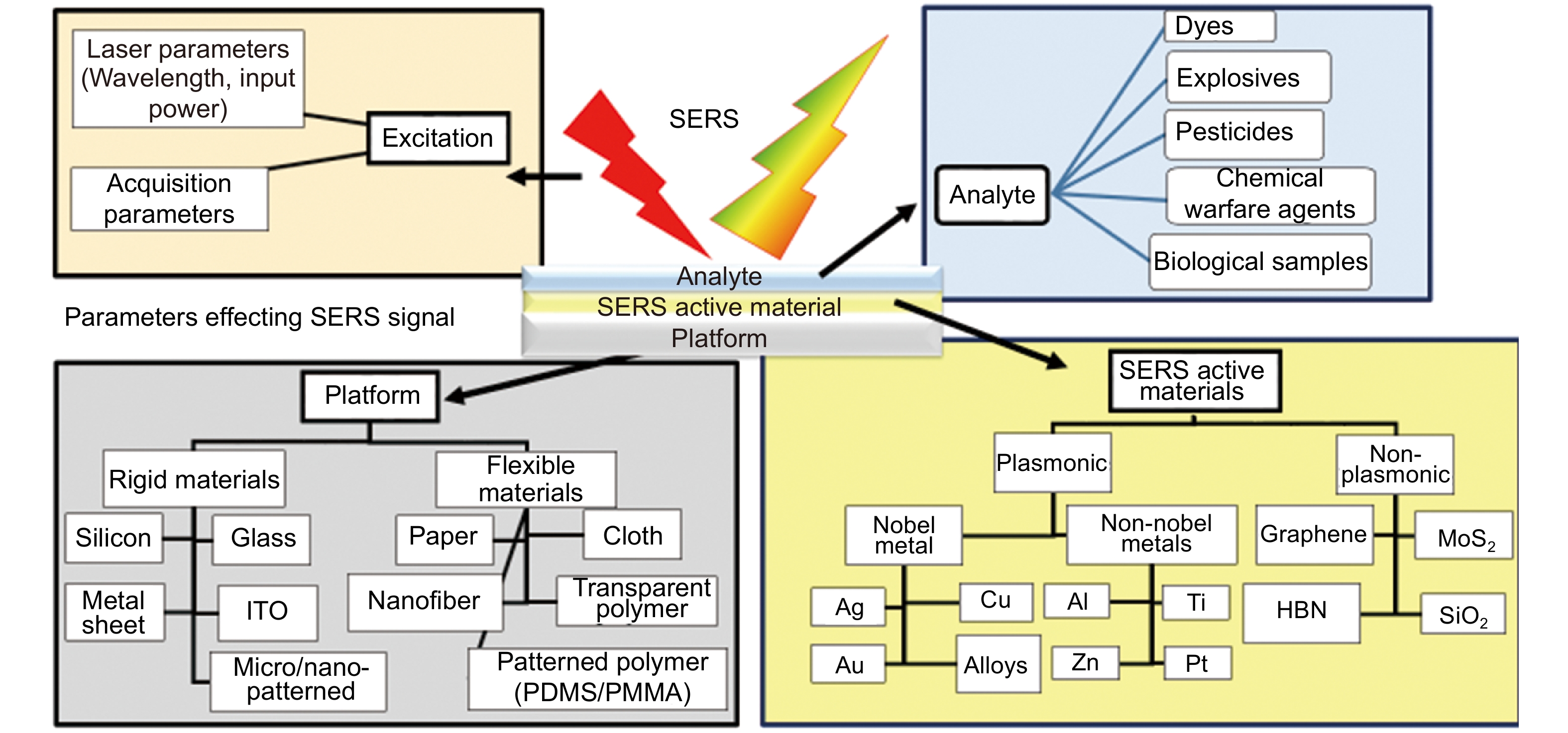
In the present-day scenario, human health, and environmental safety are the foremost concerns worldwide. Hazardous materials are referred to as those which have been determined to be capable of presenting an unreasonable risk to human health, safety, and property. The main characteristics of these materials are ignitability, corrosivity, reactivity, or toxicity. The specific categories among these materials are explosives, flammable liquids, gases, oxidizers, corrosives, flammable solids, radioactive materials, poisonous/infectious substances, and dangerous substances. We start with a short overview of various hazardous materials followed by the introduction of Raman spectroscopy and surface enhanced Rama spectroscopy/scattering (SERS) techniques. This review aims to report on the detection of hazardous materials such as explosives, pesticides, and simulants of chemical warfare agents using flexible SERS substrates.
Hazardous materials
Explosives/high energy materials (HEMs) are those materials that contain nitro groups (which are energetic) and release an enormous amount of energy in the form of light and heat when they are subjected to an external stimulus such as (a) spark (b) shock or even (c) friction. Explosives are commonly categorized as primary and secondary depending on their detonation (velocity, pressure etc.) and sensitivity parameters. Primary explosives are extremely sensitive and release enormous energy even with a small perturbation such as shock/collision. Therefore, the difficulty is generally high while handling the primary explosives. They act as boosters or initiators for detonating secondary explosives. Lead azide and mercury fulminate are a few examples of primary explosives, while 1,3,5,7-Tetranitro-1,3,5,7- tetrazocane (HMX), 1,3,5-Trinitroperhydro-1,3,5-triazine (RDX), trinitrotoluene (TNT), etc. are representative of secondary explosives secondary explosives secondary explosives seconda. Interestingly, there are few home-prepared explosives utilized in the preparation of improvised explosive devices (IEDs). These are now easily synthesized at the laboratory level from simple molecules such as ammonium nitrate (AN), dinitrotoluene (DNT), picric acid (PA), etc.. Pesticides are the chemicals used by farmers/transporters to protect the crops/vegetables/fruits from insects/pests/rodents. The overused pesticides will remain as residues in the food, which may cause risk to human health (cancer/allergies/intoxications) and the ecosystem (surface water/soil)
Therefore, rapid and reliable detection of these hazardous molecules is the primary concern of both governmental agencies and research community to reduce the risk to society. Razdan and co-workers
Raman spectroscopy and variants
Raman spectroscopy is a simple, rapid, and a non-destructive spectroscopic technique based on molecular vibrations as signatures in the spectra. The Raman spectrum of any analyte molecule provides specific information and conveys chemical/structural information. This is important in the case of explosives (in pure form or even in the mixture form) irrespective of solid, liquid, powder, or gas state
In the present times, flexible SERS substrates have received great interest due to them possessing the advantages of (a) easy sampling by swabbing/wrapping directly on any curved/rough surfaces (b) large scalability by printing/roll to roll manufacturing/electrospinning etc. and (c) low overall cost of the sensing system. The development of handy flexible substrates with compact Raman devices/smart-phones can possibly provide portable sensors in real-world sensing/safety applications and serve as a powerful analytical tool for on-field analysis. For example, the possibility of detection of ultralow concentrations [picomolar (10−12 M or pM) to femtomolar (10−15 M or fM)] of two nerve gases, VX and Tabun was reported recently by Hakonen et al
Surface-enhanced Raman scattering (SERS)
Martin Fleischmann and co-workers had reported a fortunate discovery way back in 1974, in which they observed enhanced Raman signals of a pyridine molecule adsorbed on an electrochemically roughened silver surface
![]()
Figure 1.
Reviews on different SERS studies
A variety of review reports on SERS have been published over the last decades addressing the issues concerned with fabrication techniques, applications, and their developments. For example, Fan et al
![]()
Figure 2.
Sensitivity is the biggest virtue of a good SERS substrate is the detection of molecules at very low concentrations [traces meaning parts per billion (ppb) or parts per trillion (ppt) or parts per quadrillion (ppq)]. The sensitivity is generally expressed in terms of the lowest quantity of probe molecule detection possible with a given SERS substrate. The Raman signal disappears when the molecule concentrations reach a limit value. The sensitivity of the SERS substrate varies from molecule to molecule. The sensitivity of the SERS substrate is typically represented by the enhancement factor (limit of detection for a particular vibration mode of the probe molecule). Therefore, one should be judicious with the SERS substrate and select one with a higher enhancement factor or a lower limit of detection (LOD) over a wide range of analytes. Reproducibility is related to the variation of SERS intensity of the probe molecule over the NS surface. The smaller the variation in the signal, the higher the reproducibility and it is generally reported in terms of RSD (relative standard deviation) of the SERS signal. This depends mainly on the distribution of hotspots on the substrate. Low reproducibility of any SERS substrate affects the potential usage in practical applications. It is highly challenging to produce a highly reproducible SERS platform along with a homogeneous distribution of hotspots. The fluctuations of the SERS signals are calculated statistically with RSD of the particular mode intensity in the SERS spectrum. The magnitude of %RSD, indicative of the coefficient of variation, provides uncertainty in the measurement. Lower RSD values indicate a superior substrate in terms of reproducibility. Recyclability is another essential factor to test the usage of the same SERS substrate after detecting one/two molecules followed by proper cleaning procedures
The important results from the literature survey over the last 5−10 years concerning the usage of flexible SERS substrate for various hazardous materials detection is also summarized in this article. A large number of papers have been published in this area. To demonstrate the magnitude of research, a simple search for papers published in the journals and conferences, including the title/keywords/abstract “flexible Surface Enhanced Raman Spectroscopy” or “flexible Surface Enhanced Raman Scattering” or “flexible SERS” as indexed by the Scopus search engine, resulted in typically >100 papers in 2019, >100 papers in 2020 and >40 in the year 2021 alone. The corresponding data obtained is plotted as a bar graph and is shown inFig. 3. The identification of all the developments and practical applications of flexible SERS studies in various fields will be difficult to be presented in this review. Therefore, we have acknowledged the most important recent review articles and those are listed in the Table 1 below. The readers are suggested to select and pursue the review based on their interest(s). This review is limited to the recent studies (typically during the last 3−4 years) on flexible SERS substrates used in the detection of hazardous materials, rather than including broad discussions on solid SERS substrates (nanostructures on solid targets and metal NPs suspension on the solid platform) and their developments, which is a huge field. This review is warranted because of the extremely rapid developments in the area of different nanomaterials synthesized (for SERS studies including plasmonic and non-plasmonic), novel methodologies developed for incorporating various nanoparticles in different flexible platforms, and detection of diverse analyte molecules.
![]()
Figure 3.
| S. No. | Author | Review topic | Ref. |
| 1 | Zhang et al. | Flexible SERS substrates and recent advances in food safety analysis | ref. |
| 2 | Yin et al. | Recent process of 2D materials in SERS | ref. |
| 3 | Klapec et al. | 2016–2019 published literature on the forensic related molecules and their various detection techniques using SERS | ref. |
| 4 | Li et al. | Fabrication and applications of flexible, transparent SERS substrates | ref. |
| 5 | Forbes et al. | Developed and challenges of SERS sensor in the detection of inorganic based explosives | ref. |
| 6 | Ji Sun et al. | SERS substrate developments and combination with other technologies in on-site analysis using portable Raman spectrometer | ref. |
| 7 | Jingjing et al. | Different dimensional (0D, 1D, 2D and 3D) SERS substrates for explosive detection | ref. |
| 8 | Shvalya et al. | Plasmonic NPs and 3D plasmonic NSs sensors with biological, medical, military, and chemical applications | ref. |
| 9 | To et al. | Explosive trace detection technologies and latest advances | ref. |
| 10 | Ren et al. | Qualitative and quantitative analysis; strategies of practical application of SERS substrates | ref. |
| 11 | Huang et al. | Paper SERS substrates in food safety | ref. |
| 12 | Chen et al. | 2D SERS substrates in chemical and biosensing | ref. |
| 13 | Dinesh et al. | Flexible sensor fabrication with various printing techniques | ref. |
| 14 | Xue et al. | Flexible nanofiber-based substrates fabrication and application | ref. |
| 15 | Ogundare et al. | Cellulose-based SERS substrates: fundamentals and principles | ref. |
| 16 | Zamora Sequeira et al. | Various methods for the determination of pesticides | ref. |
| 17 | Piolt et al. | Key aspects of SERS and application in the biomedical field | ref. |
| 18 | Ogundare et al. | Cellulose substrate fundamental, preparation methods, and applications | ref. |
| 19 | Lee et al. | Analyte manipulation and hybrid SERS platforms for real-world applications | ref. |
| 20 | Xu et al. | Latest advances of flexible SERS substrates in point of care diagnostic in tunable, sample swapping and in-situ SERS detection highlights | ref. |
| 21 | Zhang et al. | Electrospinning NPs based material and their sensing application | ref. |
| 22 | Restaino et al. | Plasmonic paper SERS substrates-preparation methods and sample collections | ref. |
Table 1. Important review articles on various applications of SERS that have been reported in the last three-years (2019–2021).
Flexible SERS substrates
A forthright method to achieve the SERS-active substrates is to dry the colloidal NPs (preferably plasmonic) solution on any of the glass/silicon/paper/metal surfaces.
The capabilities of flexible SERS substrates have gained tremendous research interest due to
• Inexpensive fabrication procedures making it possible to prepare large area substrates.
• Easy-to-use nature for on-site detection of a wide range of probe molecules.
• Flexibility in sample collection, i.e., possible to collect the probe molecules/sample directly from any rough surface (e.g., suitcase, bag, table surface, fruit, etc.) with the substrate by simple swabbing/swiping.
The merits of the SERS technique with the portable Raman spectrometer now widely used in national security, food safety, and environmental monitoring.
Recently explosives detection was approached by fabricating various flexible SERS substrates. Liyanage et al
![]()
Figure 4.
Paper-based SERS substrates
A detailed literature survey revealed that a variety of papers were used (as a base material) for preparing the SERS substrates such as filter paper
![]()
Figure 5.
Kim et al
![]()
Figure 6.Filter paper based SERS substrate by aggregated Ag/Au NPs for explosive molecule detection (Left side) (
Lin et al
![]()
Figure 7.(
Polymer-based SERS substrates
Nanofiber mats
Electrospinning is a method of translation of polymeric solution/melt (with or without additives) into solid nanofibers by applying the electric field
![]()
Figure 8.
The SERS performance of nanofiber depends on the properties of
• nanofibers (polymer nature, fiber diameter, the morphology of the nanofibers, and spinning time, etc.) and
• nanoparticles
• Decoration of NPs on the fiber
• The loading of NPs on the nanofiber mat leads to the NPs assembly with extremely small spacing providing scope for abundant hot spots. These play a crucial factor in SERS response.
Electrospinning polymer fibers can be used as SERS substrates by loading plasmonic NPs; similar to paper substrates, several methods are reported for embedding metal NPs onto the electrospun polymer films like dispersion of metal precursor and pre-mixing of metal NPs into the polymer solution and surface medications after electrospinning. Chamuah et al
![]()
Figure 9.(
Recently flexible polymer-based (PDMS
![]()
Figure 10.(
Fang et al
Textile based SERS substrates
The textile fabrics have also been investigated as an attractive SERS substrate (akin to paper and electrospun fiber substrate) because the fabric is naturally strong, flexible, soft, and a lightweight material. In textiles, various materials are available such as cotton, wool, silk, etc.. Comparable to other flexible substrates, the loading of NPs can be done in two ways, i.e., in-situ synthesis [soaking in different metal salts] and direct deposition of NPs [anisotropic silver nano-prisms and nano-disks to wool fabric has been reported recently
![]()
Figure 11.(
Table 2 summarizes the most important details of recently reported flexible SERS substrates including their preparation methods, materials used in those studies, and the sensitivities achieved. Such data is extremely important since the developments are occurring at a rapid pace and it is imperative to identify the strengths and weakness of each of these methodologies to come up with a viable and practical technique for making robust flexible SERS substrates. These flexible SERS substrates find niche applications in the detection of various hazardous materials in Defence, food, and environmental safety issues. Sensitivity estimations are reported in various parameters such as Molar (M), parts per billion (ppb), nanogram (ng), ng/cm2 and mg/kg. For example, in case of Thiram molecule (molecular weight of 240.44) 10 ppb is ~0.42 nM which is equivalent to ~1 pg in 10 µL; 1 ppb = 1 µg/kg; 1 ppm = 1000 ppb. Table 3 represents a summary of the commercially available SERS substrates (which is not exhaustive) and it is evident that each one of them have varied properties including the sensitivity, stability, and cost. Liu et al
| Flexible substrate type | Hazardous material type studied | Method used | SERS active material | Molecules investigated - sensitivity | Ref. |
| Paper/Cellulose | Explosives | Inkjet printing | PABT modified-Ag NPs-A4 paper | TNT- pM | ref.132 |
| In-situ | Ag NPs in agarose film supported on filter paper | TNT- 10−8 M | ref.78 | ||
| Immersion | Ag nano triangles-filter paper | PA- 10−6 M p-ATP- 10−8 M | ref.88 | ||
| Soaking | Aggregated Ag/Au NPs-filter paper | PA- 5 µM DNT- 1 µM NTO- 10 µM | ref.94 | ||
| Drop casting | Star-shaped Au NPs | PA-5 µM | ref.133 | ||
| Reduction | Ag Nanostructures- filter paper Whatman 42 | Urea nitrate- 10−6 M CV- 10−8 M | ref.134 | ||
| Drugs | Inkjet printing | Ag- chromatography paper | Organophosphate malathion –413 pg, Heroin –9 ng, Cocaine –15 ng | ref.135 | |
| Plasma assisted chemical deposition | Au-Whatman filter paper grade 1 | Cocaine- 1 ng/ml | ref.136 | ||
| Dyes | In-situ | Ag NPs-polydopamine -Filter paper | R6g- 10−10 M MG residue on Fish scales- 0.04635 pg/cm2, Crab shells- 0.06952 pg/cm2 and Shrimp skins- 0.09270 pg/cm2 | ref.137 | |
| Inkjet-printing | MoO3−x nanosheets on Chromatographic paper, printing paper, filter paper | R6g- 10–7 M CV- 10–6 M and MG- 10–6 M on fish surface | ref.74 | ||
| In-situ | Au-filter paper (Advantec #1) | MG-damped fish– 10 ppb | ref.138 | ||
| Pesticides | Silver mirror reaction | Ag- filter paper | Thiram- 10−7 M | ref.139 | |
| Pen on paper | Au NPs (15–120 nm); Au NRs (50 nm long, 14 nm thick); Ag NPs (50-80 nm) –A4 paper, Filter paper | Thiabendazole < 20 ppb | ref.73 | ||
| Airbrush spray method | Ag NPs -glass fibre paper | Enoxacin & Enrofloxacin- 10 −5 M | ref.140 | ||
| Printing | Au@Ag 30 nm Au core & 7 nm Ag shell -filter paper | Thiram- 10−9 M | ref.141 | ||
| Screen printing | Ag NPs/GO- paper | Thiram 0.26 ng cm−2Thiabendazole 28 ng cm−2Methylparathion 7.4 ng cm−2 | ref.142 | ||
| Immersion followed by APTMS | Ag NPs-PDMS sponge | Triazophos 0.79 ng Methyl Parathion 1.58 ng | ref.143 | ||
| Vacuum-assisted filtration | AuNPs- cellulose nanofiber | Thiram- 1 pM Tricyclazole- 10 pM | ref.144 | ||
| In-situ | Au NPs-pseudo-paper | Thiram- 1.1 ng/cm2 | ref.145 | ||
| Laser techniques | Au/Ag film-print paper | Fungicide mancozeb (Dithane DG) and insecticide thiamethoxam (Aktara 25 BG) | ref.146 | ||
| Immerson in NaCl solution for 5 min +dip-coating | Ag NPs- filter Paper | Melamine- 1 ppm Thiram- 1 ppm | ref.147 | ||
| Immersion | FP-Au NPs | Methyl parathion- 0.011 μg/cm2 | ref.148 | ||
| In-situ | Nanocellulose fibers-Ag NPs | Thiram- 0.05 ppm Thiabendazole- 0.09 ppm, MG 0.0014 ppm Enrofloxaci- 0.069 ppm | ref.149 | ||
| Silicon rubber mask and a vacuum filtration | Au NRs -cellulose hydrogels | Thiram- 100 fM | ref.92 | ||
| Drop casting | Quartz paper/Cellulose nanofiber/ mixture (Ag NPs+Au NSs) | Ferbam on kale leaves (50 µg/kg) | ref.150 | ||
| Vacuum filtration | Cellulose nanofibers-Au NPs | Thiram- 10−8M | ref.151 | ||
| Drop casting, inkjet printing | Au NPs-Whatman 44 FP | Benzenethiol chemical aerosol Pyridine | ref.152 | ||
| Vacuum filtration | Glass-fiber filter paper-Ag NWs coupled with polymerase chain reaction (PCR) | DNA | ref.153 | ||
| Electrochemical deposition | Mesoporous Au film@Ag NWs@cellulose nanofiber paper | R6g - 100 fM Thiram - 10 fM 2-naphthalenethiol-1 ppb | ref.154 | ||
| Self-assembling | Cellulose nanofibers -Ag@DNA/PDA (polydopamine) | Rhodamine 6G. Thiamethoxamon- 0.003 mg/kg. | ref.155 | ||
| Cotton buds | Antibiotics | In situ reduction | Ag NPs-cellulose nanocrystals-Filter paper | Phenylethanolamine A-10−9 M Metronidazole- 10−7 M | ref.93 |
| Explosives | Self-assembly & In situ | Ag NPs-cotton swab | 2,4 DNT- 5 ng | ref.156 | |
| Pesticides | Soaking, freezing, and drying | Ag NPs-chitosan foam | Triasophols Methidathion Isocrabophos | ref.157 | |
| Dipping & drying | Ag NPs-cotton swab with NaCl | Thiabendazole (TBZ), thiram, TBZ + thiram | ref.158 | ||
| 3D- sponge | Explosives | In situ | Ag NPs -polyurethane sponge | Perchlorates- 0.13 ng CChlorates- 0.13 ng Nitrates- 0. 11 ng | ref.159 |
| Nanofiber mat | Pesticides | Electrospinning | Au coated PVA nanofiber | Deltamethrin- 0.33 mg/kg Quinalphos- 0.28 mg/kg Thiacloprid- 0.26 mg/kg | ref.104 |
| CWA simulants | Electrospinning | Au NPs –PVA nanofiber | Methyl salicylate | ref.160 | |
| Dyes | Electrospinning | Ag NPs-PVA nanofiber | R6G-10−5 M | ref.161 | |
| Electrospinning and in-situ | Ag NPs-Polyimide (PI) nanofabric | p-Aminothiophenol (p-ATP)- 10−14 mol/L), | ref.162 | ||
| Fabric | Pesticides | Self-assembly/in-situ | Ag NPs- non woven fabric | Isocarbophos Sumicidin Phosgene | ref.163 |
| Dip coating | Triangular Ag nanoplates-Cotton fabric | Carbaryl- 10−5M | ref.164 | ||
| In situ | Polydopamine mediated Ag-Au NPs – cotton fabric | Carbaryl- 10−6M | ref.165 | ||
| Magneton sputtering | Ag NPs-cotton fabric | Thiram - 1 ppm | ref.127 | ||
| Magnetron sputtering | Ag-polyester fabric | R6G on cucumber, MG and Thiram | ref.166 | ||
| Photochemical deposition (254 nm) | Ag NPs on TiO2 coated polyester fiber membranes | Sodium saccharin in soft drinks- 0.3 mg/L, (cola and sprite) | ref.167 | ||
| In-situ growth | Ag NPs-Cotton fabrics | PATP-10−8 M | ref.168 | ||
| Vacuum evaporation | Ag coated (10 nm) nylon fabrics | PATP-10−9 M Thiram on cucumber surface-10−7 M | ref.169 | ||
| Dyes | Vacuum thermal evaporation and high-temperature annealing | Ag NPs-carbon fiber cloth | R6g- 10−14 mol·L−1 | ref.170 | |
| Polymers | Explosives | Oriented stacking and in-situ | Ag and Au–Ag nanoplates- PET | TNT- 10 nM RDX- 10 nM | ref.171 |
| Self-assembling | Au triangular nanoprisms on adhesive film (Scotch magic-tape) | TNT- 900 ppq RDX- 50 ppq and PETN- 50 ppq | ref.58 | ||
| Incubated overnight followed by thorough rinsing drying | Au NPs,Au NRs and Au NCs on elastomeric film (PDMS) | TNT vapor | ref.172 | ||
| Gravure printing | Ag NPs-PET | DNT vapor | ref.173 | ||
| Sol–gel method and magnetron sputtering | Ag NPs-Porous silica aerogels | NTO- 7.94×10⁻¹⁰ M | ref.174 | ||
| UV lithography and Au deposition | Ag NPs-Au coated -nanowrinkled zigzag micropattern on PDMS layer | TNT- 10−13 mol·L−1TNT residue(10−9 mol·L−1) on cloth bag | ref.175 | ||
| Dyes | Electron-beam evaporation-uniaxial stretching | Stretched Ag coated poly(ε-caprolactone) film | MG-green mussel surface- 0.1×10⁻⁶ M | ref.176 | |
| Pyramid Si template | MoS2/AgNPs/inverted pyramidal PMMA | R6G+MG | ref.177 | ||
| Pyramid Si template | GO/Ag NPs/ pyramidal PMMA | MG on shrimp | ref.178 | ||
| Ar plasma etching and Au evaporation | Worm-like Au NSs – PET film | R6G-10−9 M | ref.179 | ||
| Self-assembly and in situ chemical reduction | Raspberry-like polyamide@Ag hybrid nanoarray film | R6g-10−14 M Adenosine- 10−9 M | ref.180 | ||
| Pesticides | Drop-dry method | Au NPs (25 nm) - adhesive tape | Parathion-methyl- 2.60 ng/cm2Thiram 0.24 ng/cm2Chlorpyrifos 3.51 ng/cm2on apples, oranges, cucumbers, and green vegetables surfaces | ref.181 | |
| Spin coating and manual peeling | AgNP@AgNW network-PDMS | Thiram (0.1µM) on a leaf surface and MG (0.1µM) on a living fish scale | ref.182 | ||
| Paste and peeling of self-assembled NPs from Si | Adhesive acrylic polymer tape and polyethene terephthalate (PET) film (T/Au@Ag/PET) | Thiram on apple, tomato, and cucumber peels (5 ng/cm2) | ref.113 | ||
| Seed mediated | Gold nanobush+PDMS | Thiabendazole (TBZ) on cherry – 0.64 ng/ml Carbaryl TBZ+Carbaryl | ref.183 | ||
| Femtosecond laser induced plasma assisted ablation | Ag NPs and Au NPs FEP (fluorinated ethylene propylene) | Thiram on apple- 7.96 ng/cm2 | ref.184 | ||
| Drop casting | Ag NS with spikes-adhesive tape | Phosmet & carbaryl on apple-surface 10 −7M | ref.185 |
Table 2. Summary of the recent flexible SERS substrates, their preparation methods, materials used, and the sensitivities achieved (2014-2021).
| S. No. | Company | SERS substrate | Sensitivity | Stability | Cost | Ref |
| 1 | Cellulose with Au NPs | ~106 | 3 months | $199 (pack of 30) | ref. | |
| 2 | Glass coated with Au nanorods processed by dynamic oblique vacuum evaporation | − | − | − | ref. | |
| 3 | Electrodeposition of silver and gold nanoparticles on an ITO glass surface | ~105–106 | 4 months | 5 pcs Ag- €115
| ref. | |
| 4 | Si/Glass passivated with a thin transparent dielectic layer. | ~106 | Stable when unpacked | − | ref. | |
| 5 | Nanostructured Si deposited with Gold (Au), Silver (Ag) | − | − | 5 units €350 | ref. | |
| 6 | Au NS on polypropylene | − | 3 months
| − | ref. | |
| 7 | Ag/Au coating on silicate glass. | − | 2 months | Ag- €15
| ref. | |
| 8 | Si | − | − | 100 USD for single 2 mm × 2 mm sample. | ref. | |
| 9 | Au NSs on Si (5 mm × 5 mm) | ppb to ppm | 6 months (package)
| 2 units | ref. | |
| 10 | Ag, Au based Filter paper | − | − | − | ref. |
Table 3. A summary of the commercially available SERS substrates, their costs, sensitivities and their stability (non-exhaustive).
Conclusions and outlook
In recent years the development and applications of flexible SERS substrate has received incredible attention towards the detection of hazardous materials. In this review, we summarized the most recent research (focusing particularly on the last 3−4 years of research) on flexible based SERS substrates, including paper/cellulose, polymer nanofibers, 3D sponges, fabrics, etc., and their potential on-site detection of explosives, pesticides, chemical warfare agents, drugs for homeland security, food safety, and medical fields. There is a tremendous scope for the flexible SERS substrates in the above-mentioned fields and many others not listed here. Particularly in the field of explosive trace detection, these substrates will be highly beneficial. For example, explosives trace swiping/swabbing from luggage surfaces, clothing, vehicle surfaces, post-blast sites will be easier with such flexible substrates. These explosive molecules are sticky and leave behind small traces while handling and transporting them (on various surfaces). Such traces can be easily detected using efficient SERS substrates. Combined with a portable or handheld Raman spectrometers enriched with database/libraries of all explosive molecules, it presents a very attractive methodology for identification and prevention of terrorist activities. Similarly, testing food materials with these substrates enables prevention of easy adulteration (e.g., drinking water, milk, edible oils). Although there are several issues (e.g., further improvements in the sensitivity, long-term stability, reducing the costs) that need to be addressed for each of these methods. But there is also a huge scope for research in these areas, and we firmly believe the developments in these research areas will lead to practical devices.
Additionally, the recent developments in the understanding of SERS substrates (both plasmonic and non-plasmonic) and their potential have increased by leaps and bounds, the proof of which is evident from the number of review articles published in this area
Different real-world applications that can be envisaged with these SERS substrates include
(a) Biomedical applications, bioimaging and biosensing
(b) Inspection in food quality and safety
(c) Biochemical and medical analysis
(d) Virus detection (including COVID-19)
(e) Plant disease diagnostics
(f) Forensics
Since there are numerous methods by which SERS substrates can be fabricated
References
[1] 1
[2] What are the main sensor methods for quantifying pesticides in agricultural activities? a review. Molecules, 24, 2659(2019).
[3] 3Acute Exposure Guideline Levels for Selected Airborne Chemicals: Volume 3 (The National Academies Press, Washington, 2003).
[4] A review of chemical warfare agent simulants for the study of environmental behavior. Crit Rev Environ Sci Technol, 38, 112-136(2008).
[5] Chromo-fluorogenic sensors for chemical warfare agents in real-time analysis: journey towards accurate detection and differentiation. Chem Commun, 57, 3430-3444(2021).
[6] Laser based systems for standoff detection of CWA: a short review. IEEE Sens J, 21, 4085-4096(2021).
[7] Laser-based standoff detection of explosives: a critical review. Anal Bioanal Chem, 395, 259-274(2009).
[8] Sensor review for trace detection of explosives. Int J Sci Eng Res, 5, 337-350(2014).
[9] Sensors—an effective approach for the detection of explosives. J Hazard Mater, 144, 15-28(2007).
[10] Online remote monitoring of explosives by optical fibres. RSC Adv, 6, 103324-103327(2016).
[11] Bulk detection of explosives and development of customized metal oxide semiconductor gas sensors for the identification of energetic materials. Sens Actuators B: Chem, 258, 1252-1266(2018).
[12] Key challenges and prospects for optical standoff trace detection of explosives. TrAC Trends Anal Chem, 100, 136-144(2018).
[13] Terahertz spectroscopy of explosives and drugs. Mater Today, 11, 18-26(2008).
[14] 14Counterterrorist Detection Techniques of Explosives (Elsevier, Amsterdam, 2007).
[15] Portable Raman explosives detection. Anal Bioanal Chem, 393, 1571-1578(2009).
[16] Handheld dual-wavelength Raman instrument for the detection of chemical agents and explosives. Opt Eng, 55, 074103(2016).
[17] 17Practical Raman Spectroscopy: An Introduction (John Wiley & Sons, Chichester, 2013); http://doi.org/10.1002/9781119961284.
[18] Review of SERS substrates for chemical sensing. Nanomaterials, 7, 142(2017).
[19] Surface‐enhanced Raman spectroscopy for on‐site analysis: a review of recent developments. Luminescence, 35, 808-820(2020).
[20] Review of surface enhanced Raman scattering applications in forensic science. Anal Chem, 88, 152-169(2016).
[21] Review of explosive detection methodologies and the emergence of standoff deep UV resonance Raman. J Raman Spectros, 47, 124-141(2016).
[22] Trinitrotoluene explosive lights up ultrahigh Raman scattering of nonresonant molecule on a top-closed silver nanotube array. Anal Chem, 83, 6913-6917(2011).
[23] Detection of nerve gases using surface-enhanced Raman scattering substrates with high droplet adhesion. Nanoscale, 8, 1305-1308(2016).
[24] Raman spectra of pyridine adsorbed at a silver electrode. Chem Phys Lett, 26, 163-166(1974).
[25] Surface Raman spectroelectrochemistry: part I. Heterocyclic, aromatic, and aliphatic amines adsorbed on the anodized silver electrode. J Electroanal Chem Interfacial Electrochem, 84, 1-20(1977).
[26] Anomalously intense Raman spectra of pyridine at a silver electrode. J Am Chem Soc, 99, 5215-5217(1977).
[27] Surface roughness and the enhanced intensity of Raman scattering by molecules adsorbed on metals. J Chem Phys, 69, 4159-4161(1978).
[28] ZnO nanowire arrays decorated with titanium nitride nanoparticles as surface-enhanced Raman scattering substrates. Appl Phys A, 127, 270(2021).
[29] The origin of ultrasensitive SERS sensing beyond plasmonics. Front Phys, 16, 43300(2021).
[30] Recent progress on two-dimensional layered materials for surface enhanced Raman spectroscopy and their applications. Mater Today Phys, 18, 100378(2021).
[31] Large area few-layer hexagonal boron nitride as a Raman enhancement material. Nanomaterials, 11, 622(2021).
[32] General surface enhanced Raman spectroscopy method for actively capturing target molecules in small gaps. J Am Chem Soc, 143, 7769-7776(2021).
[33] Quantifying SERS enhancements. MRS Bull, 38, 631-640(2013).
[34] A review on the fabrication of substrates for surface enhanced Raman spectroscopy and their applications in analytical chemistry. Anal Chim Acta, 693, 7-25(2011).
[35] Paper as a platform for sensing applications and other devices: a review. ACS Appl Mater Interfaces, 7, 8345-8362(2015).
[36] A critical review of flexible and porous SERS sensors for analytical chemistry at the point-of-sample. Anal Chim Acta, 1060, 17-29(2019).
[37] Glucose sensors based on electrospun nanofibers: a review. Anal Bioanal Chem, 408, 1285-1306(2016).
[38] Explosive and chemical threat detection by surface-enhanced Raman scattering: a review. Anal Chim Acta, 893, 1-13(2015).
[39] A review of cellulose-based substrates for SERS: fundamentals, design principles, applications. Cellulose, 26, 6489-6528(2019).
[40] Recent progress in manufacturing techniques of printed and flexible sensors: a review. Biosensors, 10, 199(2020).
[41] Plasmonic tunable Ag-coated gold nanorod arrays as reusable SERS substrates for multiplexed antibiotics detection. J Mater Chem B, 9, 1123-1130(2021).
[42] Hedgehog inspired CuO nanowires/Cu2O composites for broadband visible‐light‐driven recyclable surface enhanced Raman scattering. Adv Opt Mater, 6, 1701167(2018).
[43] Advances in flexible surface-enhanced Raman scattering (SERS) substrates for nondestructive food detection: fundamentals and recent applications. Trends Food Sci Technol, 109, 690-701(2021).
[44] Interpol review of detection and characterization of explosives and explosives residues 2016-2019. Forensic Sci Int: Synergy, 2, 670-700(2020).
[45] Recent developments of flexible and transparent SERS substrates. J Mater Chem C, 8, 3956-3969(2020).
[46] Trace detection and chemical analysis of homemade fuel-oxidizer mixture explosives: emerging challenges and perspectives. TrAC Trends Anal Chem, 131, 116023(2020).
[47] Surface enhanced Raman scattering substrate for the detection of explosives: construction strategy and dimensional effect. J Hazard Mater, 387, 121714(2020).
[48] Surface-enhanced Raman spectroscopy for chemical and biological sensing using nanoplasmonics: the relevance of interparticle spacing and surface morphology. Appl Phys Rev, 7, 031307(2020).
[49] Recent developments in the field of explosive trace detection. ACS Nano, 14, 10804-10833(2020).
[50] Surface-enhanced Raman spectroscopy: benefits, trade-offs and future developments. Chem Sci, 11, 4563-4577(2020).
[51] Paper-based flexible surface enhanced Raman scattering platforms and their applications to food safety. Trends Food Sci Technol, 100, 349-358(2020).
[52] 2D materials: excellent substrates for surface-enhanced Raman scattering (SERS) in chemical sensing and biosensing. TrAC Trends Anal Chem, 130, 115983(2020).
[53] Electrospinning and electrospun nanofibers: methods, materials, and applications. Chem Rev, 119, 5298-5415(2019).
[54] A review on surface-enhanced Raman scattering. Biosensors, 9, 57(2019).
[55] Designing surface-enhanced Raman scattering (SERS) platforms beyond hotspot engineering: emerging opportunities in analyte manipulations and hybrid materials. Chem Soc Rev, 48, 731-756(2019).
[56] Toward flexible surface-enhanced Raman scattering (SERS) sensors for point-of-care diagnostics. Adv Sci, 6, 1900925(2019).
[57] Electrospinning nanoparticles-based materials interfaces for sensor applications. Sensors, 19, 3977(2019).
[58] Fabrication of a self-assembled and flexible SERS nanosensor for explosive detection at parts-per-quadrillion levels from fingerprints. Analyst, 143, 2012-2022(2018).
[59] Hybrid metal-insulator-metal structures on Si nanowires array for surface enhanced Raman scattering. Opto-Electron Eng, 44, 185-191(2017).
[60] Highly sensitive filter paper substrate for SERS trace explosives detection. Int J Spectrosc, 2012, 716527(2012).
[61] Flexible, transparent, and free-standing silicon nanowire SERS platform for in situ food inspection. ACS Sens, 2, 386-393(2017).
[62] Surface-enhanced Raman scattering detection of pesticide residues using transparent adhesive tapes and coated silver nanorods. ACS Appl Mater Interfaces, 10, 9129-9135(2018).
[63] Subnanomolar sensitivity of filter paper-based SERS sensor for pesticide detection by hydrophobicity change of paper surface. ACS Sens, 3, 151-159(2018).
[64] Stable, flexible, and high-performance SERS chip enabled by a ternary film-packaged plasmonic nanoparticle array. ACS Appl Mater Interfaces, 11, 29177-29186(2019).
[65] Recent advancements in functionalized paper-based electronics. ACS Appl Mater Interfaces, 8, 20501-20515(2016).
[66] Paper swab based SERS detection of non-permitted colourants from dals and vegetables using a portable spectrometer. Anal Chim Acta, 1090, 106-113(2019).
[67] Paper-based plasmonic platform for sensitive, noninvasive, and rapid cancer screening. Biosens Bioelectron, 54, 128-134(2014).
[68] Paper-based multiplex surface-enhanced Raman scattering detection using polymerase chain reaction probe codification. Anal Chem, 93, 3677-3685(2021).
[69] Plasmonic schirmer strip for human tear-based gouty arthritis diagnosis using surface-enhanced Raman scattering. ACS Nano, 11, 438-443(2017).
[70] Light trapping induced flexible wrinkled nanocone SERS substrate for highly sensitive explosive detection. Sens Actuators B: Chem, 314, 128081(2020).
[71] Paper-based SERS swab for rapid trace detection on real-world surfaces. ACS Appl Mater Interfaces, 2, 3429-3435(2010).
[72] Chromatographic separation and detection of target analytes from complex samples using inkjet printed SERS substrates. Analyst, 138, 3679-3686(2013).
[73] Pen on paper approach toward the design of universal surface enhanced Raman scattering substrates. Small, 10, 3065-3071(2014).
[74] Inkjet-printed paper-based semiconducting substrates for surface-enhanced Raman spectroscopy. Nanotechnology, 31, 055502(2020).
[75] SERS on paper: an extremely low cost technique to measure Raman signal. J Phys D: Appl Phys, 50, 485601(2017).
[76] Single-shot laser treatment provides quasi-three-dimensional paper-based substrates for SERS with attomolar sensitivity. Nanoscale, 7, 1667-1677(2015).
[77] Highly efficient SERS test strips. Chem Commun, 48, 5913-5915(2012).
[78] In situ silver nanoparticles synthesis in agarose film supported on filter paper and its application as highly efficient SERS test stripes. Forensic Sci Int, 237, e42-e46(2014).
[79] “Rinse, Repeat”: an efficient and reusable SERS and catalytic platform fabricated by controlled deposition of silver nanoparticles on cellulose paper. ACS Sustainable Chem Eng, 7, 14089-14101(2019).
[80] Office paper decorated with silver nanostars - an alternative cost effective platform for trace analyte detection by SERS. Sci Rep, 7, 2480(2017).
[81] Inkjet printed surface enhanced Raman spectroscopy array on cellulose paper. Anal Chem, 82, 9626-9630(2010).
[82] A three dimensional silver nanoparticles decorated plasmonic paper strip for SERS detection of low-abundance molecules. Talanta, 147, 493-500(2016).
[83] Silver‐bacterial cellulosic sponges as active SERS substrates. J Raman Spectrosc, 39, 439-443(2008).
[84] Facile fabrication of a silver nanoparticle immersed, surface-enhanced Raman scattering imposed paper platform through successive ionic layer absorption and reaction for on-site bioassays. ACS Appl Mater Interfaces, 7, 27910-27917(2015).
[85] Chloride ion assisted self assembly of silver nanoparticles on filter paper as SERS substrate. Appl Phys A, 118, 799-807(2015).
[86] Evaluation and optimization of paper-based SERS substrate for potential label-free Raman analysis of seminal plasma. J Nanomater, 2017, 4807064(2017).
[87] Immobilised gold nanostars in a paper-based test system for surface-enhanced Raman spectroscopy. Vib Spectrosc, 68, 45-50(2013).
[88] Silver nanotriangles-loaded filter paper for ultrasensitive SERS detection application benefited by interspacing of sharp edges. Sens Actuators B: Chem, 231, 357-364(2016).
[89] Highly sensitive surface enhanced Raman scattering substrates based on filter paper loaded with plasmonic nanostructures. Anal Chem, 83, 8953-8958(2011).
[90] Highly sensitive and flexible inkjet printed SERS sensors on paper. Methods, 63, 219-224(2013).
[91] Inkjet-printed fluidic paper devices for chemical and biological analytics using surface enhanced Raman spectroscopy. IEEE J Sel Top Quantum Electron, 20, 7300510(2014).
[92] Surface enhanced Raman scattering active AuNR array cellulose films for multi hazard detection. J Hazard Mater, 402, 123505(2021).
[93] Surface-modified paper-based SERS substrates for direct-droplet quantitative determination of trace substances. Cellulose, 27, 1483-1495(2020).
[94] Ag/Au nanoparticle-loaded paper-based versatile surface-enhanced Raman spectroscopy substrates for multiple explosives detection. ACS Omega, 3, 8190-8201(2018).
[95] A dual-functional PDMS-assisted paper-based SERS platform for the reliable detection of thiram residue both on fruit surfaces and in juice. Anal Methods, 12, 2571-2579(2020).
[96] A review on polymer nanofibers by electrospinning and their applications in nanocomposites. Compos Sci Technol, 63, 2223-2253(2003).
[97] A review on electrospinning design and nanofibre assemblies. Nanotechnology, 17, R89-R106(2006).
[98] 98Handbook of Synthetic Methodologies and Protocols of Nanomaterials, Liu YD, He L, Yin YD edn, 149–181 (World Scientific, 2019); http://doi.org/10.1142/9789813277847_0006.
[99] Investigating the particle to fibre transition threshold during electrohydrodynamic atomization of a polymer solution. Mater Sci Eng: C, 65, 240-250(2016).
[100] Loading of Au/Ag bimetallic nanoparticles within and outside of the flexible SiO2 electrospun nanofibers as highly sensitive, stable, repeatable substrates for versatile and trace SERS detection. Polymers, 12, 3008(2020).
[101] Fabrication of silver nanoparticles embedded into polyvinyl alcohol (Ag/PVA) composite nanofibrous films through electrospinning for antibacterial and surface-enhanced Raman scattering (SERS) activities. Mater Sci Eng: C, 69, 462-469(2016).
[102] Effective SERS detection using a flexible wiping substrate based on electrospun polystyrene nanofibers. Anal Methods, 9, 3998-4003(2017).
[103] Fabrication and formation mechanism of Ag nanoplate‐decorated nanofiber mats and their application in SERS. Chem-Asian J, 11, 86-92(2016).
[104] Gold-coated electrospun PVA nanofibers as SERS substrate for detection of pesticides. Sens Actuators B: Chem, 273, 710-717(2018).
[105] Piezoelectric electrospun nanocomposite comprising Au NPs/PVDF for nerve tissue engineering. J Biomed Mater Res Part A, 105, 1984-1993(2017).
[106] Controlled assemblies of gold nanorods in PVA nanofiber matrix as flexible free‐standing SERS substrates by electrospinning. Small, 8, 648-653(2012).
[107] Electrospun nanofibrous membranes surface-decorated with silver nanoparticles as flexible and active/sensitive substrates for surface-enhanced Raman scattering. Langmuir, 28, 14433-14440(2012).
[108] Flexible and transparent Surface Enhanced Raman Scattering (SERS)-Active Ag NPs/PDMS composites for
[109] Reliable molecular trace-detection based on flexible SERS substrate of graphene/Ag-nanoflowers/PMMA. Sens Actuators B: Chem, 249, 439-450(2017).
[110] Transparent, flexible surface enhanced Raman scattering substrates based on Ag coated structured PET (polyethylene terephthalate) for
[111] Transparent polymer-based SERS substrates templated by a soda can. Sens Actuators B: Chem, 259, 64-74(2018).
[112] Self-energized organic-inorganic hybrid composite for surface enhanced Raman spectroscopy. J Appl Phys, 129, 193102(2021).
[113] Polymer multilayers enabled stable and flexible Au@Ag nanoparticle array for nondestructive SERS detection of pesticide residues. Talanta, 223, 121782(2021).
[114] Continuous fabrication of nanostructure arrays for flexible surface enhanced Raman scattering substrate. Sci Rep, 7, 39814(2017).
[115] Femtosecond laser structuring for flexible surface-enhanced Raman spectroscopy substrates. IEEE Photonics J, 13, 6800908(2021).
[116] SERS based detection of multiple analytes from dye/explosive mixtures using picosecond laser fabricated gold nanoparticles and nanostructures. Analyst, 144, 2327-2336(2019).
[117] Ultra-sensitive reusable SERS sensor for multiple hazardous materials detection on single platform. J Hazard Mater, 407, 124353(2021).
[118] Picosecond laser fabricated Ag, Au and Ag-Au nanoparticles for detecting ammonium perchlorate using a portable Raman spectrometer. AIP Conf Proc, 1942, 050028(2018).
[119] Femtosecond laser-induced, nanoparticle-embedded periodic surface structures on crystalline silicon for reproducible and multi-utility SERS platforms. ACS Omega, 3, 18420-18432(2018).
[120] Instantaneous trace detection of nitro-explosives and mixtures with nanotextured silicon decorated with Ag–Au alloy nanoparticles using the SERS technique. Anal Chim Acta, 1101, 157-168(2020).
[121] Effect of oblique incidence on silver nanomaterials fabricated in water via ultrafast laser ablation for photonics and explosives detection. Appl Surf Sci, 303, 217-232(2014).
[122] Application of anisotropic silver nanoparticles: multifunctionalization of wool fabric. J Colloid Interface Sci, 356, 513-518(2011).
[123] Surface enhanced Raman scattering (SERS) fabrics for trace analysis. Appl Surf Sci, 386, 296-302(2016).
[124] Low-cost and large-scale flexible SERS-cotton fabric as a wipe substrate for surface trace analysis. Appl Surf Sci, 436, 111-116(2018).
[125] Coating fabrics with gold nanorods for colouring, UV-protection, and antibacterial functions. Nanoscale, 5, 788-795(2013).
[126] Advanced visible-light-driven self-cleaning cotton by Au/TiO2/SiO2 photocatalysts. ACS Appl Mater Interfaces, 2, 82-85(2010).
[127] Rapid and highly sensitive SERS detection of fungicide based on flexible “wash free” metallic textile. Appl Surf Sci, 512, 144693(2020).
[128] Flexible SERS substrate based on Ag nanodendrite–coated carbon fiber cloth: simultaneous detection for multiple pesticides in liquid droplet. Anal Bioanal Chem, 412, 1159-1167(2020).
[129] In-situ grown silver nanoparticles on nonwoven fabrics based on mussel-inspired polydopamine for highly sensitive SERS Carbaryl pesticides detection. Nanomaterials, 9, 384(2019).
[130] Evaluation of the reliability of six commercial SERS substrates. Plasmonics, 15, 743-752(2020).
[131] Detecting forensic substances using commercially available SERS substrates and handheld Raman spectrometers. Talanta, 189, 649-652(2018).
[132] Inkjet-printed silver nanoparticle paper detects airborne species from crystalline explosives and their ultratrace residues in open environment. Anal Chem, 86, 3338-3345(2014).
[133] Gold-nanoparticle-and nanostar-loaded paper-based SERS substrates for sensing nanogram-level Picric acid with a portable Raman spectrometer. Bull Mater Sci, 43, 53(2020).
[134] Fabrication of flexible, cost-effective, and scalable silver substrates for efficient surface enhanced Raman spectroscopy based trace detection. Colloids Surf A: Physicochem Eng Aspects, 619, 126542(2021).
[135] Inkjet-printed paper-based SERS dipsticks and swabs for trace chemical detection. Analyst, 138, 1020-1025(2013).
[136] Application of plasma-printed paper-based SERS substrate for cocaine detection. Sensors, 21, 810(2021).
[137] Controllable in-situ growth of silver nanoparticles on filter paper for flexible and highly sensitive SERS sensors for malachite green residue detection. Nanomaterials, 10, 826(2020).
[138] A facile wet-chemistry approach to engineer an Au-based SERS substrate and enhance sensitivity down to ppb-level detection. Nanoscale, 13, 3991-3999(2021).
[139] A novel paper rag as ‘D-SERS’substrate for detection of pesticide residues at various peels. Talanta, 128, 117-124(2014).
[140] Glass fibre paper-based test strips for sensitive SERS sensing. Anal Methods, 8, 1313-1318(2016).
[141] Highly sensitive and label-free determination of thiram residue using surface-enhanced Raman spectroscopy (SERS) coupled with paper-based microfluidics. Anal Methods, 9, 6186-6193(2017).
[142] Rapid and sensitive on-site detection of pesticide residues in fruits and vegetables using screen-printed paper-based SERS swabs. Anal Methods, 10, 4655-4664(2018).
[143] Dual functional PDMS sponge SERS substrate for the on-site detection of pesticides both on fruit surfaces and in juice. Analyst, 143, 2689-2695(2018).
[144] Low-cost, high-performance plasmonic nanocomposites for hazardous chemical detection using surface enhanced Raman scattering. Sens Actuators B: Chem, 274, 30-36(2018).
[145]
[146] Surface-enhanced Raman spectroscopy (SERS) of mancozeb and thiamethoxam assisted by gold and silver nanostructures produced by laser techniques on paper. Appl Spectros, 73, 313-319(2019).
[147] Hydrophobic paper-based SERS platform for direct-droplet quantitative determination of melamine. Food Chem, 287, 363-368(2019).
[148] Flexible paper-based SERS substrate strategy for rapid detection of methyl parathion on the surface of fruit. Spectrochim Acta Part A: Mol Biomol Spectrosc, 231, 118104(2020).
[149] Flexible Ag/nanocellulose fibers SERS substrate and its applications for in-situ hazardous residues detection on food. Appl Surf Sci, 533, 147454(2020).
[150] Development of cellulose nanofiber-based substrates for rapid detection of ferbam in kale by surface-enhanced Raman spectroscopy. Food Chem, 347, 129023(2021).
[151] Flexible nanocellulose-based SERS substrates for fast analysis of hazardous materials by spiral scanning. J Hazard Mater, 414, 125160(2021).
[152] Paper-based surface-enhanced Raman spectroscopy sensors for field applications. J Raman Spectros, 52, 563-572(2021).
[153] PCR-coupled Paper-based surface-enhanced Raman scattering (SERS) sensor for rapid and sensitive detection of respiratory bacterial DNA. Sens Actuators B: Chem, 326, 128802(2021).
[154] Mesoporous Au films assembled on flexible cellulose nanopaper as high-performance SERS substrates. Chem Eng J, 419, 129445(2021).
[155] DNA-induced assembly of silver nanoparticle decorated cellulose nanofiber: a flexible surface-enhanced Raman spectroscopy substrate for the selective charge molecular detection and wipe test of pesticide residues in fruits. ACS Sustainable Chem Eng, 9, 5217-5229(2021).
[156] Fabrication of SERS swab for direct detection of trace explosives in fingerprints. ACS Appl Mater Interfaces, 6, 21931-21937(2014).
[157] Silver-nanoparticles-loaded chitosan foam as a flexible SERS substrate for active collecting analytes from both solid surface and solution. Talanta, 191, 241-247(2019).
[158] Fabrication of sensitive silver-decorated cotton swabs for SERS quantitative detection of mixed pesticide residues in bitter gourds. New J Chem, 44, 12779-12784(2020).
[159] Mussel-inspired immobilization of silver nanoparticles toward sponge for rapid swabbing extraction and SERS detection of trace inorganic explosives. Talanta, 204, 189-197(2019).
[160] Gold nanoparticle nanofibres as SERS substrate for detection of methylene blue and a chemical warfare simulant (methyl salicylate). Bull Mater Sci, 14, 103(2021).
[161] Synthesis of polyvinyl alcohol/Ag electrospun nanofibers as highly efficient flexible SERS substrates. Vib Spectrosc, 114, 103246(2021).
[162] Highly enhanced Raman scattering with good reproducibility observed on a flexible PI nanofabric substrate decorated by silver nanoparticles with controlled size. Appl Surf Sci, 511, 145443(2020).
[163] Fabrication of non-woven fabric-based SERS substrate for direct detection of pesticide residues in fruits. J Anal Test, 1, 322-329(2017).
[164] Depositing a flexible substrate of triangular silver nanoplates onto cotton fabrics for sensitive SERS detection. Sens Actuators B: Chem, 270, 508-517(2018).
[165] Polydopamine-assisted immobilization of Ag@AuNPs on cotton fabrics for sensitive and responsive SERS detection. Cellulose, 26, 4191-4204(2019).
[166] Flexible Ag SERS substrate for non-destructive and rapid detection of toxic materials on irregular surface. Surf Interfaces, 23, 100995(2021).
[167] In situ photochemical deposition of Ag nanoparticles on polyester fiber membranes as flexible SERS substrates for sensitive detection of sodium saccharin in soft drinks. Microchem J, 164, 106003(2021).
[168] Fabrication of plasmonic cotton gauze-Ag composite as versatile SERS substrate for detection of pesticides residue. Spectrochim Acta Part A: Mol Biomol Spectrosc, 257, 119766(2021).
[169] Ag-coated nylon fabrics as flexible substrates for surface-enhanced Raman scattering swabbing applications. J Mater Res, 35, 1271-1278(2020).
[170] Flexible carbon fiber cloth decorated by Ag nanoparticles for high Raman enhancement. Opt Mater Express, 11, 1321-1333(2021).
[171] A flexible conductive film prepared by the oriented stacking of Ag and Au/Ag alloy nanoplates and its chemically roughened surface for explosive SERS detection and cell adhesion. RSC Adv, 7, 7073-7078(2017).
[172] On-demand electromagnetic hotspot generation in surface-enhanced Raman scattering substrates via “add-on” plasmonic patch. ACS Appl Mater Interfaces, 11, 37939-37946(2019).
[173] Gravure printed flexible surface enhanced Raman spectroscopy (SERS) substrate for detection of 2,4-dinitrotoluene (DNT) vapor. Sens Actuators B: Chem, 217, 129-135(2015).
[174] Flexible porous aerogels decorated with Ag nanoparticles as an effective SERS substrate for label-free trace explosives detection. Anal Methods, 12, 4123-4129(2020).
[175] A SERS stamp: multiscale coupling effect of silver nanoparticles and highly ordered nano-micro hierarchical substrates for ultrasensitive explosive detection. Sens Actuators B: Chem, 321, 128543(2020).
[176] Uniaxially stretched flexible surface plasmon resonance film for versatile surface enhanced Raman scattering diagnostics. ACS Appl Mater Interfaces, 9, 26341-26349(2017).
[177] 3D hybrid MoS2/AgNPs/inverted pyramid PMMA resonant cavity system for the excellent flexible surface enhanced Raman scattering sensor. Sens Actuators B: Chem, 274, 152-162(2018).
[178] High-performance 3D flexible SERS substrate based on graphene oxide/silver nanoparticles/pyramid PMMA. Opt Mater Express, 8, 844-857(2018).
[179] Facile fabrication of Au nanoworms covered polyethylene terephthalate (PET) film: towards flexible SERS substrates. Mater Lett, 294, 129643(2021).
[180] Raspberry like polyamide@ Ag hybrid nanoarrays with flexible cores and SERS signal enhancement strategy for adenosine detection. Chem Eng J, 422, 129983(2021).
[181] Flexible and adhesive surface enhance Raman scattering active tape for rapid detection of pesticide residues in fruits and vegetables. Anal Chem, 88, 2149-2155(2016).
[182] Improving SERS hot spots for on-site pesticide detection by combining silver nanoparticles with nanowires. J Mater Chem C, 6, 8793-8803(2018).
[183] Contrastive study of in situ sensing and swabbing detection based on SERS-active gold nanobush–PDMS hybrid film. J Agric Food Chem, 69, 1975-1983(2021).
[184] One-step fabrication of metal nanoparticles on polymer film by femtosecond LIPAA method for SERS detection. Talanta, 228, 122204(2021).
[185] Ag nanostructures with spikes on adhesive tape as a flexible sers-active substrate for in situ trace detection of pesticides on fruit skin. Nanomaterials, 9, 1750(2019).
[186] 186https://www.stellarnet.us/spectrometers-accessories/sers-substrates/.
[187] 187https://www.horiba.com/en_en/products/detail/action/show/Product/sers-substrates-1635/.
[188] 188https://www.sersitive.eu/.
[189] 189http://enspectr.com/applications/sers-analysis/.
[190] 190https://www.silmeco.com/products/sers-substrate-serstrate/.
[191] 191https://www.hamamatsu.com/jp/en/product/optical-components/sers-substrate/index.html.
[192] 192https://integratedoptics.com/products/sers-substrates.
[193] 193https://www.trademed.com/products/6451/SERS-Substrates.html.
[194] 194http://www.madatec.com/RAMAN_files/Q-SERS%20G1_data%20sheet-Madatec.pdf.
[195] 195https://www.metrohm.com/en/products/607506170.
[196] Present and future of surface-enhanced Raman scattering. ACS Nano, 14, 28-117(2020).
[197] Recent developments in quantitative SERS: Moving towards absolutequantification. Trends Anal Chem, 102, 359-368(2018).
[198] Applications of Surface-Enhanced Raman Scattering in Biochemical and Medical Analysis. Front Chem, 9, 664134(2021).
[199] Plasmonic nanoplatforms: from surface‐enhanced Raman scattering sensing to biomedical applications. J Raman Spectros, 52, 541-553(2021).
[200] Surface-enhanced Raman scattering nanotags for bioimaging. J Appl Phys, 129, 191101(2021).
[201] Evolving trends in SERS-based techniques for food quality and safety: a review. Trends Food Sci Technol, 112, 225-240(2021).
[202] SERS based lateral flow immunoassay for point-of-care detection of SARS-CoV-2 in clinical samples. ACS Appl Bio Mater, 4, 2974-2995(2021).
[203] Sensitive detection of SARS-CoV-2 using a SERS-based aptasensor. ACS Sens, 6, 2378-2385(2021).
[204] Advanced application of Raman spectroscopy and surface-enhanced Raman spectroscopy in plant disease diagnostics: a review. J Agric Food Chem, 69, 2950-2964(2021).
[205] Trends in vibrational spectroscopy offingermarks for forensicpurposes. Trends Anal Chem, 143, 116341(2021).
[206] Simple SERS substrates: powerful, portable, and full of potential. Phys Chem Chem Phys, 16, 2224-2239(2014).
[207] Surface-enhanced Raman spectroscopy: concepts and chemical applications. Angew Chem Int Ed, 53, 4756-4795(2014).
[208] The current state of the art of plasmonic nanofibrous mats as SERS substrates: design, fabrication and sensor applications. J Mater Chem B, 9, 267-282(2021).
[209] Gold Nanoparticle Nanofibers as SERS Substrate for Detection of Methylene Blue and a Chemical Warfare Simulant (Methyl Salicylate). Bull Mater Sci, 44, 103(2021).


Set citation alerts for the article
Please enter your email address

© Copyright 2018-2021 | Chinese Laser Press. All Rights Reserved 沪ICP备15018463号-20