
Yakun Le, Xiongjian Huang, Hao Zhang, Zhihao Zhou, Dandan Yang, Bozhao Yin, Xiaofeng Liu, Zhiguo Xia, Jianrong Qiu, Zhongmin Yang, Guoping Dong. Transparent glassy composites incorporating lead-free anti-perovskite halide nanocrystals enable tunable emission and ultrastable X-ray imaging[J]. Advanced Photonics, 2023, 5(4): 046002
- Advanced Photonics
- Vol. 5, Issue 4, 046002 (2023)
Abstract
1 Introduction
High-resolution and ultrastable X-ray imaging methods required in material inspection, medical diagnostics, astronomical discovery, and scientific research have stimulated extensive research on X-ray-responsive materials with high X-ray attenuation, efficient scintillation, fast light decay, and robust durability.1
Compared with lead halide perovskite materials, the configuration of anti-perovskite materials can be represented as , but electronically inverted (X is a cation, and A and B are anions or anionic groups).21,22 From the point of view of structural chemistry, anti-perovskite can accommodate a variety of elements, forming a large family of functional materials. Anti-perovskite materials have shown various interesting properties, such as magnetism,23 ionic conductivity,24 superconductivity,25 and negative thermal expansibility,26 but have rarely been reported as photoluminescent materials. One type of anti-perovskite is (A = alkali metal; M = transition metal; X = Cl, Br, I), in which the luminescence center is the tetrahedron filled in the three-dimensional (3D) octahedral anti-perovskite skeleton. This unique structure can effectively reduce the interaction of the luminescence center and increase the spatial confinement effect so that anti-perovskite materials generally have high quantum efficiency and luminescence stability.27 Recently, the specific requirements for performing X-ray imaging at high temperatures have increased dramatically. Nondestructive inspection of some high temperature industrial equipment can be done easily by X-ray imaging applications.28,29 In addition, some investigation of the effects of high temperature on a polymer electrolyte fuel cell also needs high temperature X-ray imaging technology.30,31 What is more, high-temperature X-ray microtomographic imaging can supply a new technique for studying mechanical behavior of multiphase composites.32 Most importantly, the specific requirements for detectors used in space aimed at achieving stunning elevated temperature stability.33,34 Therefore, it would be highly attractive to explore and design an anti-perovskite material for X-ray imaging applications in a high-temperature environment.
Here, we demonstrate state-of-the-art high-resolution and ultrastable X-ray imaging in a high-temperature and humid environment using the designed lead-free anti-perovskite nanocrystal (NC)-embedded glass. ions are transferred into through in situ crystallization in the glass matrix during annealing, therefore displaying tunable luminescence color from red to green controlled by the annealing schedule. With the protection of the transparent glass matrix, NCs show excellent optical properties, good machinability, and high stability. As expected, the NC-embedded glass exhibits X-ray detection limit of , a high X-ray imaging spatial resolution of and a high X-ray dose irradiation stability at . More importantly, this transparent NC–glass composite enables high-resolution X-ray imaging even in a high-temperature and humid environment. Our results would have strong implications for the development of next-generation X-ray imaging devices.
Sign up for Advanced Photonics TOC. Get the latest issue of Advanced Photonics delivered right to you!Sign up now
2 Results and Discussion
2.1 Synthesis and Structural Characterization of Cs3MnBr5 NCs in the Glass
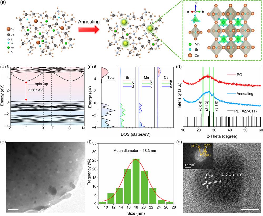
A precursor glass (PG) containing cesium, manganese, and bromine elements is designed and fabricated by the melt-quenching method, and are crystallized in the glass matrix by annealing above the glass transition () temperature (Fig. S1 in the Supplemental Material), as shown in Fig. 1(a). As a lead-free anti-perovskite material, the crystal consists of tetrahedrons filled in the (3D) octahedral skeleton [Fig. 1(a)]. From the density functional theory (DFT) calculations of band structures and corresponding density of states [Figs. 1(b) and 1(c)], the direct bandgap of crystal is , contributing to the absorption band in the ultraviolet (UV) region and high optical transmittance in the visible region.35 The diffraction peaks at 22.783 deg, 26.914 deg, and 29.454 deg corresponding to (004), (213), and (310) crystal facets of (PDF#27-0117) are observed in the X-ray diffraction (XRD) pattern after the glass was annealed at 570°C for 5 h [Fig. 1(d)]. In addition, X-ray photoelectron spectroscopy (XPS) was performed on the glass sample before and after annealing (Fig. S2 in the Supplemental Material). The characteristic peaks of Mn (653.4 eV) and (642.1 eV) are observed, which are higher than that in manganese halide (), indicating that the electron density and covalent bond ratio of -embedded glass are increased.36 This may result in stronger ionic bonding between manganese ions and bromine ions, which could enhance the stability under high-energy-ray irradiation.17 The electron paramagnetic resonance (EPR) spectra show a wide EPR signal with a line width of 609.4 G instead of six fine structures because of the high concentration in the glass (Fig. S3 in the Supplemental Material).37 The signal is more obvious after annealing, which is due to the stronger magnetic coupling caused by the decrease in the spacing of ions after the NCs crystallized in the glass.38 A transmission electron microscope (TEM) image shows dispersed NCs with an average size of 18.3 nm, indicating the formation of NCs with good crystal quality in the glass matrix [Figs. 1(e) and 1(f)]. The crystal lattice fringes with a spacing of 0.305 nm can be seen in the high-resolution transmission electron microscope (HRTEM) image, which corresponds to the (310) crystal facet of [Fig. 1(g)]. The above results confirm the successful precipitation of NCs in the glass after annealing.
![]()
Figure 1.Structural properties of
2.2 Photoluminescence Properties of Cs3MnBr5 NCs in the Glass
As a typical transition metal ion, the optical properties of ions are influenced by the strong interactions between electrons in their outermost d orbitals and their ligands, which can be described by the crystal field strength (10Dq) and the Racah parameter (), respectively.39,40 The Tanabe–Sugano diagram in Fig. 2(a) reveals the change in the energy level of as the electronic configuration of is sensitive to local perturbations. The 10Dq and for Mn (IV) in NCs are calculated to be 8192 and , and for Mn (VI) in glass are 11,306 and , respectively (see note 1 in the Supplemental Material). These calculations confirm that the electronic configuration of doped in the glass was significantly changed after the crystallization of the glass.
![]()
Figure 2.PL properties of
The absorption spectra of the PG and the glass samples annealing at different temperatures are recorded in the wavelength region of 300 to 800 nm [Fig. 2(b)]. The narrow peak at 413 nm is attributed to the electronic transition of , of ions. Under the excitation of 365 nm UV light, a green emission band peaking at 523 nm emerges after crystallization, which originates from in the tetrahedron of NCs [Fig. 2(c)]. It is worth noting that there are two luminescence centers in the glass after annealing, attributed to NCs and the remaining ions in the glass matrix. Thus, the luminescence color can also be adjusted by controlling the excitation wavelength due to the two different luminescence centers, as shown in Fig. 2(d) and Figs. S4(a, b) in the Supplemental Material. The variation of two different emission bands in the time-resolved emission spectrum (1 to 10 ms) shows two different coordination environments around in the annealed glass sample (Fig. S5 in the Supplemental Material). With the increase of annealing temperature, the fluorescence lifetimes of the green and red emission change a little [Figs. 2(e) and 2(f)]. This is because is in a stable tetragonal and hexagonal field environment, respectively. Due to the crystal coordination environment of NCs, the green emission lifetime reaches the microsecond range, which is often required for dynamical X-ray imaging.
To further increase the concentration of NCs, we also investigated the effect of annealing duration on the luminescence properties of the samples. The glass samples were treated at 570°C for 5 to 40 h, respectively, and a series of photoluminescence (PL) spectra and lifetime decay curves were recorded [Figs. S6(a–d) in the Supplemental Material]. It can be found that with the increase in annealing duration, the green emission of the samples is gradually enhanced due to the increase in the concentration of NCs. However, the scattering caused by the increase in crystal size also leads to a large optical loss of the glass samples. Thus, the sample treated at 570°C for 10 h has the highest PL quantum yield of 35.5% (Fig. S7 in the Supplemental Material). Depending on the annealing temperature and duration, the emission color of the glass samples can be precisely adjusted from red to green (Fig. S8 in the Supplemental Material). In contrast to the typical ( = Cl, Br, I) materials, which have poor thermal and optical stability, the stability of NC-embedded glass has been greatly improved. At 503 K, the green emission of the sample retains 66% of the intensity recorded at room temperature [Fig. S9(a, b) in the Supplemental Material]. In addition, under the continuous irradiation of a 375 nm laser with for 60 min, the PL intensity of remained unchanged (Fig. S10(a, b) in the Supplemental Material). These characteristics can be attributed to the unique restriction of the high-density luminescent center in the anti-perovskite configuration and their larger binding energies.
2.3 Radioluminescence Properties of Cs3MnBr5 NCs in the Glass
The tunable PL, excellent stability, and large Stokes shift (negligible self-absorption) demonstrate the large potential of NC-embedded glass for applications in the field of X-ray detection applications. In contrast to the PL mechanism, the radioluminescence (RL) mechanism involves an additional photoelectron conversion process that converts high-energy electrons to low-energy ones for radiative recombination [Fig. 3(a)].15 The X-ray is absorbed by heavy atoms, such as Cs, Mn, Ge from NC-embedded glass through photoelectric effect and Compton scattering. Then, a large number of hot electrons are released and captured by the luminescence center after being thermalized. We investigate the X-ray absorption coefficient of the NC-embedded glass from 1 to 1000 keV. The absorption coefficient of NC-embedded glass is comparable with the typical scintillators, such as (BGO) and [Fig. 3(b)].41 The light yield of NC-embedded glass is estimated by using commercial scintillator BGO as a standard sample (Fig. S11 in the Supplemental Material).42 Due to the dual-emission centers, the glass samples demonstrate RL with an adjustable red–green ratio under X-ray excitation by adjusting the annealing temperature and duration [Fig. 3(c) and Fig. S12 in the Supplemental Material]. It is shown in Fig. 3(c) that with the increase of annealing duration, the emission color of glass samples changes from light yellow to yellow-green under X-ray irradiation. The NC-embedded glass shows the similar PL and RL spectra, indicating that they originate from the same radiative recombination channel upon X-ray and UV excitation. Interestingly, from the RL spectra recorded under X-ray excitation with different dose rates, we observe that NCs have a better linear response to the dose rate of X-rays than that of ions in the glass sample (Fig. S13 in the Supplemental Material). In Fig. 3(d), we show the RL spectra of a series of low-dose X-ray excitations. For scintillation materials, the detection limit is one of the most important parameters for medical examination. Here, the detection limit of the X-ray dose rate is for NC-embedded glass when the signal-to-noise ratio is 3. This value is significantly lower than the dose rate used in X-ray medical diagnosis ( dose rate), as shown in Fig. 3(e).43 Therefore, these merits of NC-embedded glass make it possible to achieve high-performance X-ray detection and real-time X-ray imaging.
![]()
Figure 3.RL properties of
2.4 X-Ray Imaging Performance of Cs3MnBr5 NCs in the Glass
Due to the high optical transparency of glass (Fig. S14 in the Supplemental Material) and the excellent RL properties, this NC-embedded glass is used for high-resolution X-ray imaging. Here, we construct a self-made X-ray imaging system [Fig. 4(a)], and a series of objects, such as an AI chip, a charging cable, and a circuit board, were imaged by NC-embedded glass under X-ray and captured by a commercial digital camera. As shown in Fig. 4(b), the internal structures are directly observed, indicating that NC-embedded glass is promising for electronics inspection and damage imaging. To further demonstrate the X-ray imaging capability of NC-embedded glass, images of the standard X-ray resolution pattern plate with different thicknesses of glasses were used (Fig. S15 in the Supplemental Material). The highest resolution of the X-ray image was achieved while the glass thickness is 0.6 mm (Fig. S16 in the Supplemental Material). The observation down-limit is between 18 and , which is consistent with the calculated results of modulation transfer functions (MTFs) according to the slanted-edge method [Fig. 4(d) and Fig. S17 in the Supplemental Material]. Due to the high transparency of the glass, the high X-ray luminescence efficiency and the negligible self-absorption effect of NCs, the spatial resolution of our glass sample reaches at , which exceeds most recently reported materials for X-ray imaging [Fig. 4(e)]. In addition, real-time radiography was successfully performed by recording the rotation procedure of an iron spring with an angular velocity of . As shown in Fig. 4(f) and Video 1, we can see the image of the rotary spring, further confirming the high-quality and rapid X-ray imaging based on the NC-embedded glass.
![]()
Figure 4.Demonstrations for real-time radiography. (a) The schematic of the X-ray imaging system. (b) Photographs of an AI chip (left), charging cable (middle), and circuit board (right) under daylight and X-ray irradiation. Scale bars, 1 cm. (c) Bright-field and X-ray images of the standard X-ray resolution pattern plate with the
2.5 Ultrastable X-Ray Imaging Application
Owing to the encapsulation by the glass medium and the stability of the anti-perovskite structures, the NC-embedded glass is expected to be used for X-ray imaging applications in medical fields and space stations. Moreover, we are surprised to find that the NC-embedded glass has very stable X-ray-excited optical properties when tested in a variety of harsh environments. The temperature-dependent RL spectra of NC-embedded glass are shown in Figs. 5(a) and 5(b). It can be found that at temperature up to 563 K, the RL intensity of red emission from in the glass drops sharply, while the green emission from NCs retains 73% of the room temperature intensity. In addition, the high thermal stability is further supported by the periodic change in emission intensity under repeated heating/cooling from 303 to 563 K for six cycles, as shown in Fig. 5(c). The excellent RL stability of NC-embedded glass in high-temperature environment exceeds most recently reported materials for X-ray imaging (Table S1 in the Supplemental Material). These results indicate that NC-embedded glass possesses strong thermal stability.
![]()
Figure 5.Ultrastable X-ray imaging. (a) Temperature-dependent RL spectra and (b) emission mapping of the glass sample under X-ray irradiation with a dose rate of
We continue with a series of demonstrations of the imaging performance of the NC-embedded glass in different environments. First, an iron spring is encapsulated in an ABS cylindrical resin and placed together with the NC-embedded glass in a colorimetric dish filled with dimethyl silicone oil [Fig. 5(d)]. The spring inside is not visible under daylight but can be imaged by NC-embedded glass under X-ray irradiation. In addition, the X-ray images of the spring can be detected at different temperatures, benefited by the stable thermal RL property of NC-embedded glass [Fig. 5(e)]. In this experiment, the temperature of the NC-embedded glass is detected by using an infrared camera. The X-ray image at the temperature up to 121.6°C is still clear, indicating that the NC-embedded glass can be used for high-temperature X-ray imaging. In another experiment, we place an integrated circuit chip and the NC-embedded glass in deionized water and recorded the underwater X-ray imaging for 24 h [Fig. S18(a, b) in the Supplemental Material]. After the NC-embedded glass was immersed in deionized water for 0, 5, 15, and 24 h, the X-ray images of the internal structure of the chip are still clear. Moreover, the underwater X-ray images remain unchanged at different temperatures (Fig. S19 in the Supplemental Material). Figure 5(f) presents the RL intensity of NC-embedded glass recorded under repeated high-dose X-ray () excitation. It can be seen that after 120 on–off cycles, the RL intensity remains unchanged, showing good long-term X-ray irradiation stability. Moreover, even under a higher dose rate up to , its luminescence was only slightly affected for a short time (Fig. S20 in the Supplemental Material). In addition, the X-ray images from NC-embedded glass can still be observed after continuous X-ray irradiation for 2 h at a high dose rate up to [Fig. 5(g)]. Considering the above X-ray imaging demonstrations in different environments, the NC-embedded glass is expected to be applied for the next generation of scintillation materials.
3 Conclusion
In summary, we have successfully prepared an ultrastable monolithic scintillator based on a lead-free anti-perovskite NC-embedded glass. The experimental results show that the NC-embedded glass has high optical transmittance, excellent tunable optical properties, and durable stability. Therefore, the NC-embedded glass can achieve an X-ray detection limit of , high X-ray imaging spatial resolution of , and excellent stability under high-dose X-ray irradiation. The NC-embedded glass can be made into optical fibers, so the X-ray imaging performance and resolution may be further improved by using the pixelated dual tapered fiber arrays method.47 More importantly, with the protection of the glass matrix and the stability of the anti-perovskite structure, we demonstrate high-resolution X-ray imaging under a high-temperature and humidity environment by using NC-embedded glass. Our findings not only provide an effective strategy for achieving ultrastable X-ray-excited luminescence in a harsh environment but also broaden the applications of lead-free anti-perovskite materials in the applications of advanced X-ray radiography.
4 Appendix A: Sample Preparation
The glass samples were fabricated by using the melting-quenching method. Reagent-grade raw materials including , , ZnO, , NaBr, , and MnO (glass composition: ) were mixed and then melted at 1100°C. The PG was made by pouring the melt into a mold. Then the glass was annealed at 350°C for 5 h and cooled for 10 h to room temperature to release the thermal stress. The PG samples were cut after removal from the mold and well-polished for crystallization of anti-perovskite NCs by annealing at 490°C, 530°C, and 570°C.
5 Appendix B: Sample Characterization
Differential scanning calorimetry was measured by an STA449C Jupiter (Netzsch, Bavaria, Germany) in an air atmosphere with a heating rate of . The XRD patterns were recorded by a D8 ADVANCE X-ray diffractometer (Bruker, Faellanden, Switzerland) with () radiation. Absorption spectra were measured on a Lambda 900 (Perkin Elmer, Waltham, MA) spectrophotometer. An Edinburgh FLS 920 instrument (Edinburgh Instruments Ltd., Livingston, United Kingdom) equipped with a photomultiplier tube for light detection (Hamamatsu, Japan) was used to measure the PL spectra, excitation spectra, and lifetime decay of bulk glass sample. A 450 W ozone‐free xenon lamp and a microsecond‐pulsed xenon flash lamp were used as excitation sources during PL and lifetime decay measurement, respectively. For thermal and optical stability tests, PL spectra were excited by a 375 nm laser and recorded by an Ocean Optics HR4000 spectrometer. XPS measurements were performed on an Axis Ultra DLD XPS instrument (Kratos, England) with a monochromatic Al source (1486.6 eV). The morphology and size distribution of NCs were measured by transmission electron microscopy (TEM, JEM-2100F, JEOL, Japan). The glass sample after crystallization was put into an agate mortar and ground to powder in ethanol for 20 min, and then dispersed in ethanol with ultrasonic treatment for 10 min. Finally, three to five drops of supernatant were dropped on the copper net for the TEM image test. The model of the electric rotary displacement table used for real-time imaging was Thorlabs PRM1Z8. FLS1000 spectrofluorometer (Edinburgh Instruments Ltd., United Kingdom) equipped with an X-ray tube (MAGPRO, Moxtek, target and tube voltage 50 kV) is used to measure the RL spectra. The digital camera used for X-ray imaging is a Nikon d610.
6 Appendix C: Calculation of the X-Ray Attenuation Coefficients
The detail of calculation is fully described in the subsection entitled “Supplementary Note 2” in the Supplemental Material.
Yakun Le is pursuing his PhD under the supervision of Prof. Guoping Dong, at the School of Materials Science and Engineering from the South China University of Technology (SCUT). His research focuses on metal halide nanocrystal-in-glass composite and their advanced optical applications.
Guoping Dong received his MS (2007) and PhD (2010) from Wuhan University of Technology and Shanghai Institute of Optics and Fine Mechanics (CAS), respectively. Then he worked in South China University of Technology (SCUT) as a research assistant (2010) and associate professor (2011). He is currently a full professor (2014) in State Key Laboratory of Luminescent Materials and Devices at SCUT. His research focuses on design, preparation, and optoelectronic properties of optical functional materials and devices.
Biographies of the other authors are not available.
References
[1] J. A. Rowlands. Material change for X-ray detectors. Nature, 550, 47-48(2017).
[6] Q. Chen et al. All-inorganic perovskite nanocrystal scintillators. Nature, 561, 88-93(2018).
[25] N. Hoffmann et al. Superconductivity in antiperovskites. NPJ Comput. Mater., 8, 150(2022).
[46] X. Ou et al. High-resolution X-ray luminescence extension imaging. Nature, 590, 410-415(2021).

Set citation alerts for the article
Please enter your email address
© Copyright 2018-2021 | Chinese Laser Press. All Rights Reserved 沪ICP备15018463号-20