
Li Wang, Yufeng Han, Hongchen Wang, Yaojie Han, Jinhua Liu, Gang Lu, Haidong Yu. A MXene-functionalized paper-based electrochemical immunosensor for label-free detection of cardiac troponin I[J]. Journal of Semiconductors, 2021, 42(9): 092601
- Journal of Semiconductors
- Vol. 42, Issue 9, 092601 (2021)
Abstract
1. Introduction
Acute myocardial infarction (AMI) is one of the most life-threatening diseases[
Paper-based sensors have attracted tremendous attention from researchers since George Whitesides fabricated a low-cost, low-fluid consumption, and portable microfluidic paper-based glucose sensor in 2007[
MXenes are emerging 2D transition metal carbide and nitride materials with the formula Mn+1XnTx[
In this work, a cost-efficient and portable paper-based electrochemical immunosensor was successfully constructed by wax printing and screen-printing. The MXene nanosheets were used not only for improving the conductivity of SPEs, but also for immobilizing a large number of antibodies. The paper-based immunosensor with MXene-modified SPEs (MXene/SPEs) exhibited high sensitivity and selectivity towards cTnI with a low limit of detection (LOD) of 0.58 ng/mL, as well as good repeatability. Our paper-based immunosensor demonstrates great potential in highly sensitive detection of cTnI, which has a broad application prospect in clinical AMI diagnosis.
2. Experimental
2.1. Chemicals
Whatman chromatography paper (No. 1) was acquired from Zhengcheng Experimental Instrument Co., Ltd (Shanghai). Bovine serum albumin (BSA), Tween-20, and ethanol (99.5%) were supplied by Macklin Biochemical Co., Ltd (Shanghai). Phosphate buffered saline (PBS, 0.01 mol/L, pH = 7.4) and the PBS solution doped with 0.05% Tween-20 (PBST) were both freshly prepared. Human heart-type cardiac troponin I (cTnI) and capture monoclonal antibodies (anti-cTnI) were acquired from Linc-Bio Science Co., Ltd (Shanghai). The commercialized ELISA kit used to detect cTnI was acquired from Enzyme-linked Biotechnology Co., Ltd (Shanghai). All chemicals used in this work are analytical grade and used directly.
2.2. Characterizations
The morphology of MXene sheets was characterized with a transmission electron microscopy (TEM, JEM-1400PLUS, JEOL, Japan). The X-ray diffraction (XRD) patterns were collected on an X-ray diffractometer (SmartLab 3, Rigaku, Japan) with Cu Kα radiation (λ = 1.5418 Å). The atomic force microscopy (AFM) was performed on Park XE-70 (Korea). The cyclic voltammetry (CV) and differential pulse voltammetry (DPV) were conducted on CHI630E (CH Instrument, China). PalmSens4 (PalmSens, Netherlands) was used to measure the electrochemical impedance spectroscopy (EIS).
2.3. Synthesis of Ti3C2-MXene
Briefly, an etching solution was obtained by mixing 1 g of LiF and 5 mL of DI water with 15 mL of 12 mol/L HCl. Then, 1 g Ti3AlC2 (MAX) was added slowly into the etching solution, which was stirred for 24 h to remove the aluminum (Al) layer. Afterward, the mixed suspension was transferred to a centrifuge tube, which was then centrifuged at 3500 rpm for five minutes. The centrifugal product was repeatedly washed with DI water until the pH of supernatant was ≥6. Subsequently, the cleaned product was mixed with DI water again, followed by being sonicated for one hour. Finally, the Ti3C2 (MXene) nanosheets were collected by centrifugation at 3500 rpm for 5 min.
2.4. Silanization of MXene sheets
The (3-aminopropyl) triethoxysilane (APTES) was used for the functionalization of the as-prepared Ti3C2-MXene nanosheets. In detail, 1 mL of APTES was gradually added to the mixed suspension that contains 100 mg Ti3C2-MXene nanosheets and 50 mL ethanol, and the suspension was stirred (500 rpm) for 48 h at room temperature. For the removal of unbound APTES molecules, the resulting product was washed three times with a mixture of water and ethanol (1 : 1). Then, the resulting product was dried at 50 °C in a vacuum overnight to obtain the APTES-functionalized Ti3C2-MXene (f-Ti3C2-MXene), which was then finely pulverized into powder and then sonicated in a 0.1% Nafion solution in ethanol and DI water for one hour to obtain a 1 mg/mL suspension.
2.5. Fabrication of paper-based electrochemical immunosensor
The paper-based electrochemical sensor was fabricated in the following four steps as described by Wang and coworkers[
2.6. Modification of the immunosensor
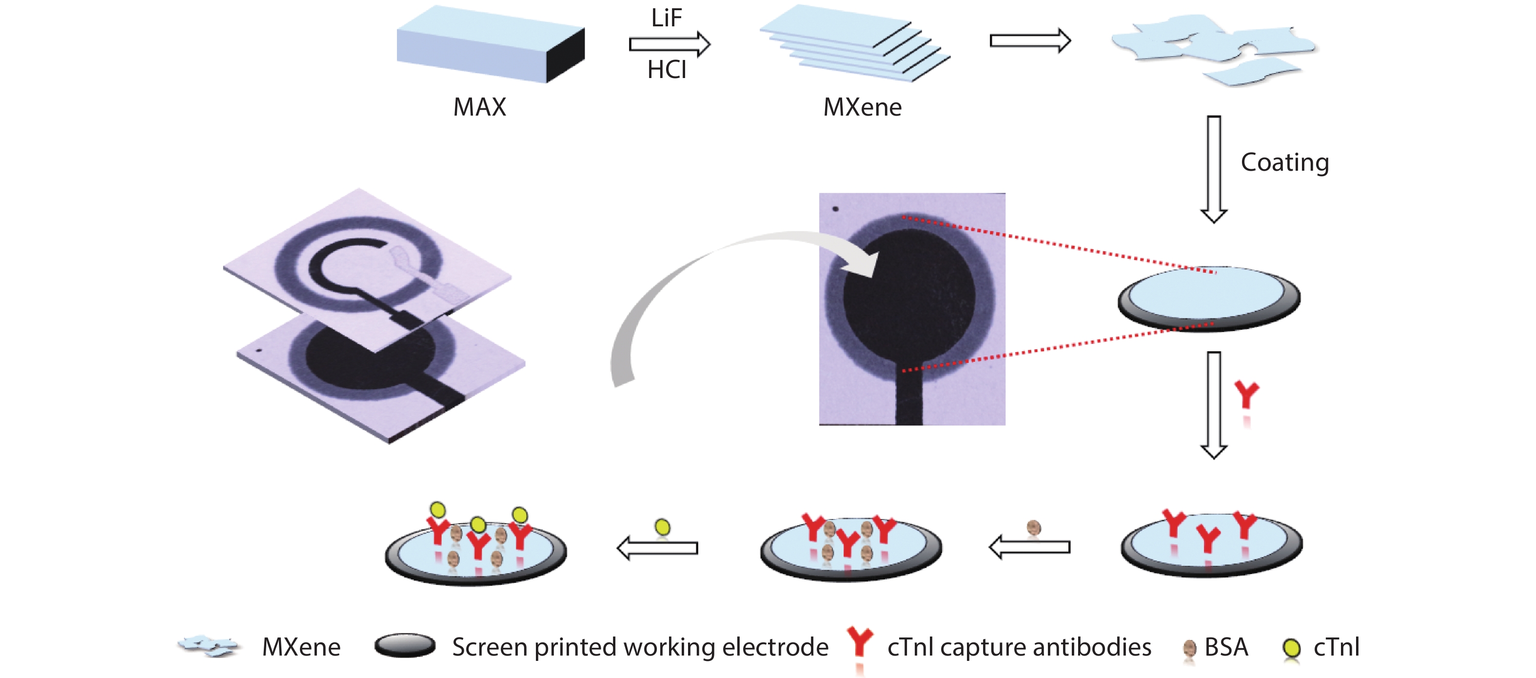
The modified paper-based electrochemical sensor was prepared according to the process shown in Fig. 1. First, f-Ti3C2-MXene was coated on the surface of the SPE, followed by drying at room temperature. Then, 60 μg/mL anti-cTnI was dropped on the MXene-modified electrode and incubated for 30 min. Subsequently, 10 μL 1% BSA solution was dropped to minimize the non-specific adsorption by blocking the remaining active sites. Finally, the immunosensors were stored at 4 °C before usage.
![]()
Figure 1.(Color online) Schematic diagram illustrating the fabrication of the paper-based cTnI immunosensor and their usage in detection of cTnI.
2.7. Electrochemical measurements
The DPV measurement was performed in the range of –0.2 to 0.7 V, while the scan rate of CV was 50 mV/s. EIS experiments were performed in the frequency range of 0.1–10 000 Hz and AC amplitude of 5 mV. The electrochemical tests were performed in a mixed solution of 0.1 mol/L KCl and 0.005 mol/L K3[Fe(CN)6]. The electrochemical detection, selectivity, and repeatability tests were performed in a PBS buffer (0.01 mol/L, pH = 7.4) containing K3[Fe(CN)6]/K4[Fe(CN)6] (0.005 mol/L) and KCl (0.1 mol/L).
3. Result and discussion
3.1. Characterization of MXene nanosheets
The Ti3C2-MXene was prepared by etching the Al layers in the MAX (M = metal, A = Al, X = C or N) precursor in a mixed solution of hydrochloride (HCl) and lithium fluoride (LiF). The obtained MXene nanosheets were well dispersed in aqueous solution, and the color of the suspension was dark green (Fig. 2(a)). The obtained Ti3C2-MXene nanosheet was thin and highly transparent to the electron beam in the TEM image (Fig. 2(b)). From the X-ray diffraction (XRD) pattern, the (104) peak of the MAX phase disappeared and the (002) peak left-shifted for 2.9 degrees (Fig. 2(c)), suggesting that the Ti3C2-MXene was successfully prepared. As shown in the atomic force microscopy (AFM) image (Fig. 2(d)), the thickness of the MXene nanosheets was only 4 nm, indicating that the obtained MXene nanosheets were only a few atomic layers thick. Moreover, the Fourier transform infrared (FT-IR) spectra of the pristine MXene and f-MXene (Fig. S1) were compared to investigate the salinization of Ti3C2-MXene with APTES. The bands at 567 and 1633 cm–1 were presented in the Ti3C2-MXene sample, and these two bands are ascribed to the vibration modes of the Ti–O and C=O bonds[
![]()
Figure 2.(Color online) Characterizations of MXene. (a) Scheme showing the crystal structure of the MXene and a photograph of the colloid solution of the MXene. (b) TEM image of the MXene. (c) XRD patterns of the etched MXene (black line) and original MAX phase (red line). (d) AFM image of the Ti3C2 MXene. The insert is the line profile of the white line in (d).
3.2. Electrochemical characteristics of the modified electrodes
The synthesized MXene sheets were functionalized with APTES and then adsorbed on the paper-based SPEs. The interfacial properties of the modified electrodes were characterized by EIS. The impedance spectrum comprises a semicircular and a linear portion. The semicircular diameter at high frequency corresponds to the value of the charge transfer resistance (Rct), while the slope of the linear part at low frequency corresponds to the value of the diffusion resistance[
![]()
Figure 3.(Color online) Surface modifications of the electrodes. (a) EIS of the modified electrodes measured in a mixed solution of KCl (0.1 mol/L) and K3[Fe(CN)6] (0.005 mol/L). (b) CV of the modified electrodes, measured at a scan rate of 50 mV/s in a mixed solution of KCl (0.1 mol/L) and K3[Fe(CN)6] (0.005 mol/L).
3.3. Optimization of the immunoassay
To achieve the best analytical performance, the fabrication of the MXene-modified paper-based electrochemical immunosensor was optimized by tuning the pH value, anti-body immobilization period, and PBST washing times. The pH value of buffer influences the properties of a biosensor greatly, due to the fact that the antibody may be inactivated in acidic or alkaline conditions[
![]()
Figure 4.(Color online) Optimization of the immunosensor. (a) The effect of pH value of PBS (5.5, 6, 6.5, 7, 7.4, 8, 8.5, and 9) on the current response of the immunosensor. (b) The effect of the immobilization period (10, 20, 30, 40, 50, and 60 min) of antibodies on the current response of the immunosensor, error bar = standard deviation (
3.4. Electrochemical detection of cTnI
In optimal conditions, the obtained immunosensor was used for the detection of cTnI at different concentrations, and the results were evaluated by DPV. The intensity of the peak DPV current decreased linearly with the increase in cTnI concentration within the range of 5–100 ng/mL, and a low LOD of 0.58 ng/mL was observed (Fig. 5(a)), which is due to the fact that the antibody layer forms a barrier layer on the electrode, impeding the electron transfer between the redox probe ([Fe(CN)6]3–/4–) and electrode surface. Hence, cTnI could be quantitatively detected by monitoring the degree of the decreased peak DPV current. The linear regression equation was y = −0.06876x + 31.49 (R2 = 0.9745), where y is the peak DPV current and x is the concentration of cTnI. Moreover, compared with other cTnI biosensors from Table 1, the obtained immunosensor can achieve a relatively lower detection limit, suggesting that the obtained paper-based electrochemical cTnI immunosensor has great application potential for measuring the cTnI in clinical diagnosis.
![]()
Figure 5.(Color online) Electrochemical detection of cTnI. (a) DPV responses of the immunosensor to cTnI at concentrations of 0–100 ng/mL. (b) Calibration curve toward different concentrations of cTnI, error bar = standard deviation (
As a product made of natural materials, paper has been commonly used in portable and disposable devices, owing to its intrinsic flexibility, abundance, and low cost[
3.5. Selectivity and repeatability of the immunosensor
Selectivity is a key factor to ensure the accuracy of biosensing. To estimate the selectivity of our paper-based immunosensor to cTnI, non-specific proteins (50 ng/mL) including bovine serum albumin (BSA), glucose oxidase (GOx), immunoglobulins G (IgG), α-fetoprotein (AFP), carcinoembryonic antigen (CEA), neuron-specific enolase (NSE), human chorionic gonadotropin (HCG), and the mixed solution (including cTnI and these non-specific proteins) were tested for control studies. Fig. 6(a) shows that a marked decrease in peak current was found only in the cTnI solution and the mixed solution containing cTnI, indicating that the developed immunosensor has a high selectivity toward cTnI. Additionally, the reproducibility of our immunosensor was further estimated. Five paper-based immunosensors prepared with the same parameters were used to detect 50 ng/mL cTnI by DPV in the same optimal conditions. Fig. 6(b) shows that the obtained relative standard deviation (RSD) was only 6.3%, suggesting a high reproducibility of our immunosensor.
![]()
Figure 6.(Color online) Characterization of the repeatability and selectivity of the immunosensor. (a) The selectivity to cTnI against non-target protein molecules: BSA, AFP, CEA, GOx, NSE, HCG, and Blank. (b) Reproducibility of the proposed immunosensors in the detection of cTnI.
4. Conclusion
In summary, a label-free, highly sensitive and selective paper-based electrochemical immunosensor was successfully fabricated by functionalizing SPEs with two-dimensional Ti3C2-MXene nanosheets. The f-MXene nanosheets improve the conductivity of the SPEs greatly and also facilitate the immobilization of antibodies. Therefore, the sensitivity of the sensor was largely improved. The immunosensor exhibited high sensitivity and selectivity towards cTnI within the range of 5–100 ng/mL. A low LOD of 0.58 ng/mL was achieved. Moreover, the sensor also showed high repeatability. Owing to the advantages of low cost, fast response, high selectivity and sensitivity, our MXene-functionalized paper-based immunosensor demonstrates a great potential in label-free determination of cTnI in clinical diagnosis. Moreover, the inexpensive and environment-friendly nature of paper, as well as the simple fabrication procedure, make our paper-based immunosensor a promising candidate for green and flexible next-generation electronics.
Acknowledgements
This work was financially supported by the National Key R&D Program of China (2017YFA0204700), the Joint Research Funds of Department of Science & Technology of Shaanxi Province and Northwestern Polytechnical University (2020GXLH-Z-021), the China–Sweden Joint Mobility Project (51811530018), and the Fundamental Research Funds for the Central Universities.
Appendix A. Supplementary materials
Supplementary materials to this article can be found online at https://doi.org/1674-4926/42/9/092601.
References
[1] M I Mohammed, M P Y Desmulliez. Lab-on-a-chip based immunosensor principles and technologies for the detection of cardiac biomarkers: a review. Lab Chip, 11, 569(2011).
[2] S Ko, B Kim, S S Jo et al. Electrochemical detection of cardiac troponin I using a microchip with the surface-functionalized poly(dimethylsiloxane) channel. Biosens Bioelectron, 23, 51(2007).
[3] X Y Guo, L J Zong, Y C Jiao et al. Signal-enhanced detection of multiplexed cardiac biomarkers by a paper-based fluorogenic immunodevice integrated with zinc oxide nanowires. Anal Chem, 91, 9300(2019).
[4] C Zhang, P F Du, Z J Jiang et al. A simple and sensitive competitive bio-barcode immunoassay for triazophos based on multi-modified gold nanoparticles and fluorescent signal amplification. Anal Chim Acta, 999, 123(2018).
[5] H J Kim, W L Shelver, E C Hwang et al. Automated flow fluorescent immunoassay for part per trillion detection of the neonicotinoid insecticide thiamethoxam. Anal Chim Acta, 571, 66(2006).
[6] M H Wang, J J Liu, X L Qin et al. Electrochemiluminescence detection of cardiac troponin I based on Au-Ag alloy nanourchins. Analyst, 145, 873(2020).
[7] Z J Yang, J Shen, J Li et al. An ultrasensitive streptavidin-functionalized carbon nanotubes platform for chemiluminescent immunoassay. Anal Chim Acta, 774, 85(2013).
[8] Y J Zhao, X H Liu, J Li et al. Microfluidic chip-based silver nanoparticles aptasensor for colorimetric detection of thrombin. Talanta, 150, 81(2016).
[9] L Gao, Q F Yang, P Wu. Recent advances in nanomaterial-enhanced enzyme-linked immunosorbent assays. Analyst, 145, 4069(2020).
[10] A W Martinez, S T Phillips, M J Butte et al. Patterned paper as a platform for inexpensive, low-Volume, portable bioassays. Angew Chem Int Ed, 46, 1318(2007).
[11] Y C Jiao, C Du, L J Zong et al. 3D vertical-flow paper-based device for simultaneous detection of multiple cancer biomarkers by fluorescent immunoassay. Sens Actuators B, 306, 127239(2020).
[12] R R Nair. Organic electrochemical transistor on paper for the detection of halide anions in biological analytes. Flex Print Electron, 5, 045004(2020).
[13] W Yuan, X Z Wu, W B Gu et al. Printed stretchable circuit on soft elastic substrate for wearable application. J Semicond, 39, 015002(2018).
[14] L J Zong, Y C Jiao, X Y Guo et al. Paper-based fluorescent immunoassay for highly sensitive and selective detection of norfloxacin in milk at picogram level. Talanta, 195, 333(2019).
[15] L J Zong, Y F Han, L Gao et al. A transparent paper-based platform for multiplexed bioassays by wavelength-dependent absorbance/transmittance. Analyst, 144, 7157(2019).
[16] D R Wang, Y F Mei, G S Huang. Printable inorganic nanomaterials for flexible transparent electrodes: from synthesis to application. J Semicond, 39, 011002(2018).
[17] Z Y Li, X Huang, G Lu. Recent developments of flexible and transparent SERS substrates. J Mater Chem C, 8, 3956(2020).
[18] M Z Zou, Y Ma, X Yuan et al. Flexible devices: from materials, architectures to applications. J Semicond, 39, 011010(2018).
[19] H Wang, H Li, Y Huang et al. A label-free electrochemical biosensor for highly sensitive detection of gliotoxin based on DNA nanostructure/MXene nanocomplexes. Biosens Bioelectron, 142, 111531(2019).
[20] M Naguib, M Kurtoglu, V Presser et al. Two-dimensional nanocrystals produced by exfoliation of Ti3AlC2. Adv Mater, 23, 4248(2011).
[21] Z H Ren, D C Qi, P Sonar et al. Flexible sensors based on hybrid materials. J Semicond, 41, 040402(2020).
[22] M Naguib, V N Mochalin, M W Barsoum et al. 25th anniversary article: MXenes: a new family of two-dimensional materials. Adv Mater, 26, 992(2014).
[23] M Naguib, O Mashtalir, J Carle et al. Two-dimensional transition metal carbides. ACS Nano, 6, 1322(2012).
[24] G P Neupane, T Yildirim, L L Zhang et al. Emerging 2D MXene/organic heterostructures for future nanodevices. Adv Funct Mater, 30, 2005238(2020).
[25] R Xiao, C X Zhao, Z Y Zou et al. In situ fabrication of 1D CdS nanorod/2D Ti3C2 MXene nanosheet Schottky heterojunction toward enhanced photocatalytic hydrogen evolution. Appl Catal B, 268, 118382(2020).
[26] Y Meng, J C Ho. MXene-based wearable biosensor. J Semicond, 40, 110202(2019).
[27] Z Li, Y Wu. 2D early transition metal carbides (MXenes) for catalysis. Small, 15, 1804736(2019).
[28] B Ahmed, A EI Ghazaly, J Rosen. i-MXenes for energy storage and catalysis. Adv Funct Mater, 30, 2000894(2020).
[29] K Huang, Z J Li, J Lin et al. Two-dimensional transition metal carbides and nitrides (MXenes) for biomedical applications. Chem Soc Rev, 47, 5109(2018).
[30] H X Zhang, Z H Wang, Q X Zhang et al. Ti3C2 MXenes nanosheets catalyzed highly efficient electrogenerated chemiluminescence biosensor for the detection of exosomes. Biosens Bioelectron, 124, 184(2019).
[31] Q Jiang, N Kurra, M Alhabeb et al. All pseudocapacitive MXene-RuO2 asymmetric supercapacitors. Adv Energy Mater, 8, 1703043(2018).
[32] Q Y Yang, Z Xu, B Fang et al. MXene/graphene hybrid fibers for high performance flexible supercapacitors. J Mater Chem A, 5, 22113(2017).
[33] Y T Du, X Kan, F Yang et al. MXene/graphene heterostructures as high-performance electrodes for Li-ion batteries. ACS Appl Mater Inter, 10, 32867(2018).
[34] B Ahmed, D H Anjum, Y Gogotsi et al. Atomic layer deposition of SnO2 on MXene for Li-ion battery anodes. Nano Energy, 34, 249(2017).
[35] B Ahmed, D H Anjum, M N Hedhili et al. H2O2 assisted room temperature oxidation of Ti2C MXene for Li-ion battery anodes. Nanoscale, 8, 7580(2016).
[36] Y Wang, J P Luo, J T Liu et al. Electrochemical integrated paper-based immunosensor modified with multi-walled carbon nanotubes nanocomposites for point-of-care testing, of 17 beta-estradiol. Biosens Bioelectron, 107, 47(2018).
[37] G L Zhang, T C Wang, Z H Xu et al. Synthesis of amino-functionalized Ti3C2T
[38] L D White, C P Tripp. Reaction of (3-Aminopropyl)dimethylethoxysilane with amine catalysts on silica surfaces. J Colloid Interf Sci, 232, 400(2000).
[39] J M Lei, X M Chen. RuO2/MnO2 composite materials for high-performance supercapacitor electrodes. J Semicond, 36, 083006(2015).
[40] S P Wang, J J Wang, Y F Zhu et al. Cantilever with immobilized antibody for liver cancer biomarker detection. J Semicond, 35, 104008(2014).
[41] L Y Miao, L Jiao, Q R Tang et al. A nanozyme-linked immunosorbent assay for dual-modal colorimetric and ratiometric fluorescent detection of cardiac troponin I. Sens Actuators B, 288, 60(2019).
[42] S Lee, S H Kang. Quenching effect on gold nano-patterned cardiac troponin I chip by total internal reflection fluorescencemicroscopy. Talanta, 104, 32(2013).
[43] F Torabi, F H R Mobini, B Danielsson et al. Development of a plasma panel test for detection of human myocardial. Biosens Bioelectron, 22, 1218(2007).
[44] M M Gong, D Sinton. Turning the page: advancing paper-based microfluidics for broad diagnostic application. Chem Rev, 117, 8447(2017).
[45] E Noviana, C P McCord, K M Clark et al. Electrochemical paper-based devices: sensing approaches and progress toward practical applications. Lab Chip, 20, 9(2019).

Set citation alerts for the article
Please enter your email address
© Copyright 2018-2021 | Chinese Laser Press. All Rights Reserved 沪ICP备15018463号-20