• Photonics Research
  • Vol. 9, Issue 12, 2309 (2021)
Mantas Žurauskas1、2, Aneesh Alex2、3, Jaena Park1, Steve R. Hood2、4, and Stephen A. Boppart1、2、5、6、7、8、*
Author Affiliations
  • 1Beckman Institute for Advanced Science and Technology, University of Illinois at Urbana-Champaign, Urbana, Illinois 61801, USA
  • 2GSK Center for Optical Molecular Imaging, University of Illinois at Urbana-Champaign, Urbana, Illinois 61801, USA
  • 3GlaxoSmithKline, Collegeville, Pennsylvania 19426, USA
  • 4GlaxoSmithKline, Stevenage, Hertfordshire SG1 2NY, UK
  • 5Department of Electrical and Computer Engineering, University of Illinois at Urbana-Champaign, Urbana, Illinois 61801, USA
  • 6Department of Bioengineering, University of Illinois at Urbana-Champaign, Urbana, Illinois 61801, USA
  • 7Cancer Center at Illinois, University of Illinois at Urbana-Champaign, Urbana, Illinois 61801, USA
  • 8Carle Illinois College of Medicine, University of Illinois at Urbana-Champaign, Urbana, Illinois 61801, USA
  • show less
    DOI: 10.1364/PRJ.434236 Cite this Article Set citation alerts
    Mantas Žurauskas, Aneesh Alex, Jaena Park, Steve R. Hood, Stephen A. Boppart. Fluorescent nanodiamonds for characterization of nonlinear microscopy systems[J]. Photonics Research, 2021, 9(12): 2309 Copy Citation Text show less
    Structure of a nanodiamond based imaging target. (a) The imaging target combines a laser-etched grid with embedded fluorescent nanodiamonds along with a 2D layer of nanodiamonds deposited on a coverslip. A standard microscope slide provides structural support. (b) Photograph of an assembled target. The grid facilitates finding the physical location of the same particles during repeated longitudinal measurements. (c) Laser machined structure is visible in transmission widefield imaging mode. (d) Nanodiamonds embedded in the grid facilitate view finding in fluorescence imaging mode. (e), (f) Magnified images of a portion of the grid. (g)–(i) Images of the layer with sparse randomly distributed 100 nm nanodiamond particles, captured (g) immediately after assembling the phantom, (h) after one month of storage, and (i) after six months of storage. The measured fluorescence intensity for individual particles varied by less than 2%.
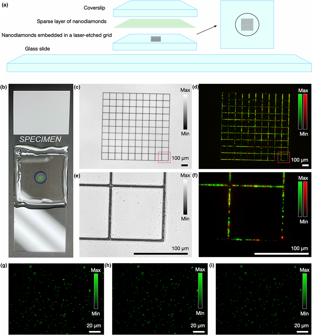
    Fig. 1. Structure of a nanodiamond based imaging target. (a) The imaging target combines a laser-etched grid with embedded fluorescent nanodiamonds along with a 2D layer of nanodiamonds deposited on a coverslip. A standard microscope slide provides structural support. (b) Photograph of an assembled target. The grid facilitates finding the physical location of the same particles during repeated longitudinal measurements. (c) Laser machined structure is visible in transmission widefield imaging mode. (d) Nanodiamonds embedded in the grid facilitate view finding in fluorescence imaging mode. (e), (f) Magnified images of a portion of the grid. (g)–(i) Images of the layer with sparse randomly distributed 100 nm nanodiamond particles, captured (g) immediately after assembling the phantom, (h) after one month of storage, and (i) after six months of storage. The measured fluorescence intensity for individual particles varied by less than 2%.
    Simultaneous four-channel multiphoton imaging of different types of calibration targets. (a) A dense lawn of 100 nm nanodiamond particles with NV color centers. (b) Locator grid with embedded 100 nm nanodiamonds is strongly visible in purple as well as in red channels. (c) Sparse nanodiamond particles deposited on a coverslip. (d) Locator grid with embedded 700 nm nanodiamond particles.
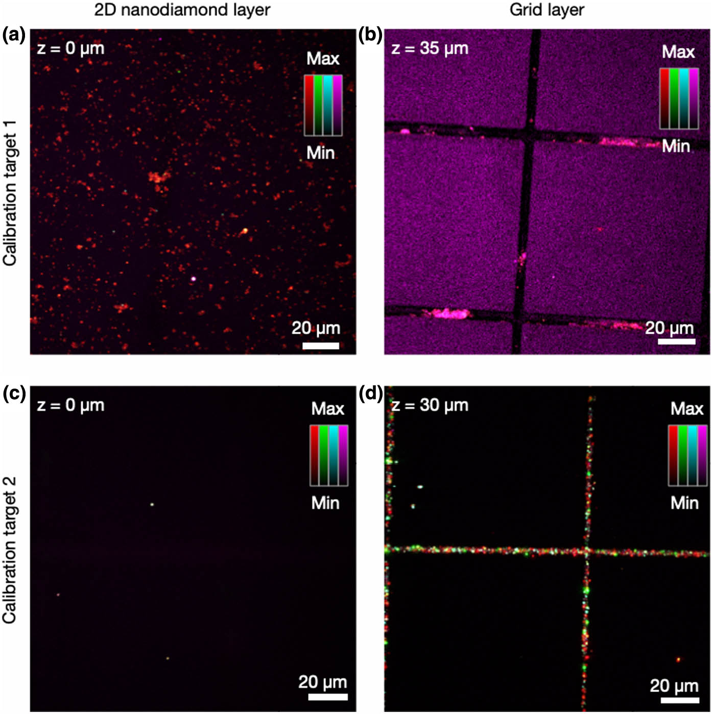
    Fig. 2. Simultaneous four-channel multiphoton imaging of different types of calibration targets. (a) A dense lawn of 100 nm nanodiamond particles with NV color centers. (b) Locator grid with embedded 100 nm nanodiamonds is strongly visible in purple as well as in red channels. (c) Sparse nanodiamond particles deposited on a coverslip. (d) Locator grid with embedded 700 nm nanodiamond particles.
    Fluorescence lifetime measurements. (a) A layer of nanodiamond particles with fluorescent centers deposited on glass can be used as a reference for assessing the fluorescence lifetime imaging performance of microscopy systems. (b) Overall fluorescence decay from all nanoparticles across the field of view. The inset in (b) shows representative examples of decay curves measured for individual particles.
    Fig. 3. Fluorescence lifetime measurements. (a) A layer of nanodiamond particles with fluorescent centers deposited on glass can be used as a reference for assessing the fluorescence lifetime imaging performance of microscopy systems. (b) Overall fluorescence decay from all nanoparticles across the field of view. The inset in (b) shows representative examples of decay curves measured for individual particles.
    Bleaching in fluorescent beads and in fluorescent nanodiamonds. Fluorescence microscopy images of (a) fluorescent beads and (c) fluorescent nanodiamonds deposited on the glass. (b) Fluorescent dyes in beads decay exponentially as a function of time, due to photobleaching. (d) Fluorescence of color centers in the diamond particles remains stable over time.
    Fig. 4. Bleaching in fluorescent beads and in fluorescent nanodiamonds. Fluorescence microscopy images of (a) fluorescent beads and (c) fluorescent nanodiamonds deposited on the glass. (b) Fluorescent dyes in beads decay exponentially as a function of time, due to photobleaching. (d) Fluorescence of color centers in the diamond particles remains stable over time.
    Dense random lawn of nanodiamonds for assessing imaging system performance. (a) A dense layer of subresolution 100 nm nanodiamond particles with NV centers deposited on glass can be used for rapid assessment of imaging performance of microscopy systems. (b) FFT of (a) reveals the broad frequency content of the image with no directionally ordered features. (c) The radial profile of (b) provides a rapid proxy for modulation transfer function measurements. The inset in (c) shows a schematic example representing a drop of imaging quality in a compromised optical system; (d) and (e) show lateral and axial slices, respectively, from a captured volume containing a single particle.
    Fig. 5. Dense random lawn of nanodiamonds for assessing imaging system performance. (a) A dense layer of subresolution 100 nm nanodiamond particles with NV centers deposited on glass can be used for rapid assessment of imaging performance of microscopy systems. (b) FFT of (a) reveals the broad frequency content of the image with no directionally ordered features. (c) The radial profile of (b) provides a rapid proxy for modulation transfer function measurements. The inset in (c) shows a schematic example representing a drop of imaging quality in a compromised optical system; (d) and (e) show lateral and axial slices, respectively, from a captured volume containing a single particle.
    Modulation of nanodiamond fluorescence intensity with a magnetic field. (a), (b) A miniature electromagnetic coil can be positioned near the nanodiamond layer. (c) Fluorescence intensity, as measured with a widefield fluorescence microscope, is significantly reduced in the presence of a magnetic field. (d) The modulation is not observed when the same field is applied to the fluorescent beads.
    Fig. 6. Modulation of nanodiamond fluorescence intensity with a magnetic field. (a), (b) A miniature electromagnetic coil can be positioned near the nanodiamond layer. (c) Fluorescence intensity, as measured with a widefield fluorescence microscope, is significantly reduced in the presence of a magnetic field. (d) The modulation is not observed when the same field is applied to the fluorescent beads.
    Mantas Žurauskas, Aneesh Alex, Jaena Park, Steve R. Hood, Stephen A. Boppart. Fluorescent nanodiamonds for characterization of nonlinear microscopy systems[J]. Photonics Research, 2021, 9(12): 2309
    Download Citation