
Yuchen CHANG, Ziyang LIN, Xin XIE, Zhangfan WU, Aihua YAO, Song YE, Jian LIN, Deping WANG, Xu CUI. An Injectable Composite Bone Cement Based on Mesoporous Borosilicate Bioactive Glass Spheres [J]. Journal of Inorganic Materials, 2020, 35(12): 1398
- Journal of Inorganic Materials
- Vol. 35, Issue 12, 1398 (2020)
Abstract
In the treatment of bone defect and fracture, injectable bone cements have been widely used due to their excellent plasticity and formability, which make them easy to match irregular bone defects and avoid gaps between the implant and the bone[
Injectable cements composed of borate bioactive glass are a new class of bone substitutes which have good mechanical and handling properties[
With a higher specific surface area and reactivity, mesoporous borosilicate bioactive glass nanoparticles process a higher Ca2+ release, which may accelerate the ionic cross-linking reaction between borosilicate glass and sodium alginate solution[
1 Materials and methods
1.1 Materials
Tributyl borate (TBB), tetraethyl orthosilicate (TEOS), cetyltrimethyl ammonium bromide (CTAB), triethylphosphate (TEP), calcium nitrate tetrahydrate (CN), absolute ethanol, deionized water, ammonia solution (25wt% NH3 in water), sodium hydrogen phosphate (Na2HPO4), D-gluconic acid d-lactone (GDL) and sodium alginate (SA) were purchased from Sinopharm Chemical Reagent Co., Ltd (Shanghai, China). All chemical reagents were chemical grade.
1.2 Preparation and characterization of mesoporous borosilicate bioactive glass spheres (MBGS)
The mesoporous borosilicate bioactive glass spheres were synthesized by Sol-Gel method using CTAB as template. 0.14 g CTAB was dissolved in a mixture containing 40 mL absolute ethanol and 60 mL deionized water under stirring at 40 ℃ until CTAB completely dissolved. Then a certain amount of TBB and TEOS were then added into the solution. Subsequently 0.50 mL ammonia, 0.30 mL TEP and 1.913 g CN were separately added into the solution at 30 min intervals. After stirred for another 1.5 h, the resulting solution was centrifuged to collect the white precipitate. Thereafter, the white precipitate was further washed three times with absolute ethanol and deionized water and dried at 60 ℃ for 24 h. The MBGS powders were eventually obtained after removal of remaining organics by sintering at 450 ℃ for 3 h (1 ℃/min). Three MBGS samples with various B2O3/SiO2 ratios were designated as MBGS-20, MBGS-30, and MBGS-40, and their corresponding compositions were shown in Table1.
The phase composition and microstructure of the MBGS were analyzed using X-ray diffractometer (XRD, D/max2550, Rigaku Co., Japan), and transmission electron microscope (TEM, JEM2100, JEOL Co., Japan). N2 adsorption-desorption isotherms were performed at 77 K using a NOVA 2200e (Quantachrome Instruments Co., America). The specific surface area and pore volume were calculated from N2 adsorption data in the partial relative pressure range of 0.05-0.35 using the Brunauere- Emmette-Teller (BET) method[
1.3 Fabrication of MBGS/SA composite bone cements
The SA liquid phase was firstly prepared by dissolving 2 g SA, 1 g D-gluconic acid d-lactone and 3 g sodium hydrogen phosphate in 100 mL deionized water. The MBGS-20, MBGS-30, and MBGS-40 powders were separately mixed with the liquid phase at a liquid/solid ratio of 0.75 mL/g to fabricate MBGS/SA composite bone cements. The resultant three bone cement samples were designated as BC-20, BC-30, and BC-40, respectively.
1.4 Evaluation of the workability of the MBGS/SA composite bone cements
The injectability of the MBGS/SA composite bone cements were evaluated in vitro using a procedure described previously[
where M0 is the original mass of the paste; M1 is the mass of the paste extruded from the syringe.
| Glass | B2O3 | SiO2 | CaO | P2O5 |
|---|---|---|---|---|
| MBGS-20 | 20 | 40 | 36 | 4 |
| MBGS-30 | 30 | 30 | 36 | 4 |
| MBGS-40 | 40 | 20 | 36 | 4 |
Table 1.
The compositions of MBGS/mol%
The initial setting time of the bone cement paste determined according to ISO9917-1. A PMMA mold containing five cylindrical holes (ϕ6 mm×12 mm) was placed in a water bath at 37.8 ℃. Then the cement paste (prepared as described earlier) was injected into the mold cavity. The setting time of the paste was determined using a Vicat’s apparatus (1.13 mm dimeter and 400 g weight)[
For compressive strength test, the bone cement pastes were cast into cylindrical Teflon mold and kept in an incubator at 37 ℃ and 100% relative humidity for 24 h to obtain cylindrical bone cement samples (ϕ6 mm×12 mm). Subsequently the compressive tests were performed using Electronic universal testing machine (CTM2500, Xie Qiang Instrument Manufacturing (Shanghai) Co. ltd, China) at a constant cross head speed of 0.5 mm/min. Five samples of each cement were tested and the compressive strength was determined as a mean ± SD.
The anti-washout property of the bone cements was measured by testing the remaining weight percentage after being shaken in phosphate buffer solution (PBS) for different times. The mass of the as-prepared cement paste was firstly weighted as M0. The sample was then carefully placed in a cup filled with PBS. The cup was put into a shaker and vibrated at a speed of 180 r/min and 37 ℃ for 2, 5, 12, 24 h. At each time, the amount of cement paste remaining was collected, freeze-dried and weighed as Mn (n=2, 5, 12 and 24). The remaining weight percentage was used to evaluate the anti-washout ability according to the following equation[
1.5 Evaluation of biomineralization in vitro
The bioactivity of the MBGS/SA composite bone cements was studied by immersion in simulate body fluid (SBF) at 37 ℃. Cylindrical samples of each cement (ϕ6 mm × 12 mm) were set for 4 h and immersed in 10 mL SBF in polyethylene containers. After 14 d of immersion, the samples were removed, washed with deionized water, dried at 90 ℃ and weighed. The phase compositions of the composite bone cements after being immersed in SBF were analyzed by X-ray diffraction (XRD) (D/max- 2500VB2+/PC X-ray diffractometer) using graphite monochromatized CuKα radiation (λ=0.15406 nm) at a scanning rate of 10 (°)/min (in the range of 10°-80°). The morphological features of the composite bone cements before and after immersion in SBF were examined in a field emission scanning electron microscope (FESEM, Hitachi S-4700). The samples were sputter-coated with gold prior to examination.
2 Results and discussion
2.1 Textural properties of MBGS
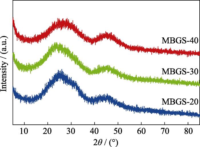
The phase composition, morphology and microstructure of the as-prepared MBGS were determined using XRD, FT-IR, TEM and nitrogen adsorption and desorption isotherm analyses. The B2O3/SiO2 ratio in the studied range had no effect on the phase composition (Fig. 1) and morphology (Fig. 2) of MBGS, and all three samples remained amorphous and display uniform and regular spherical morphologies. However, there was an increase in their specific surface area, average pore diameter and total pore volume with increasing boron content, as evidenced by the data in Table 2. Additionally, it was found from TEM images that mesoporous structure became more apparent for the samples with high boron content.
![]()
Figure 1.XRD patterns of the as-prepared MBGS
![]()
Figure 2.TEM images of the as-prepared MBGS
| Sample | Specific surface area/(m2∙g-1) | Average pore diameter/nm | Total pore |
|---|---|---|---|
| MBGS-20 | 161.71 | 13.33 | 0.052 |
| MBGS-30 | 176.98 | 13.97 | 0.057 |
| MBGS-40 | 214.28 | 15.11 | 0.079 |
Table 2.
Specific surface area, average pore diameter and total pore volume of the three groups of MBGS
The microstructure of the MBGs with various B2O3/SiO2 was determined by FT-IR spectra in Fig. 3. The main resonances consisted of the vibrational modes of the [SiO4] and [BO3] groups in the borosilicate glass network. The resonance at 1350-1450 cm-1 and at 690-720 cm-1 were assigned to the B-O stretching and bending modes of BO3 groups, respectively, while the resonances at 800 and 460 cm-1 were assigned to symmetric stretching mode and bending mode of Si-O-Si[
![]()
Figure 3.FT-IR spectra of the as-prepared MBGS
The mesoporous structure of the MBGS was confirmed by nitrogen adsorption and desorption isotherms in Fig. 4. All three samples represented type IV isotherms which was typical for mesoporous materials[
![]()
Figure 4.(a) N2 adsorption-desorption isotherms and (b) pore size distributions of MBGS-20, MBGS-30, and MBGS-40
2.2 Microstructures of the MBGS/SA bone cements
To confirm the favorable roles of MBGS textual properties (specific surface area and pore volume), the surface and cross-sectional morphologies of the MBGS/SA bone cements were firstly observed. As shown in Fig. 5 (a-c), the three samples presented distinctly different surface morphologies. The bone cements with higher B2O3/SiO2 ratio (BC-30 and BC-40) had a significantly larger number of interconnected pores, whereas BC-20 displayed relatively smooth surface. As indicated in our previous study[
![]()
Figure 5.SEM images of (a, d) BC-20, (b, e) BC-30, and (c, f) BC-40 bone cement specimens
The increased B2O3/SiO2 ratio caused poor chemical stability of the glass network and promoted the rapid degradation of MBGS. In our previous study, B ions accumulative concentration released from borosilicate bioactive bone cement with B2O3/SiO2 ratio increasing during immersion was increased[
2.3 Setting properties
As shown in Fig. 6, since the B2O3/SiO2 ratio was increased, the setting time was shortened from 21 min to 9 min, which was comparable to other cements and suitable for clinical applications. The compressive strength of the bone cements is slightly increased from (3.4±0.2) MPa to (4.1±0.3) MPa as the B2O3/SiO2 ratio increased from 1 : 2 to 2 : 1, of which bone cement with B2O3/SiO2 ratio at 2 : 1 is 2 times higher than that of the non-mesoporous- derived bone cements produced under the same conditions in our previous study[
![]()
Figure 6.(a) Setting time and (b) compressive strength of BC-20, BC-30, and BC-40, respectively* indicates significant difference between groups,
2.4 Injectability
Data from the injectability test showed that both BC-20 and BC-30 pastes could be extruded easily as confirmed by Fig. 7 and their injectability remained about 90% during the first 6 min, indicating their appropriate viscosity and excellent injectability. In comparison, larger force was required for the extrusion of the BC-40 paste, yet only 76wt% fraction was extruded. Additionally, since the viscosity of the BC-40 paste was too high to completely remove the incorporated bubbles, an abrupt drop in the extrusion force was observed on curve C in Fig. 7. The three pastes remained continuous and regular shape after extrusion as shown in Fig. 8. BC-20 and BC-30 pastes were quite soft and sticky, while the BC-40 paste was relatively dry, further indicating its high viscosity and poor injectability. The difference in the injectability of bone cements was due to the different texture structures of the Sol-Gel-derived borosilicate glasses. The higher specific surface area of MBGS-40 and subsequent rapid ions release resulted in a hydrogel network with higher cross- linked degree, which increases the viscosity of the paste, thereby encountering larger extrusion resistance.
![]()
Figure 7.(a) Force-displacement curves and (b) injectabilities of mesoporous borosilicate bone cements for different B2O3 contents* indicates significant difference between groups,
![]()
Figure 8.Photographs of the mesoporous borosilicate bone cement extruded from the syring
2.5 Anti-washout property
The anti-washout property is an important parameter to evaluate the ability of bone cements to keep the initial shape in a dynamic aqueous environment[
![]()
Figure 9.Anti-washout properties of mesoporous borosilicate bone cements
![]()
Figure 10.Morphologies of mesoporous borosilicate bone cement specimens after being shaken in PBS at 37 ℃ for 2, 5, 12, and 24 h, respectively
2.6 Biomineralization ability in vitro
The mineralization ability of the bone cements was confirmed by using XRD, SEM and EDS analyses. In Fig. 11, after 3 d of immersion in SBF, weak diffraction peaks in XRD patterns corresponding to HA crystalline were presented, then the peaks became sharper and more defined over time, indicating the progressive crystallization of HA. The result demonstrated significantly more rapid HA formation compared with the bone cements produced from non-mesoporous borosilicate glass, attributable to the high surface area and porosity of the present MBGs coupled with rapid ion release. Wang, et al.[
![]()
Figure 11.XRD patterns of bone cement specimens after being immersed in SBF for 3, 7 and 14 d, respectively
![]()
Figure 12.SEM images and EDS data of (a, d) BC-20, (b, e) BC-30, (c, f) BC-30 after being immersed in SBF for 14 d
Consequently, BC-30 bone cement had appropriate setting time, injectability, anti-washout properties and mineralization ability in vitro among these three samples and can be regarded as the optimal composition. As mentioned previously, the solidification of the composite bone cement was a result of the Ca2+ induced gelation process of sodium alginate. The low chemical durability of the Sol-Gel-derived borosilicate bioactive glasses and resultant rapid Ca2+ release, therefore, provided a prerequisite for the gelation. In addition to the glass composition, their excellent textural properties including high specific surface area and pore volume played an essential role in enhancing the mechanical properties and workability of the bone cements. Also, both factors favored their bioactive response, thus a rapid mineralization rate was observed. These desirable properties indicated that the composite bone cements would be promising candidates for the use in minimally invasive surgery for treatment of bone defects. Furthermore, the exceptional textural properties of the MBGS enable the loading of diverse drugs, thereby achieving a multifunctional task in bone tissue engineering applications. In the future research, improving the compressive strength of mesoporous borosilicate bioactive glass bone cement should be continuously studied to meet the requirement of clinical application.
3 Conclusions
In this study, mesoporous borosilicate bioactive glass spheres with various B2O3/SiO2 ratios were prepared by Sol-Gel template method. The resultant glass spheres were then combined with alginate to fabricate injectable composite bone cements. The results indicated that an increase in glass boron content resulted in decreased network connectivity, and thus achieving higher specific surface area and larger pore volume of MBGS, which further accelerated the glass degradation and Ca2+ release. It is also confirmed that compared to non-mesoporous bioactive glass used in our previous study, the good textural properties of the mesoporous borosilicate bioactive glass spheres endow the composite bone cements with mproved workability, including clinically appropriate setting time (10-20) min, excellent injectability and anti- washout property, as well as good bioactivity. What’s more, BC-30 was the optimal composition for bone cement. Therefore, the present bone cement is a more appropriate option as bone filling and regeneration materials for minimally invasive applications.
References
[1] S VERRIER, L HUGHES, A ALVES et al. Evaluation of the in vitro cell-material interactions and in vivo osteo-integration of a spinal acrylic bone cement. European Spine Journal, 21, S800-S809(2012).
[2] J D KRETLOW, L KLOUDA, A G MIKOS. Injectable matrices and scaffolds for drug delivery in tissue engineering. Adv. Drug Delivery Rev, 59, 263-273(2007).
[4] M V CABANAS, L M RODRIGUEZ-LORENZO, M VALLET-REGI. Setting behavior and in vitro bioactivity of hydroxyapatite/ calcium sulfate cements. Chem. Mater, 14, 3550-3555(2002).
[11] H B LI, D P WANG, Y Y WU et al. Effect of citric acid concentration on the properties of borate glass bone cement. Journal of Inorganic Materials, 32, 831-836(2017).
[12] X CUI, W H HUANG, Y D ZHANG et al. Evaluation of an injectable bioactive borate glass cement to heal bone defects in a rabbit femoral condyle model. Materials Science & Engineering C-Materials for Biological Applications, 73, 585-595(2017).
[13] X CUI, Y D ZHANG, H WANG et al. An injectable borate bioactive glass cement for bone repair: preparation, bioactivity and setting mechanism. Journal of Non-Crystalline Solids, 432, 150-157(2016).
[14] Y Y WU, S YE, A H YAO et al. Effect of gas-foaming porogen- nahco3 and citric acid on the properties of injectable macroporous borate bioactive glass cement. Journal of Inorganic Materials, 32, 777-784(2017).
[15] R F MOSEMAN. Chemical disposition of boron in animals and humans. Environmental Health Perspectives, 102, 113-117(1994).
[18] A H YAO, D P WANG, W H HUANG et al. In vitro bioactive characteristics of borate-based glasses with controllable degradation behavior. Journal of The American Ceramic Society, 90, 303-306(2007).
[19] Q A FU, M N RAHAMAN, H L FU et al. Silicate, borosilicate, and borate bioactive glass scaffolds with controllable degradation rate for bone tissue engineering applications. I. Preparation and in vitro degradation. Journal of Biomedical Materials Research Part A, 95A, 164-171(2010).
[20] Q A FU, M N RAHAMAN, B S BAL et al. Silicate, borosilicate, and borate bioactive glass scaffolds with controllable degradation rate for bone tissue engineering applications. II. In vitro and in vivo biological evaluation. Journal of Biomedical Materials Research Part A, 95A, 172-179(2010).
[24] C SCHERDEL, G REICHENAUER, M WIENER. Relationship between pore volumes and surface areas derived from the evaluation of N2-sorption data by DR-, BET- and t-plot. Microporous Mesoporous Mater, 132, 572-575(2010).
[25] J LANDERS, G Y GOR, A V NEIMARK. Density functional theory methods for characterization of porous materials. Colloids and Surfaces a-Physicochemical and Engineering Aspects, 437, 3-32(2013).
[26] X XIE, L B PANG, A H YAO et al. Nanocement produced from borosilicate bioactive glass nanoparticles composited with alginate. Australian Journal of Chemistry, 72, 354-361(2019).
[37] Y WANG, T LIAO, M SHI et al. Facile synthesis and in vitro bioactivity of radial mesoporous bioactive glasses. Materials Letters, 206, 205-209(2017).


Set citation alerts for the article
Please enter your email address

© Copyright 2018-2021 | Chinese Laser Press. All Rights Reserved 沪ICP备15018463号-20