• Laser & Optoelectronics Progress
  • Vol. 55, Issue 12, 121102 (2018)
Lu Song1 and Yuanhua Feng2、*
Author Affiliations
  • 1 Guangdong Provincial Key Laboratory of Optical Fiber Sensing and Communications, Institute of Photonics Technology, Jinan University, Guangzhou, Guangdong 510632, China
  • 2 College of Information Science and Technology, Jinan University, Guangzhou, Guangdong 510632, China
  • show less
    DOI: 10.3788/LOP55.121102 Cite this Article Set citation alerts
    Lu Song, Yuanhua Feng. Ultrafast Flow Cytometric Quantitative Phase Imaging System Based on Optical Coherent Detection Technology[J]. Laser & Optoelectronics Progress, 2018, 55(12): 121102 Copy Citation Text show less
    Schematic of the time stretch imaging system
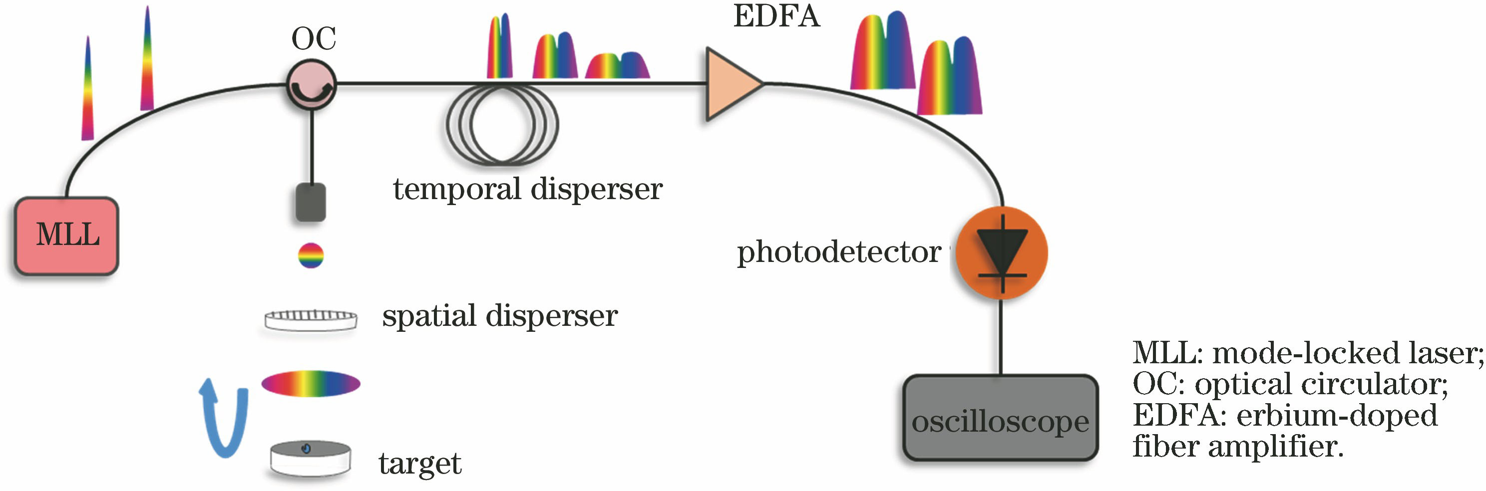
    Fig. 1. Schematic of the time stretch imaging system
    Structure schematic of optical coherent receiver
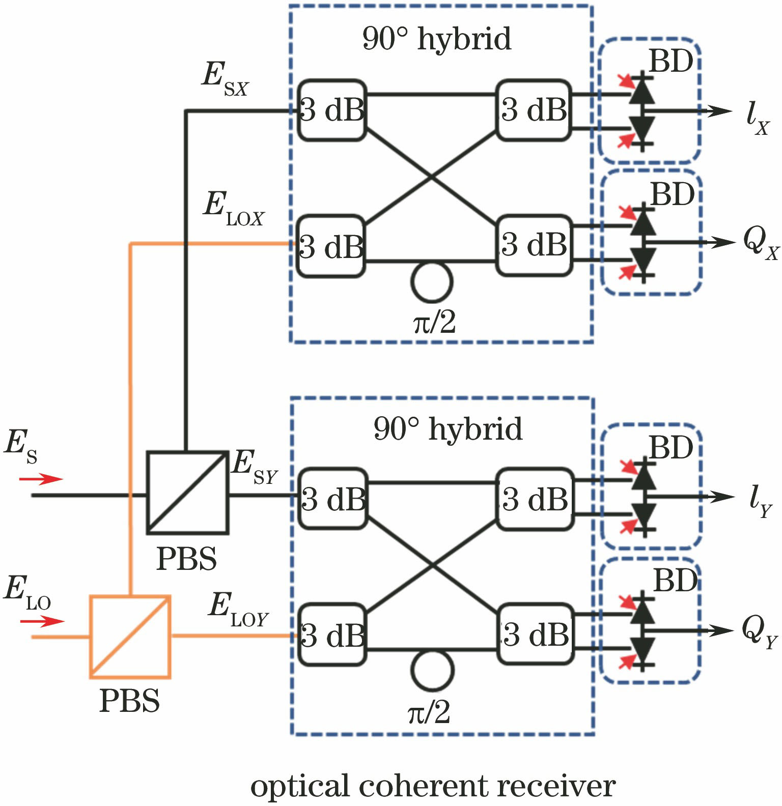
    Fig. 2. Structure schematic of optical coherent receiver
    Relationship between resolution limit and dispersion
    Fig. 3. Relationship between resolution limit and dispersion
    Schematic of the time-stretch-based ultrafast polarization cytometric imaging system.(a) Time domain dispersion; (b) spatial dispersion and imaging; (c) optical coherent detection
    Fig. 4. Schematic of the time-stretch-based ultrafast polarization cytometric imaging system.(a) Time domain dispersion; (b) spatial dispersion and imaging; (c) optical coherent detection
    (a) Image of resolution target; (b) time domain waveform captured by the detector; (c) differential and Gauss fitting curves
    Fig. 5. (a) Image of resolution target; (b) time domain waveform captured by the detector; (c) differential and Gauss fitting curves
    Phase contrast microscope images and phase images. (a) Phase contrast microscope image of normal nasopharyngeal carcinoma cell slice; (b) phase image of normal nasopharyngeal carcinoma cell slice; (c) phase contrast microscope image of nasopharyngeal carcinoma cell slice treated with growth inhibition drugs; (d) phase image of normal nasopharyngeal carcinoma cell slice treated with growth inhibition drugs
    Fig. 6. Phase contrast microscope images and phase images. (a) Phase contrast microscope image of normal nasopharyngeal carcinoma cell slice; (b) phase image of normal nasopharyngeal carcinoma cell slice; (c) phase contrast microscope image of nasopharyngeal carcinoma cell slice treated with growth inhibition drugs; (d) phase image of normal nasopharyngeal carcinoma cell slice treated with growth inhibition drugs
    Phase images (scale bar: 10 μm). (a) Three-dimensional phase image of single nasopharyngeal carcinoma cell; (b) phase images of nasopharyngeal carcinoma cells; (c) phase images of normal nasopharyngeal epithelial cells
    Fig. 7. Phase images (scale bar: 10 μm). (a) Three-dimensional phase image of single nasopharyngeal carcinoma cell; (b) phase images of nasopharyngeal carcinoma cells; (c) phase images of normal nasopharyngeal epithelial cells
    Lu Song, Yuanhua Feng. Ultrafast Flow Cytometric Quantitative Phase Imaging System Based on Optical Coherent Detection Technology[J]. Laser & Optoelectronics Progress, 2018, 55(12): 121102
    Download Citation