• Laser & Optoelectronics Progress
  • Vol. 58, Issue 8, 0810021 (2021)
Junxie Chen1 and Yipeng Liao2、*
Author Affiliations
  • 1College of Artificial Intelligence, Yango University, Fuzhou, Fujian 350015, China
  • 2College of Physics and Information Engineering, Fuzhou University, Fuzhou, Fujian 350108, China
  • show less
    DOI: 10.3788/LOP202158.0810021 Cite this Article Set citation alerts
    Junxie Chen, Yipeng Liao. Edge Detection of Noisy Images in NSCT Domain Based on Fractional Differentiation[J]. Laser & Optoelectronics Progress, 2021, 58(8): 0810021 Copy Citation Text show less
    Tiansi operator
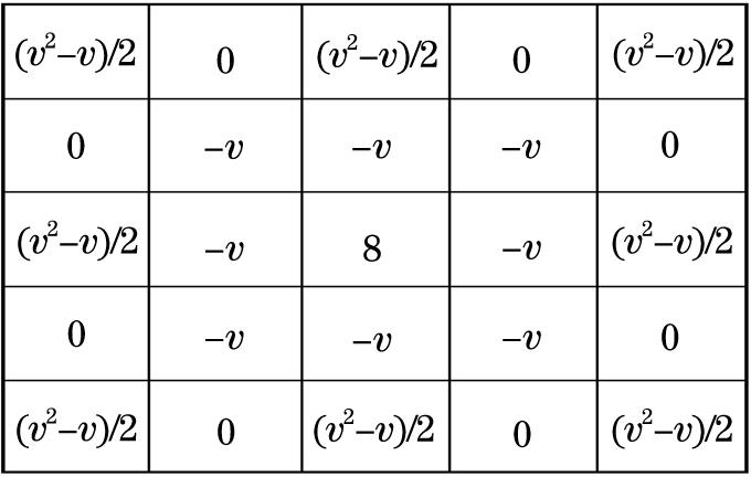
    Fig. 1. Tiansi operator
    Decomposing process of NSCT
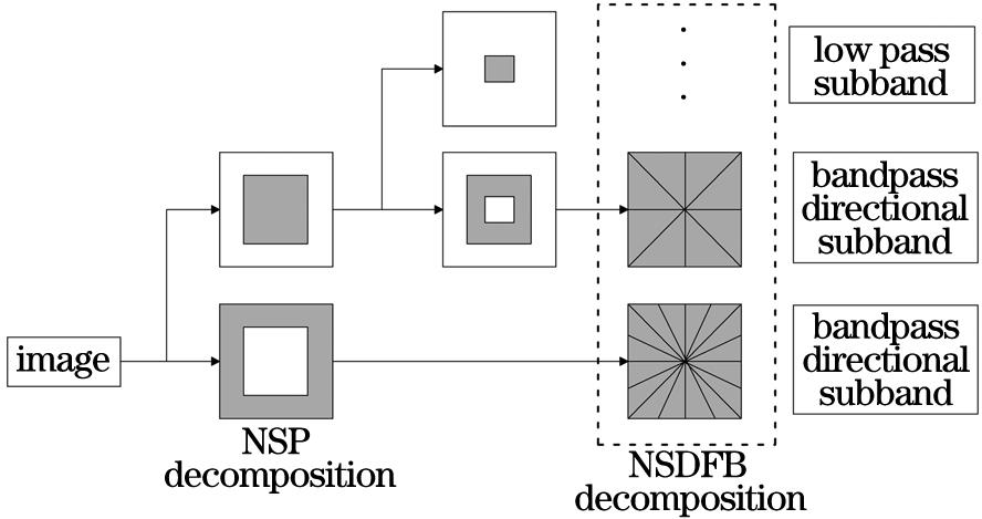
    Fig. 2. Decomposing process of NSCT
    Flow chart of the proposed method
    Fig. 3. Flow chart of the proposed method
    Original images for experiment. (a) Lena; (b) cartoon image
    Fig. 4. Original images for experiment. (a) Lena; (b) cartoon image
    Edge detection results of Lena image. (a) Low frequency; (b) high frequency scale 1; (c) high frequency scale 2; (d) high frequency; (e) fusion result
    Fig. 5. Edge detection results of Lena image. (a) Low frequency; (b) high frequency scale 1; (c) high frequency scale 2; (d) high frequency; (e) fusion result
    Edge detection results of cartoon image. (a) Low frequency; (b) high frequency scale 1; (c) high frequency scale 2; (d) high frequency; (e) fusion result
    Fig. 6. Edge detection results of cartoon image. (a) Low frequency; (b) high frequency scale 1; (c) high frequency scale 2; (d) high frequency; (e) fusion result
    Edge extraction results of Lena image. (a) Original image; (b) image with 10% noise; (c) extraction result with 10% noise; (d) image with 50% noise; (e) extraction result with 50% noise
    Fig. 7. Edge extraction results of Lena image. (a) Original image; (b) image with 10% noise; (c) extraction result with 10% noise; (d) image with 50% noise; (e) extraction result with 50% noise
    Edge extraction results of medical image 1. (a) Original image; (b) image with 10% noise; (c) extraction result with 10% noise; (d) image with 50% noise; (e) extraction result with 50% noise
    Fig. 8. Edge extraction results of medical image 1. (a) Original image; (b) image with 10% noise; (c) extraction result with 10% noise; (d) image with 50% noise; (e) extraction result with 50% noise
    Edge extraction results of medical image 2. (a) Original image; (b) image with 10% noise; (c) extraction result with 10% noise; (d) image with 50% noise; (e) extraction result with 50% noise
    Fig. 9. Edge extraction results of medical image 2. (a) Original image; (b) image with 10% noise; (c) extraction result with 10% noise; (d) image with 50% noise; (e) extraction result with 50% noise
    Extracted results of methods for Lena image. (a) Canny; (b) method in Ref. [7]; (c) method in Ref. [15]; (d) method in Ref. [20]; (e) proposed method
    Fig. 10. Extracted results of methods for Lena image. (a) Canny; (b) method in Ref. [7]; (c) method in Ref. [15]; (d) method in Ref. [20]; (e) proposed method
    Extracted results of methods for cartoon image. (a) Canny; (b) method in Ref. [7]; (c) method in Ref. [15]; (d) method in Ref. [20]; (e) proposed method
    Fig. 11. Extracted results of methods for cartoon image. (a) Canny; (b) method in Ref. [7]; (c) method in Ref. [15]; (d) method in Ref. [20]; (e) proposed method
    Processing results of different methods for noisy Lena image. (a) Method in Ref. [7]; (b) method in Ref. [15]; (c) method in Ref. [16]; (d) method in Ref. [20]; (e) proposed method
    Fig. 12. Processing results of different methods for noisy Lena image. (a) Method in Ref. [7]; (b) method in Ref. [15]; (c) method in Ref. [16]; (d) method in Ref. [20]; (e) proposed method
    Processing results of different methods for noisy medical image 1. (a) Method in Ref. [7]; (b) method in Ref. [15]; (c) method in Ref. [16]; (d) method in Ref. [20]; (e) proposed method
    Fig. 13. Processing results of different methods for noisy medical image 1. (a) Method in Ref. [7]; (b) method in Ref. [15]; (c) method in Ref. [16]; (d) method in Ref. [20]; (e) proposed method
    Processing results of different methods for noisy medical image 2. (a) Method in Ref. [7]; (b) method in Ref. [15]; (c) method in Ref. [16]; (d) method in Ref. [20]; (e) proposed method
    Fig. 14. Processing results of different methods for noisy medical image 2. (a) Method in Ref. [7]; (b) method in Ref. [15]; (c) method in Ref. [16]; (d) method in Ref. [20]; (e) proposed method
    Comparison of results of different methods for different noise. (a) Lena image; (b) medical image 1; (c) medical image 2
    Fig. 15. Comparison of results of different methods for different noise. (a) Lena image; (b) medical image 1; (c) medical image 2
    IndexCannyMethod in Ref. [7]Method in Ref. [15]Method in Ref. [20]Proposed method
    α50641754687295329841
    β6103267880361052910547
    R0.8300.6550.8550.9050.933
    Table 1. Comparison of extracted results of Lena image by different methods
    IndexCannyMethod in Ref. [7]Method in Ref. [15]Method in Ref. [20]Proposed method
    α77623378103781321315693
    β92845211120531450116802
    R0.8360.6480.8610.9110.934
    Table 2. Comparison of extracted results for cartoon image by different methods
    Junxie Chen, Yipeng Liao. Edge Detection of Noisy Images in NSCT Domain Based on Fractional Differentiation[J]. Laser & Optoelectronics Progress, 2021, 58(8): 0810021
    Download Citation