• Photonics Research
  • Vol. 11, Issue 11, 1838 (2023)
Yu-Xuan Ren1、2, Joshua Lamstein2、3, Chensong Zhang2、4, Claudio Conti5, Demetrios N. Christodoulides6, and Zhigang Chen2、7、*
Author Affiliations
  • 1Institute for Translational Brain Research, MOE Frontiers Center for Brain Science, Fudan University, Shanghai 200032, China
  • 2Department of Physics and Astronomy, San Francisco State University, San Francisco, California 94132, USA
  • 3Center for Systems and Therapeutics, Gladstone Institutes, San Francisco, California 94158, USA
  • 4Division of CryoEM and Bioimaging, Stanford Synchrotron Radiation Lightsource, SLAC National Accelerator Laboratory, Menlo Park, California 94025, USA
  • 5Department of Physics, University Sapienza, I-00185 Roma, Italy
  • 6Department of Electrical Engineering, University of Southern California, Los Angeles, California 90089, USA
  • 7TEDA Applied Physics Institute and School of Physics, Nankai University, Tianjin 300457, China
  • show less
    DOI: 10.1364/PRJ.496013 Cite this Article Set citation alerts
    Yu-Xuan Ren, Joshua Lamstein, Chensong Zhang, Claudio Conti, Demetrios N. Christodoulides, Zhigang Chen. Biophotonic rogue waves in red blood cell suspensions[J]. Photonics Research, 2023, 11(11): 1838 Copy Citation Text show less
    (a) Experimental setup for observing wave dynamics in RBC suspensions. Laser, 532 nm wavelength; HW, half-wave plate; PBS, polarizing beam splitter; L1 through L4, lenses; M1, M2, reflective mirrors; CCD, charge-coupled device. (b)–(d) Illustrations of (b) linear propagation of a plane wave illumination, (c) linear diffraction of a focused beam at low power, and (d) nonlinear self-focusing at high power.
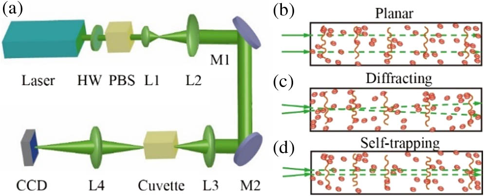
    Fig. 1. (a) Experimental setup for observing wave dynamics in RBC suspensions. Laser, 532 nm wavelength; HW, half-wave plate; PBS, polarizing beam splitter; L1 through L4, lenses; M1, M2, reflective mirrors; CCD, charge-coupled device. (b)–(d) Illustrations of (b) linear propagation of a plane wave illumination, (c) linear diffraction of a focused beam at low power, and (d) nonlinear self-focusing at high power.
    Observation of biophotonic RWs in an RBC suspension. (a) Speckle-like pattern observed in an RBC suspension, in which rare spikes with a giant intensity appear. (b) 3D intensity profile of the zoomed region marked by a white square in (a) demonstrates one rogue event (an intensity “hot spot”). (c) The intensity histogram suggests long-tail statistics where the RW threshold is marked by dashed blue line (a.u., arbitrary unit). (d) The 1D spatial spectrum of the speckle-like pattern shows a broad Gaussian distribution with a bandwidth of ∼0.1 μm−1 (solid line represents the Gaussian fit). (e) A typical temporal trace monitoring a single pixel on the CCD camera demonstrates that the RW appears at 38.5 s. (f) Return time statistics, where the solid curve shows the fit to exponential model exp(−τ/τc), with a characteristic return time of 9.57±0.26 s.
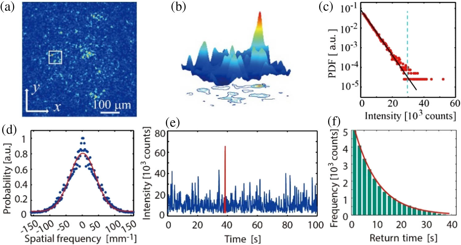
    Fig. 2. Observation of biophotonic RWs in an RBC suspension. (a) Speckle-like pattern observed in an RBC suspension, in which rare spikes with a giant intensity appear. (b) 3D intensity profile of the zoomed region marked by a white square in (a) demonstrates one rogue event (an intensity “hot spot”). (c) The intensity histogram suggests long-tail statistics where the RW threshold is marked by dashed blue line (a.u., arbitrary unit). (d) The 1D spatial spectrum of the speckle-like pattern shows a broad Gaussian distribution with a bandwidth of 0.1  μm1 (solid line represents the Gaussian fit). (e) A typical temporal trace monitoring a single pixel on the CCD camera demonstrates that the RW appears at 38.5 s. (f) Return time statistics, where the solid curve shows the fit to exponential model exp(τ/τc), with a characteristic return time of 9.57±0.26  s.
    RW event probability regulated by cell concentrations. (a) The spatial output of a 532 nm laser beam passing through the RBC suspension in isotonic buffer. (b) The corresponding spatial spectrum suggests a broadband distribution. (c) The FWHM for the spatial spectrum varies with the cell concentration. (d) RW probability as a function of cell concentration. The incident powers for each curve are labeled in the legend. Scale bars: (a) 15 μm, (b) ∼0.1 μm−1.
    Fig. 3. RW event probability regulated by cell concentrations. (a) The spatial output of a 532 nm laser beam passing through the RBC suspension in isotonic buffer. (b) The corresponding spatial spectrum suggests a broadband distribution. (c) The FWHM for the spatial spectrum varies with the cell concentration. (d) RW probability as a function of cell concentration. The incident powers for each curve are labeled in the legend. Scale bars: (a) 15 μm, (b) 0.1  μm1.
    Measured event probability and maximum strength of RWs in RBC suspensions under different osmotic conditions. (a) An isotonic RBC has a biconcave shape and diverges the beam. (b) A hypotonic or hypertonic RBC assumes a spherical shape and converges the beam. (c) RW probability with RBC suspensions in hypotonic (black), isotonic (red), and hypertonic (pink) buffer. (d) Maximum strength of RW events in RBC suspensions under different osmotic conditions.
    Fig. 4. Measured event probability and maximum strength of RWs in RBC suspensions under different osmotic conditions. (a) An isotonic RBC has a biconcave shape and diverges the beam. (b) A hypotonic or hypertonic RBC assumes a spherical shape and converges the beam. (c) RW probability with RBC suspensions in hypotonic (black), isotonic (red), and hypertonic (pink) buffer. (d) Maximum strength of RW events in RBC suspensions under different osmotic conditions.
    RW events measured in polystyrene (spherical bead) suspensions for comparison. (a) Typical frame for light scattering in a suspension of 5 μm polystyrene beads showing two (“hot-spot”) rogue events. (b) RW probability varies with time for beads with different sizes, where three traces for different sizes are measured separately and concatenated for better visualization. (c) The ratio of the maximum over average intensities changes with time from the video series. Traces in (b) and (c) are for polystyrene beads with diameter 2 μm (black), 5 μm (red), and 11 μm (pink).
    Fig. 5. RW events measured in polystyrene (spherical bead) suspensions for comparison. (a) Typical frame for light scattering in a suspension of 5 μm polystyrene beads showing two (“hot-spot”) rogue events. (b) RW probability varies with time for beads with different sizes, where three traces for different sizes are measured separately and concatenated for better visualization. (c) The ratio of the maximum over average intensities changes with time from the video series. Traces in (b) and (c) are for polystyrene beads with diameter 2 μm (black), 5 μm (red), and 11 μm (pink).
    RW events observed with a plane wave illumination in isotonic RBC suspensions. (a) RW event captured in the nearly linear propagation regime. (b) Rogue event identified in the time-course signal taken at the chosen pixel. The duration of the RW is ∼0.27 s. (c) RW probability as a function of the laser wavelength.
    Fig. 6. RW events observed with a plane wave illumination in isotonic RBC suspensions. (a) RW event captured in the nearly linear propagation regime. (b) Rogue event identified in the time-course signal taken at the chosen pixel. The duration of the RW is 0.27  s. (c) RW probability as a function of the laser wavelength.
    Influence of focusing nonlinearity on RW statistics in RBC suspensions. (a), (b) Linear case when the laser beam shows no self-action at 10 mW. (c), (d) Nonlinear case when the laser beam shows self-focusing at 500 mW. The solid straight line in (b) shows an exponential fit, while the curved line in (d) represents a fit using a dual-exponential distribution function. (e)–(h) are the corresponding simulation for weak and strong nonlinear cases (a.u., arbitrary unit).
    Fig. 7. Influence of focusing nonlinearity on RW statistics in RBC suspensions. (a), (b) Linear case when the laser beam shows no self-action at 10 mW. (c), (d) Nonlinear case when the laser beam shows self-focusing at 500 mW. The solid straight line in (b) shows an exponential fit, while the curved line in (d) represents a fit using a dual-exponential distribution function. (e)–(h) are the corresponding simulation for weak and strong nonlinear cases (a.u., arbitrary unit).
    Yu-Xuan Ren, Joshua Lamstein, Chensong Zhang, Claudio Conti, Demetrios N. Christodoulides, Zhigang Chen. Biophotonic rogue waves in red blood cell suspensions[J]. Photonics Research, 2023, 11(11): 1838
    Download Citation