1Collaborative Innovation Center of Light Manipulations and Applications in Universities of Shandong School of Physics and Electronics, Shandong Normal University, Jinan 250014, China
2Shandong Key Laboratory of Medical Physics and Image Processing & Shandong Provincial Engineering and Technical Center of Light Manipulations, Jinan 250014, China
3School of Information Science and Engineering, Shandong University, Qingdao 266237, China
4Shandong Key Laboratory of Biophysics, Institute of Biophysics, Dezhou University, Dezhou 253023, China
Can Li, Jinjuan Gao, Muhammad Shafi, Runcheng Liu, Zhipeng Zha, Dejun Feng, Mei Liu, Xuejian Du, Weiwei Yue, Shouzhen Jiang, "Optical fiber SPR biosensor complying with a 3D composite hyperbolic metamaterial and a graphene film," Photonics Res. 9, 379 (2021)
Copy Citation Text
In the present study, an optical fiber surface plasmon resonance (SPR) biosensor was developed for measuring time- and concentration-dependent DNA hybridization kinetics. Its design complies with a 3D multilayer composite hyperbolic metamaterial (HMM), a graphene film, and a D-shaped plastic optical fiber. According to the numerical simulation and the experimental demonstration, the SPR peak of the designed biosensor can be effectively altered in the range of visible to near-infrared by varying the HMM structure. The sensitivity of the appliance was shown to achieve a value of up to 4461 nm/RIU, allowing its applicability for bulk refractive index sensing. Furthermore, a biosensor designed in this work displayed high-resolution capability (ranging from 10 pM to 100 nM), good linearity, and high repeatability along with a detection limit down to 10 pM, thus suggesting a vast potential for medical diagnostics and clinical applications.
1. INTRODUCTION
Surface plasmon resonance (SPR) currently turns out to be one of the most feasible techniques for developing biological sensors for label-free, point-of-care, and high-throughput applications [1–7]. The resonance results from the coherent free electron oscillations that are propagated along the metal/dielectric interface when the wave vector satisfies the momentum matching conditions [8]. Typical metals used for SPR applications are gold and silver [9–11]. Owing to their highly sensitive electromagnetic field associated with the mentioned oscillations, the SPR exhibits a noticeable sensitivity to the refractive index (RI) of the surrounding dielectric material.
Conventional SPR sensors possess the built-in Kretschmann configuration, which strongly depends on large optical components, thereby resulting in high operational costs and difficulty in miniaturization in the fields of precision medicine and system biology [12–14]. In this regard, the emerging optical fiber that exhibits numerous prominent features (e.g., low cost, lightweight, ease of use, long-distance sensing, and small dimensions) can be employed for developing state-of-the-art SPR sensors [5,6,15–17]. Thus far, meaningful works have been performed to improve the already existing optical fiber SPR sensor configuration by applying optical fibers. Given the detection area morphology, fiber-optic SPR sensors can be D-shaped [18], U-shaped [19], tapered [20], or take other forms [21]. Among these, the D-shaped sensing probe has a range of benefits (e.g., high sensitivity, easy functional modification, and wide detection area). As for optical fiber materials, plastic optical fiber (POF) has aroused attention due to its super machinability and handling along with low cost [22–24].
So far, tremendous effort has been made to boost the development of optical fiber SPR sensors. In 2018, Gong et al. proposed a novel D-shaped optical fiber SPR sensor based on the graphene/Au hybrid structure with a sensitivity of 1227 nm/RIU for DNA hybridization detection [25]. In 2019, by depositing a Cu-nanoparticle/carbon nanotube composite film onto the gold film, Zhang et al. obtained a reflective fiber optic SPR sensor for simultaneous measurement of nitrate concentration and temperature [21]. In 2020, seizing the advantages of photonic crystal fibers, Wang et al. developed an ultra-wide detection range refractive-index SPR sensor with a sensing range of 1.29–1.49 [26]. In 2020, using glucose oxidase as a sensitive film covalently bonded on the gold film, Zheng et al. designed a highly sensitive and reflective glucose SPR sensor to measure glucose concentrations [27]. However, a leap forward in clinical applications toughened requirements for the sensitivity and stability of optical fiber SPR sensors.
Sign up for Photonics Research TOC. Get the latest issue of Photonics Research delivered right to you!Sign up now
Over the past few years, rapid progress in the micro/nano processing technology enabled the experimental demonstration of various artificial optical metamaterials, whose magnetic properties were improved compared to conventional materials [28]. In this respect, hyperbolic metamaterials (HMMs) have rapidly gained a central role due to their capability to access and manipulate the near field of a light scatterer or a light emitter, which derives directly from the excitation, within HMMs, of coupled surface plasmons [8,29]. HMMs refer to uncommon artificial electromagnetic metamaterials that display hyperbolic dispersion with bulk 3D sub-wavelength structures that are anisotropic in permittivity. Recently, extreme sensitivity has been achieved by employing HMMs [30–32]. In 2009, Kabashin et al. proposed a plasmonic nanorod metamaterial for high-sensitivity plasmonic biosensors with sensitivity to RI variations of more than 30,000 nm/RIU using a prism-coupling mechanism [33]. In 2016, Kandammathe et al. developed a miniaturized plasmonic biosensor platform based on a hyperbolic metamaterial of a three-dimensional multilayer metal () and unconventional two-dimensional gold diffraction gratings, allowing sensitivity of up to 30,000 nm/RIU [34]. Furthermore, the angular scans with extraordinary angular sensitivity of 7000° per RIU were achieved in a visible-to-near-infrared (NIR) wavelength range [35]. However, the complex system configurations bring some limitations to the mentioned biosensors, such as insufficiency to address high costs and difficulty in miniaturization.
In this respect, the present work is aimed at designing an SPR optical fiber biosensor based on a 3D composite HMM, a graphene film, and D-shaped plastic optical fiber (D-POF) (G/HMM/D-POF). The performance of the setup was assessed via numerical simulation and experimental demonstration. Compared with a conventional fiber optic SPR sensor, introducing the multilayer HMM was shown to considerably improve the sensitivity of the appliance under consideration. Furthermore, graphene was proved to enhance the performance of a biosensor in three aspects: sensitivity, molecular affinity, and stability [36–39]. Under the D-type fiber structure, the position of the SPR resonance peak could be effectively tuned in the range of visible to NIR by altering the structure of HMM. The G/HMM/D-POF sensor proposed in this study exhibits a set of advantages, such as simple fabrication and low cost along with high sensitivity (4461 nm/RIU), good stability, linearity, and repeatability. Furthermore, in the aspect of biological detection, the SPR biosensor was successfully applied for measurement of time- and concentration-dependent DNA hybridization kinetics, which demonstrated excellent performance.
2. MATERIALS AND METHODS
A. Materials
The graphene-oxide dispersion was provided by Xianfeng Nano Technology Co., Ltd. A 50% hydrazine hydrate solution () and poly(allylamine hydrochloride) (PAH, ) were purchased at China Pharmaceutical Co., Ltd. In addition, the 1-pyrenebutanoic acid succinimidyl ester (PBASE), dimethyl sulfoxide (DMSO), and phosphate-buffered saline (PBS, P5368-10PAK) were offered by Sigma-Aldrich Co. (Shanghai, China). The DNA (Table 1) was purchased at Sangon Biotech Inc. (Shanghai, China). Finally, the probe aptamer, mis_DNA, and t_DNA solutions with different concentrations were produced and diluted in a PBS buffer solution to be applied, as well.
DNA Applied in This Study
Type
Sequences (15-mers)
Probe aptamer
Target DNA
Mis_DNA
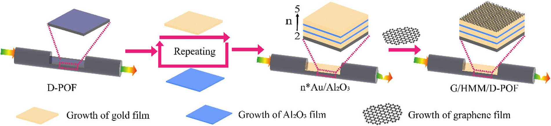
B. Preparation of D-POF
A 1-mm-thick plastic optical fiber working in a wavelength range of 450–840 nm was adopted as the probe substrate of the SPR sensor. First, it was cut into 15-cm-long strips. Second, in the middle of the optical fiber section, the cladding and part of the fiber core were removed by the side polishing to obtain sensing areas with 1.5-cm lengths. Last, the D-POF was cleaned with de-ionized (DI) water to remove surface impurities.
In turn, a multilayer graphene structure was capped with the HMM/D-POF surface through the 1-h immersion in PAH solution (), and subsequent reactions with a graphene-oxide dispersion (; flake diameters of 50–150 nm) for 5 h and hydrazine (, 50%) for 1 h [45,46]. After that, the obtained G/HMM/D-POF structure was washed with DI water and dried with nitrogen.
D. Modification and DNA Detection
In order to modify the G/HMM/D-POF for DNA detection, the obtained sensor was immersed in a 1 μM (1 M = 1 mol/L) PBASE solution for 4 h [38,47]. The pyrene group of the PBASE could undergo stacking with graphene and succinimide conjugate with an group-modified aptamer probe. The unmodified PBASE was removed by triple washing with DMSO and de-ionized water. After that, on the G/HMM/D-POF surface, the probe aptamer with was fixed by inserting the sensor into the solution (1 μM) for 4 h. Subsequently, the probe was rinsed with a PBS solution and de-ionized water to remove the unreacted probe aptamer. Finally, the modified G/HMM/D-POF sensor could be employed to perform the detection of DNA hybridization. The performance of the sensor proposed in this study was assessed using t_DNA and mis_DNA solutions of various concentrations. Before each detection, the sensor was cleaned with a PBS solution. For the DNA dissociation dynamics, the relevant solution was replaced with a pure PBS buffer to remove target DNAs. The dissociation was ended via a 60-s immersion of 10-mM aqueous sodium hydroxide solution and rinsing with PBS solution. Furthermore, the entire reaction was monitored in the real-time mode through the transmission spectrum.
E. Experimental Setup
Figure 2.Schematic of an experimental setup based on the G/HMM/D-POF sensor.
Figure 3.Simulation results. (a) Real and imaginary parts of the in-plane and out-of-plane permittivity components for an structure with . (b) Critical wavelength for structures as a function of value. (c) Simulation setup of the structure (). (d) Normalized transmission spectra of Au/D-POF and () for RI increasing from 1.340 to 1.352, respectively. (e) SPR peak wavelengths at the RI of 1.340 and their redshifts at RIs varying from 1.340 to 1.352 for Au/D-POF and (). (f) Electric field distributions within Au/D-POF and () structures at the resonance wavelengths for the RI of 1.340. (g) NormE values for Au/D-POF and ().
Furthermore, the finite difference time domain (FDTD) method was employed to demonstrate the different sensitivities to RI variations of HMMs with different values. For comparison, this study collected the performance of conventional SPR sensors exhibiting the structure of only one 50-nm gold film (Au/D-POF). The simulation setup is illustrated in Fig. 3(c). TM-polarized light with a 72° angle of incidence was adopted as the excitation source. Figure 3(d) displays the normalized transmission spectra of Au/D-POF and systems for the bulk RIs ranging from 1.340 to 1.352. As seen, compared with a transmission spectrum of Au/D-POF at the RI of 1.340, the resonant peak values associated with structures exhibit a pronounced redshift, and the morphology was broadening with increasing value. According to the quantitative assessments from Fig. 3(d), the redshift observed for with changing RI from 1.340 to 1.352 increases from 44 to 54 nm and the resonance peak wavelength at the RI of 1.340 is altered from 683 to 723 nm [Fig. 3(e)]. The full width at half-maximum for Au/D-POF and () at the RI of 1.340 in Fig. 3(d) is 57.52, 57.76, 58.22, 59.26, and 60.96 nm, respectively. The broadening of the resonance peak with increasing can be explained in terms of the imaginary part of the dielectric constant [Fig. 3(a)]. For pure gold, a monotonic increase in the imaginary part for the wavelengths above 470 nm caused a narrow SPR resonance at lower wavelengths.
For , a feature similar to a concave protuberance existed in the spectrum of the imaginary part in the dielectric constant, thereby allowing more wavelengths to approach the SPR resonance and causing the broadening and redshifting of the relevant resonance peaks in Fig. 3(d). Furthermore, a periodical stacking of sub-wavelength metal/dielectric layers makes the electromagnetic fields bounded at the individual plasmonic interfaces further contribute to a collective response, thus acting as the source of HMMs’ super sensitive property [48,51]. In this respect, the electric field distributions within Au/D-POF and structures at the resonance wavelengths for the RI of 1.340 are illustrated in Fig. 3(f). The emergence of periodic oscillations along with a substantial increase of the resulting electric field with increasing is likely due to the coupling between the electromagnetic waves bounded at the individual plasmonic interfaces. Subsequently, the alteration curves of the electric field norm with physical depth in each distribution were integrated [Fig. 3(g)].
Figure 4.(a) Optical graph of Au/D-POF and (). (b) Top SEM image of . (c) SEM cross-sectional image of a 50-nm-thick gold structure. (d) SEM cross-sectional image of . (e) EDS spectra of . (f)–(h) EDS element distribution maps from (f) Al, (g) O, (h) Au on .
Figure 5.(a)–(e) Normalized transmission spectra of Au/D-POF and () in the ethanol solution with RIs ranging from 1.340 to 1.352, respectively. (f) SPR peak wavelengths for Au/D-POF and () in the ethanol solution at the RI (1.340) and the redshifts caused by increasing RI (from 1.340 to 1.352).
Figure 6.(a) Front SEM image of G/HMM/D-POF in the sensing area. (b) Raman spectra of graphene oxide and chemically produced graphene. The inset shows the peak position distribution within a scanned area of μμ. (c) Normalized transmission spectra of G/HMM/D-POF in the ethanol solutions with RIs from 1.340 to 1.352. (d) Resonance wavelength redshift of G/HMM/D-POF as a function of RI. (e) Normalized transmission spectra of G/HMM/D-POF at the RI of 1.352, recorded in a cycling mode (up to 10 cycles). (f) Typical response-recovery characteristic curve of G/HMM/D-POF in ethanol solution (RI of 1.352) at a resonance wavelength of 648 nm.
Figure 7.(a) Normalized transmission spectra of the G/HMM/D-POF sensor during DNA modification. (b) Normalized transmission spectra of G/HMM/D-POF in t_DNA solutions with concentrations varying from 10 pM to 100 nM after being sufficiently complementary. (c) Redshift and SPR peak wavelength versus t_DNA concentration.
Figure 8.(a) Normalized transmission spectra before and after adding mis_DNA. (b) Timing detection curve for t_DNA solutions with concentrations from 10 pM to 100 nM. (c) Normalized transmission spectra of G/HMM/D-POF in the 0.1 nM t_DNA solution during the binding process. (d) Normalized transmission spectra of G/HMM/D-POF in the 0.1 nM t_DNA solution during the dissociation. (e) Real-time redshift for the binding process and dissociation in the 0.1 nM t_DNA solution. (f) Real-time SPR wavelength shift for G/HMM/D-POF in mis_DNA and t_DNA solutions with concentrations from 10 pM to 100 nM.
To a more specific analysis, a 0.1-nM DNA solution was proposed as a particular example. In this respect, Figs. 8(c) and 8(d) display the normalized transmission spectra recorded during the binding and dissociation processes, respectively. In Fig. 8(c), the SPR peak exhibits a noticeable redshift. The binding process occurs nearly 14 min before the dynamic equilibrium is attained, with only a slight deviation. After sufficient complementarity, the dissociation began. The SPR peak blueshifted with the dissociation and gradually returned to a pre-hybridization position, tending again to the dynamic equilibrium [Fig. 8(d)]. The DNA dissociation was ended by the 60-s immersion of a sensor in a 10-mM aqueous sodium hydroxide solution and its rinsing with PBS solution. The recovery of a resonance peak position at nearly 736.15 nm indicated the complete regeneration for the next binding experiment. As found out afterward, the PBASE and probe aptamer modified on a sensor surface were not washed away. Figure 8(e) summarizes the redshift data for the hybridization and dissociation processes. Here, the resonance peak wavelength before hybridization is taken as the reference point and the entire variation in the SPR resonant wavelength appears as the time-dependent nonlinear curve. The process can be properly described with the Langmuir model [38]. The initial reaction speed of the binding process was the fastest. With the increasing density of the bonded molecules, the hybridization was gradually decelerated and eventually reached equilibrium. In turn, dissociation appears as a reverse reaction of binding. Furthermore, the resonance peak redshift curves associated with the DNA hybridization were compared at different solution concentrations (10 pM–100 nM) in the same time range. As shown in Fig. 8(f), the initial speed of the hybridization process increases with the concentration of the t_DNA solution. When the t_DNA and probe aptamer are sufficiently complementary, the density of the bonded molecules is associated with the t_DNA solution concentration. In other words, an increase in the t_DNA concentration increases the expected final density of binding.
Thus, the theoretical and experimental results obtained for a G/HMM/D-POF biosensor reveal its high-resolution capability, good linearity, fast response-recovery speed, and high-sensitivity DNA detection with a detection limit down to 10 pM for short DNA (15-mers).
4. CONCLUSION
An optical fiber SPR biosensor complying with a 3D HMM composite structure and a graphene film was proposed in this study. According to a comprehensive theoretical and experimental analysis, the SPR resonance peak position could be able to be tuned within a visible-to-near-infrared range by altering the number of Au layers in the HMM system. Besides this, the sensor exhibited high sensitivity (4461 nm/RIU) and excellent linearity, as well as good stability and repeatability for bulk refractive index detection. Furthermore, according to the time- and concentration-dependent DNA hybridization kinetics data, the proposed sensor exhibited prominent performance with a detection limit of as low as 10 pM for short DNA (15-mers). These results made this appliance promising for a broad range of potential applications such as genetic screening, transcriptional profiling, biomarkers, and discovery of single-nucleotide variants. Thus, the combination of HMMs and 2D materials was shown to have great potential for SPR fiber optic sensors, making them promising for a broad range of medical diagnostics and clinical applications.
Can Li, Jinjuan Gao, Muhammad Shafi, Runcheng Liu, Zhipeng Zha, Dejun Feng, Mei Liu, Xuejian Du, Weiwei Yue, Shouzhen Jiang, "Optical fiber SPR biosensor complying with a 3D composite hyperbolic metamaterial and a graphene film," Photonics Res. 9, 379 (2021)