• Acta Optica Sinica
  • Vol. 41, Issue 3, 0322001 (2021)
Longhui Li1, Chen Zhang2、3、*, Ge Jin1, Weimin Yuan2、3, Zhen Li1, Yan Gu1, Zhen Zhang1, Zhao Xu1, Bowen Jiang1, Yidai Liao1, Chao Wu1, Yufei Li1, Yan Guo1, Tian Qin1, Zhixing Ling2、3, and Donghua Zhao2、3
Author Affiliations
  • 1North Night Vision Technology Co., Ltd., Nanjing, Jiangsu 211106, China
  • 2National Astronomical Observatories, Chinese Academy of Sciences, Key Laboratory of Space Astronomy and Technology, Chinese Academy of Sciences, Beijing 100012, China
  • 3College of Astronomy and Space Science, University of Chinese Academy of Sciences, Beijing 100049, China
  • show less
    DOI: 10.3788/AOS202141.0322001 Cite this Article Set citation alerts
    Longhui Li, Chen Zhang, Ge Jin, Weimin Yuan, Zhen Li, Yan Gu, Zhen Zhang, Zhao Xu, Bowen Jiang, Yidai Liao, Chao Wu, Yufei Li, Yan Guo, Tian Qin, Zhixing Ling, Donghua Zhao. Test and Analysis of Micropore Statistical Characteristics of Angel Type Planar Lobster Eye Optical Device[J]. Acta Optica Sinica, 2021, 41(3): 0322001 Copy Citation Text show less
    Focused imaging diagram of planar lobster eye optical device. (a) Diagram of structure; (b) principle of focused imaging
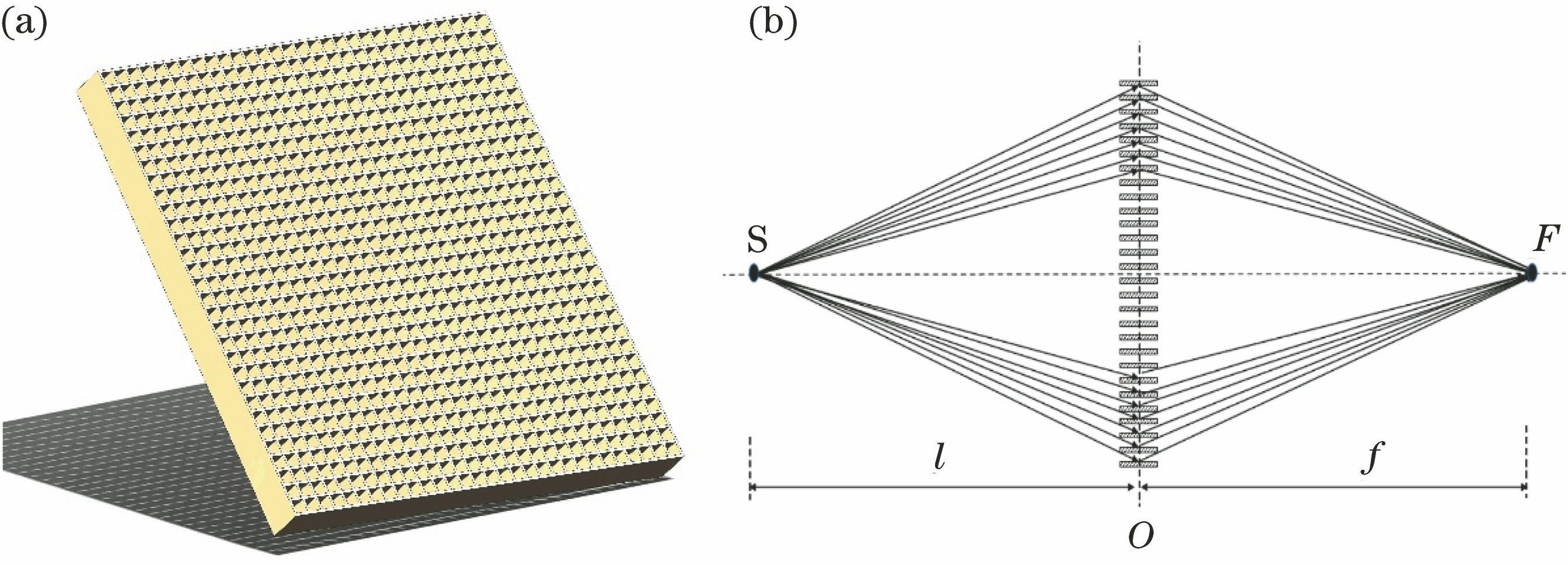
    Fig. 1. Focused imaging diagram of planar lobster eye optical device. (a) Diagram of structure; (b) principle of focused imaging
    Diagrams of planar lobster eye X-ray vacuum test equipment. (a) Diagram of equipment; (b) physical map of test chamber; (c) clamping fixture with low stress
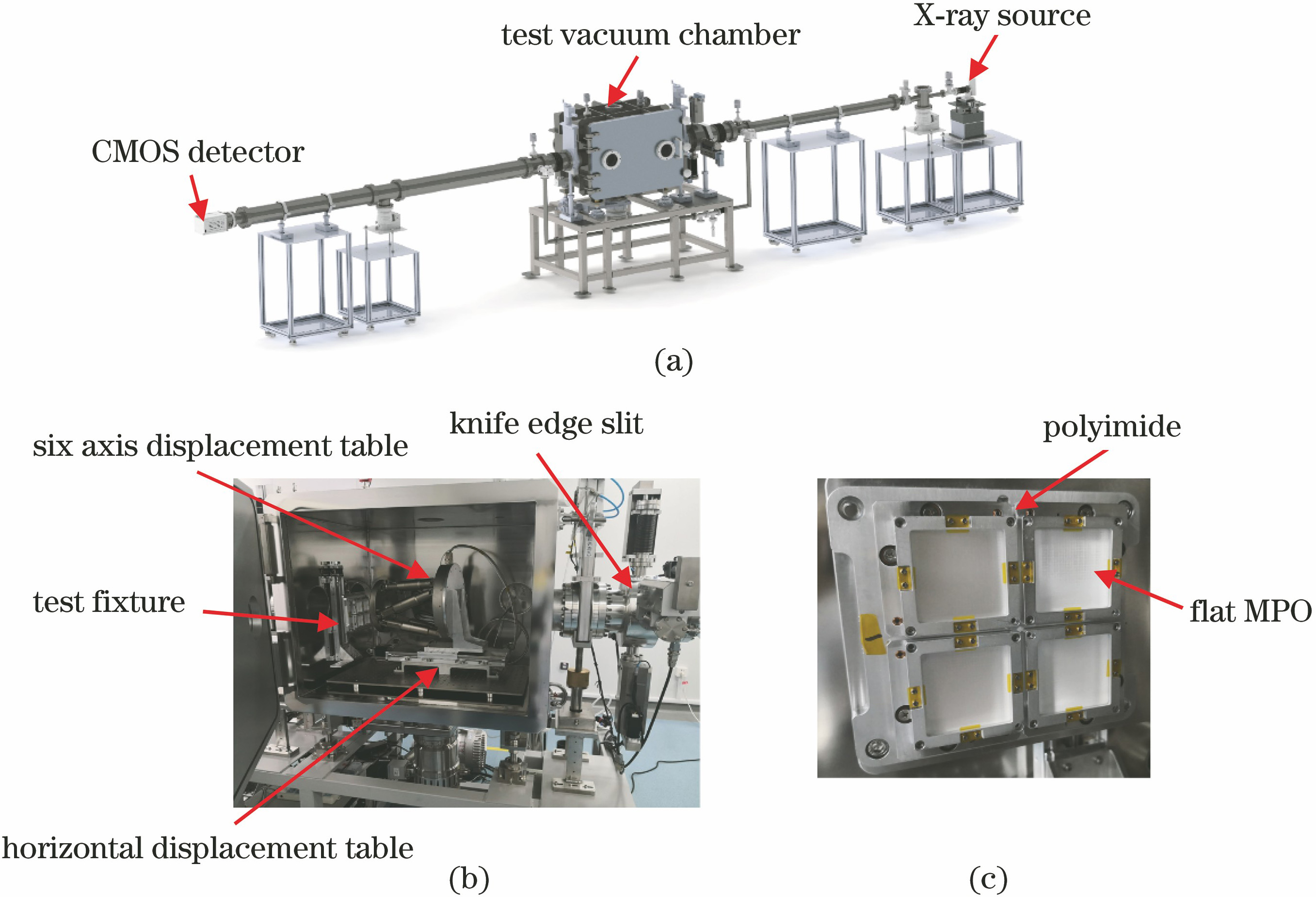
    Fig. 2. Diagrams of planar lobster eye X-ray vacuum test equipment. (a) Diagram of equipment; (b) physical map of test chamber; (c) clamping fixture with low stress
    Focused imaging results of flat MPO at different focusing positions. (a) 3600 mm; (b) 3620 mm; (c) 3650 mm; (d) 3700 mm
    Fig. 3. Focused imaging results of flat MPO at different focusing positions. (a) 3600 mm; (b) 3620 mm; (c) 3650 mm; (d) 3700 mm
    Contour distributions of focused imaging. (a) Partial enlargement map of secondary focal spot; (b) contour map corresponding to secondary focal spot
    Fig. 4. Contour distributions of focused imaging. (a) Partial enlargement map of secondary focal spot; (b) contour map corresponding to secondary focal spot
    Results of repeated test of angular resolution
    Fig. 5. Results of repeated test of angular resolution
    Focused images in central region of 13 mm×13 mm. (a) Focused imaging results; (b) contour map of secondary focal spot
    Fig. 6. Focused images in central region of 13 mm×13 mm. (a) Focused imaging results; (b) contour map of secondary focal spot
    Schematic diagrams of tilt type defect structure. (a) Ideal condition; (b) square channel deviating from ideal position with deviating angle of θ
    Fig. 7. Schematic diagrams of tilt type defect structure. (a) Ideal condition; (b) square channel deviating from ideal position with deviating angle of θ
    Imaging results of flat MPO in different areas. (a) p1; (b) p2; (c) p3; (d) p4; (e) p5; (f) p6; (g) p7; (h) p8; (i) p9
    Fig. 8. Imaging results of flat MPO in different areas. (a) p1; (b) p2; (c) p3; (d) p4; (e) p5; (f) p6; (g) p7; (h) p8; (i) p9
    Simulation results by Monte Carlo. (a) 1st group; (b) 2nd group
    Fig. 9. Simulation results by Monte Carlo. (a) 1st group; (b) 2nd group
    Longhui Li, Chen Zhang, Ge Jin, Weimin Yuan, Zhen Li, Yan Gu, Zhen Zhang, Zhao Xu, Bowen Jiang, Yidai Liao, Chao Wu, Yufei Li, Yan Guo, Tian Qin, Zhixing Ling, Donghua Zhao. Test and Analysis of Micropore Statistical Characteristics of Angel Type Planar Lobster Eye Optical Device[J]. Acta Optica Sinica, 2021, 41(3): 0322001
    Download Citation