• Chinese Optics Letters
  • Vol. 15, Issue 9, 090002 (2017)
O. Semyachkina-Glushkovskaya1, A. Abdurashitov1, A. Pavlov1、2、*, A. Shirokov3, N. Navolokin4, O. Pavlova1, A. Gekalyuk1, M. Ulanova1, N. Shushunova1, A. Bodrova1, E. Saranceva1, A. Khorovodov1, I. Agranovich1, V. Fedorova1, M. Sagatova1, A. E. Shareef1, C. Zhang5、6, D. Zhu5、6, and V. Tuchin1、7、8
Author Affiliations
  • 1Saratov National Research State University, Saratov 410012, Russia
  • 2Yuri Gagarin State Technical University of Saratov, Saratov 410054, Russia
  • 3Institute of Biochemistry and Physiology of Plants and Microorganisms, Russian Academy of Sciences, Saratov 410049, Russia
  • 4Saratov State Medical University, Saratov 410012, Russia
  • 5Britton Chance Center for Biomedical Photonics, Wuhan National Laboratory for Optoelectronics (WNLO), Huazhong University of Science and Technology (HUST), Wuhan 430074, China
  • 6Key Laboratory for Biomedical Photonics, HUST, Ministry of Education, Wuhan 430074, China
  • 7National Research Tomsk State University, Tomsk 634050, Russia
  • 8Institute of Precision Mechanics and Control, Russian Academy of Sciences, Saratov 410028, Russia
  • show less
    DOI: 10.3788/COL201715.090002 Cite this Article Set citation alerts
    O. Semyachkina-Glushkovskaya, A. Abdurashitov, A. Pavlov, A. Shirokov, N. Navolokin, O. Pavlova, A. Gekalyuk, M. Ulanova, N. Shushunova, A. Bodrova, E. Saranceva, A. Khorovodov, I. Agranovich, V. Fedorova, M. Sagatova, A. E. Shareef, C. Zhang, D. Zhu, V. Tuchin. Laser speckle imaging and wavelet analysis of cerebral blood flow associated with the opening of the blood–brain barrier by sound[J]. Chinese Optics Letters, 2017, 15(9): 090002 Copy Citation Text show less
    (Color online) (A) Two ROIs are overlaid onto LSCI image and (B) normalized histograms of these ROIs.
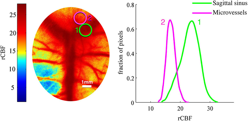
    Fig. 1. (Color online) (A) Two ROIs are overlaid onto LSCI image and (B) normalized histograms of these ROIs.
    Spectrofluorometric assay of EB extravasation from the blood into the brain parenchyma suggesting the increase in the BBB permeability to EB in 90 min after sound with afterward-rapid normalization of BBB leakage in 4 h. Asterisks indicate significant changes (p<0.05).
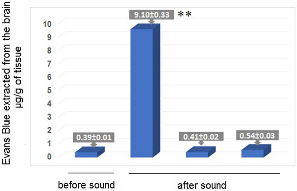
    Fig. 2. Spectrofluorometric assay of EB extravasation from the blood into the brain parenchyma suggesting the increase in the BBB permeability to EB in 90 min after sound with afterward-rapid normalization of BBB leakage in 4 h. Asterisks indicate significant changes (p<0.05).
    (Color online) Confocal imaging of BBB permeability to dextran 70 kDa in mice before and after sound: (A) no extravasation of dextran 70 kDa before sound; (B) and (C) extravasation of dextran 70 kDa in 90 min and 24 h after sound, respectively [defined as red clouds around the sagittal sinus, which is a main cerebral vein (B)], and a group of microvessels, including venules, draining the blood into the cerebral vein (C).
    Fig. 3. (Color online) Confocal imaging of BBB permeability to dextran 70 kDa in mice before and after sound: (A) no extravasation of dextran 70 kDa before sound; (B) and (C) extravasation of dextran 70 kDa in 90 min and 24 h after sound, respectively [defined as red clouds around the sagittal sinus, which is a main cerebral vein (B)], and a group of microvessels, including venules, draining the blood into the cerebral vein (C).
    Histological analysis of BBB permeability to solutes of small molecular weight: (A) before sound, no solute extravasation; (B) in 90 min after sound, the vasogenic edema observed suggests the high BBB leakage for the water and other solutes with their accumulation in space between cerebral microvessels and the brain parenchyma; (C) in 24 h after sound-stress, the vasogenic edema is still observed.
    Fig. 4. Histological analysis of BBB permeability to solutes of small molecular weight: (A) before sound, no solute extravasation; (B) in 90 min after sound, the vasogenic edema observed suggests the high BBB leakage for the water and other solutes with their accumulation in space between cerebral microvessels and the brain parenchyma; (C) in 24 h after sound-stress, the vasogenic edema is still observed.
    (Color online) LSCI for monitoring of the CBF of the cerebral veins and microvessels in four groups: control–(without sound); 90 min after sound—the reduction of venous CBF associated with the opening BBB; 4 h after sound—normalization of the venous CBF with recovery of the BBB; 24 h after sound—the repeated decrease in the venous CBF associated again with the opening BBB. No significant changes in the CBF on the level of microcirculation in different times after sound.
    Fig. 5. (Color online) LSCI for monitoring of the CBF of the cerebral veins and microvessels in four groups: control–(without sound); 90 min after sound—the reduction of venous CBF associated with the opening BBB; 4 h after sound—normalization of the venous CBF with recovery of the BBB; 24 h after sound—the repeated decrease in the venous CBF associated again with the opening BBB. No significant changes in the CBF on the level of microcirculation in different times after sound.
    (Color online) Wavelet-based analysis of the changes of the CBF in the cerebral veins (orange) and microvessels (violet) before and after sound: the control (without sound); in 90 min and 24 h elapsed after sound-stress, the disruption of BBB function is seen; in 4 h, the recovery of the BBB function is seen. Asterisk indicates significant changes (p<0.05).
    Fig. 6. (Color online) Wavelet-based analysis of the changes of the CBF in the cerebral veins (orange) and microvessels (violet) before and after sound: the control (without sound); in 90 min and 24 h elapsed after sound-stress, the disruption of BBB function is seen; in 4 h, the recovery of the BBB function is seen. Asterisk indicates significant changes (p<0.05).
    O. Semyachkina-Glushkovskaya, A. Abdurashitov, A. Pavlov, A. Shirokov, N. Navolokin, O. Pavlova, A. Gekalyuk, M. Ulanova, N. Shushunova, A. Bodrova, E. Saranceva, A. Khorovodov, I. Agranovich, V. Fedorova, M. Sagatova, A. E. Shareef, C. Zhang, D. Zhu, V. Tuchin. Laser speckle imaging and wavelet analysis of cerebral blood flow associated with the opening of the blood–brain barrier by sound[J]. Chinese Optics Letters, 2017, 15(9): 090002
    Download Citation