
Jia-nan LÜ, Jun-sheng LI, Guo-xia HUANG, Liu-juan YAN, Ji MA. Spectroscopic Analysis on the Interaction of Chrysene With Herring Sperm DNA and Its Influence Factors[J]. Spectroscopy and Spectral Analysis, 2022, 42(1): 210
- Spectroscopy and Spectral Analysis
- Vol. 42, Issue 1, 210 (2022)
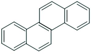
Fig. 1. Chemical structure of CHR
![UV-Visible spectra of CHR in the absence and presence of DNA at 25 ℃ at pH 7.40[CHR]=9.96×10⁻⁶ mol·L-1, a—i: [DNA]=(0, 0.14, 0.27, 0.41, 0.54, 0.68, 0.81, 0.94, 1.07)×10-6 mol·L-1](/richHtml/gpxygpfx/2022/42/1/210/img_2.png)
Fig. 2. UV-Visible spectra of CHR in the absence and presence of DNA at 25 ℃ at pH 7.40
[CHR]=9.96×10⁻⁶ mol·L-1,a —i : [DNA]=(0, 0.14, 0.27, 0.41, 0.54, 0.68, 0.81, 0.94, 1.07)×10-6 mol·L-1
[CHR]=9.96×10⁻⁶ mol·L-1,
Fig. 3. RLS spectra of CHR with and without DNA at 25 ℃ at pH 7.40
[CHR]=2.59×10⁻⁶ mol·L-1,a —k : [DNA]=(0, 0.35, 0.70, 1.05, 3.42, 1.39, 1.73, 2.07, 2.41, 2.75, 3.08)×10-6 mol·L-1
[CHR]=2.59×10⁻⁶ mol·L-1,
Fig. 4. (a) Changes of RLS intensity of CHR-DNA at different temperature; (b) Changes of RLS intensity of CHR-DNA at different pH at 30 ℃ (p <0.05)
[CHR]=2.59×10⁻⁶ mol·L-1, [DNA]=1.65×10⁻⁶ mol·L-1
[CHR]=2.59×10⁻⁶ mol·L-1, [DNA]=1.65×10⁻⁶ mol·L-1
Fig. 5. (a), (b), (c), (d) was respectively effect of different concentration of NaCl, CaCl2, Vitamin C, SLS on RLS intensity of CHR-DNA (p <0.05)
[CHR]=2.59×10⁻⁶ mol·L-1, [DNA]=1.65×10⁻⁶ mol·L-1
[CHR]=2.59×10⁻⁶ mol·L-1, [DNA]=1.65×10⁻⁶ mol·L-1
|
Table 1. Saturation value binding with DNA of CHR

Set citation alerts for the article
Please enter your email address
© Copyright 2018-2021 | Chinese Laser Press. All Rights Reserved 沪ICP备15018463号-20