
Tianyu Zhao, Zhaojun Wang, Kun Feng, Yansheng Liang, Minru He, Xue Yun, Ming Lei. High-Speed Structured Illumination Microscopy and Its Applications[J]. Laser & Optoelectronics Progress, 2020, 57(24): 240001
- Laser & Optoelectronics Progress
- Vol. 57, Issue 24, 240001 (2020)
![Roadmap of significant theoretical and technical breakthroughs of SIM[29-43]](/richHtml/lop/2020/57/24/240001/img_1.jpg)
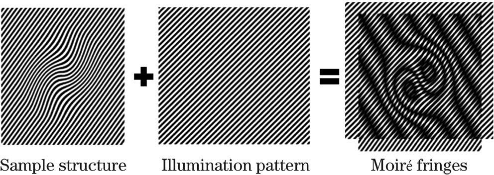
Fig. 2. Generation of Moiré fringes. If sample structure is multiplied by structural light, beat pattern (Moiré fringes) will appear
Fig. 3. Optical path of laser interference SIM
Fig. 4. Schematic diagram of iterative method for searching grating patterns displayed on SLM[56]
Fig. 5. Grating searching algorithm[56]. (a) Pixelated grating. White and gray represent on state and off state, respectively, and lattice points are pixels marked in red; (b)(c) grating with two steps of 2π/3 phase shift in y direction. Lattice point as a reference is marked in violet
Fig. 6. Pizza polarizer and zero-order vortex half-wave retarder. (a) Pizza polarizer[38];(b) zero-order vortex half-wave retarder[61]
Fig. 7. Principle of polarization control by using FLC[54]
Fig. 8. Timing sequence of SIM
Fig. 9. Results of three initial phase estimation methods. (a) Deconvolution wide-field image; (b)--(d) super-resolution image reconstructed by three phase estimation algorithms; (e)--(h) magnified views. Scale bar: (a)--(d) 5μm; (e)--(h) 500nm
Fig. 10. Densely packed cellular actin skeleton structures obtained by Hessian reconstruction[42]. (a) Wide-field image, traditional Wiener filtering result, and Hessian deconvolution result. Scale bar: 2μm; (b) magnified images reconstructed by different deconvolution methods. Scale bar: 500nm
Fig. 11. Flow chart of data processing by real-time display SIM[74]
Fig. 12. Research on morphological function of mouse brain neurons[75]. (a) Deconvolved widefield and SIM images of cell membrane labeled as ChR2-GFP. Scale bar: 5μm; (b) OTFs of SIM and deconvolved widefield images in Fig. 12 (a); (c) deconvolved widefield and SIM images of labeled cytoplasm. Scale bar: 5μm; (d) OTFs of SIM and deconvolved widefield images in Fig. 12 (c); (e) time-
Fig. 13. Commercial SIM products[83-85]. (a) Elyra 7 from Zeiss, Germany; (b) N-SIM from Nikon, Japan; (c) GE DeltaVision OMX from General Electric, America
Fig. 14. SIM with aberration correction by deformable mirror[87]
Fig. 15. Orientation of protein molecule determined by polarized information and SIM[43]. (a) Wide-field and (b) SIM images of microtubulin of U2OS cell. Scale bar: 10μm;(c) time-lapse SIM images showing the dynamic process. Scale bar: 1μm;(d)--(g) orientations of α-tubulin determined by model based on polarized information
|
Table 1. Partial parameters of commercial SIM products

Set citation alerts for the article
Please enter your email address
© Copyright 2018-2021 | Chinese Laser Press. All Rights Reserved 沪ICP备15018463号-20