[1] Notea A. The Ge (Li) spectrometer as a point detector[J]. Nuclear Instruments and Methods, 91, 513-515(1971).
[2] Debertin K, Ren J P. Measurement of the activity of radioactive samples in Marinelli beakers[J]. Nuclear Instruments and Methods in Physics Research Section A: Accelerators, Spectrometers, Detectors and Associated Equipment, 278, 541-549(1989).
[3] Presler O, Peled O, German U et al. Off-center efficiency of HPGe detectors[J]. Nuclear Instruments and Methods in Physics Research Section A: Accelerators, Spectrometers, Detectors and Associated Equipment, 484, 444-450(2002).
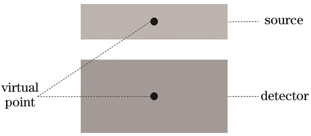
[4] Presler O, German U, Pelled O et al. The validity of the virtual point detector concept for absorbing media[J]. Applied Radiation and Isotopes, 60, 213-216(2004).
[5] Tian Z N, Liu W B, Chen W et al. Virtual point source efficiency calibration technology based on virtual point detector[J]. High Power Laser and Particle Beams, 30, 034001(2018).
[6] Xiong W B, Qiu C H, Duan T Y et al. Peak efficiency calibration of HPGe detectors for volume sources based on virtual point detector model[J]. Atomic Energy Science and Technology, 45, 999-1004(2011).
[7] Nowicki A, Gambin B. Ultrasonic synthetic apertures: review[J]. Archives of Acoustics, 39, 427-438(2015).
[8] Lü X G, Wang M Q, Li G Y. Application of synthetic aperture focusing technique in ultrasonic detection of concrete[J]. Journal of Graphics, 35, 946-949(2014).
[9] Li L, Wei W, Xia W. Coal-rock ultrasonic imaging and testing research based on synthetic aperture focusing technology[J]. Journal of China University of Mining & Technology, 47, 727-734(2018).
[10] Sutcliffe M, Charlton P, Weston M. Multiple virtual source aperture imaging for non-destructive testing[J]. Insight-Non-Destructive Testing and Condition Monitoring, 56, 75-81(2014).
[11] Chaix J F, Garnier V, Corneloup G. Concrete damage evolution analysis by backscattered ultrasonic waves[J]. NDT & E International, 36, 461-469(2003).
[12] Passmann C, Ermert H. A 100 MHz ultrasound imaging system for dermatologic and ophthalmologic diagnostics[J]. IEEE Transactions on Ultrasonics, Ferroelectrics and Frequency Control, 43, 545-552(1996).
[13] Nikolov S, Jensen J. Velocity estimation using synthetic aperture imaging[C](2001).
[14] Misaridis T, Jensen J A. Use of modulated excitation signals in medical ultrasound. Part III: high frame rate imaging[J]. IEEE Transactions on Ultrasonics, Ferroelectrics, and Frequency Control, 52, 208-219(2005).
[15] O’Donnell M, Thomas L J. Efficient synthetic aperture imaging from a circular aperture with possible application to catheter-based imaging[J]. IEEE Transactions on Ultrasonics, Ferroelectrics and Frequency Control, 39, 366-380(1992).
[16] Trahey G E, Nock L F. Synthetic receive aperture imaging with phase correction for motion and for tissue inhomogeneities. II. Effects of and correction for motion[J]. IEEE Transactions on Ultrasonics, Ferroelectrics and Frequency Control, 39, 496-501(1992).
[17] Bell A G. Upon the production and reproduction of sound by light[J]. Journal of the Society of Telegraph Engineers, 9, 404-426(1880).
[18] Gao R K, Xu Z Q, Song L et al. Breaking acoustic limit of optical focusing using photoacoustic-guided wavefront shaping[J]. Laser & Photonics Reviews, 15, 2000594(2021).
[19] Xu M H, Wang L V. Universal back-projection algorithm for photoacoustic computed tomography[J]. Proceedings of SPIE, 5697, 251-254(2005).
[20] Xu M H, Wang L H V. Photoacoustic imaging in biomedicine[J]. Review of Scientific Instruments, 77, 041101(2006).
[21] Zhou Y, Yao J J, Wang L V. Tutorial on photoacoustic tomography[J]. Journal of Biomedical Optics, 21, 061007(2016).
[22] Wang L V, Yao J. A practical guide to photoacoustic tomography in the life sciences[J]. Nature Methods, 13, 627-638(2016).
[23] Jeon S, Kim J, Lee D et al. Review on practical photoacoustic microscopy[J]. Photoacoustics, 15, 100141(2019).
[24] Attia A B E, Balasundaram G, Moothanchery M et al. A review of clinical photoacoustic imaging: current and future trends[J]. Photoacoustics, 16, 100144(2019).
[25] Hu S, Gonzales E, Soetikno B et al. Optical-resolution photoacoustic microscopy of ischemic stroke[J]. Proceedings of SPIE, 7899, 789906(2011).
[26] Kim C, Cho E C, Chen J Y et al. In vivo molecular photoacoustic tomography of melanomas targeted by bioconjugated gold nanocages[J]. ACS Nano, 4, 4559-4564(2010).
[27] Wang X D, Pang Y J, Ku G et al. Noninvasive laser-induced photoacoustic tomography for structural and functional in vivo imaging of the brain[J]. Nature Biotechnology, 21, 803-806(2003).
[28] Zhang H F, Maslov K, Wang L V. In vivo imaging of subcutaneous structures using functional photoacoustic microscopy[J]. Nature Protocols, 2, 797-804(2007).
[29] Qiu C, Bai Y Y, Yin T H et al. Targeted imaging of orthotopic prostate cancer by using clinical transformable photoacoustic molecular probe[J]. BMC Cancer, 20, 419(2020).
[30] Yao J J, Maslov K I, Zhang Y et al. Label-free oxygen-metabolic photoacoustic microscopy in vivo[J]. Journal of Biomedical Optics, 16, 076003(2011).
[31] Schwarz M, Buehler A, Aguirre J et al. Three-dimensional multispectral optoacoustic mesoscopy reveals melanin and blood oxygenation in human skin in vivo[J]. Journal of Biophotonics, 9, 55-60(2016).
[32] Zhang C B, Gao R K, Zhang L L et al. Design and synthesis of a ratiometric photoacoustic probe for in situ imaging of zinc ions in deep tissue in vivo[J]. Analytical Chemistry, 92, 6382-6390(2020).
[33] Ning B, Kennedy M J, Dixon A J et al. Simultaneous photoacoustic microscopy of microvascular anatomy, oxygen saturation, and blood flow[J]. Optics Letters, 40, 910-913(2015).
[34] Manohar S, Vaartjes S E, van Hespen J C G et al. Initial results of in vivo non-invasive cancer imaging in the human breast using near-infrared photoacoustics[J]. Optics Express, 15, 12277-12285(2007).
[35] Wang L V, Hu S. Photoacoustic tomography: in vivo imaging from organelles to organs[J]. Science, 335, 1458-1462(2012).
[36] Deán-Ben X L, Razansky D. Functional optoacoustic human angiography with handheld video rate three dimensional scanner[J]. Photoacoustics, 1, 68-73(2013).
[37] Zhou H C, Chen N B, Zhao H X et al. Optical-resolution photoacoustic microscopy for monitoring vascular normalization during anti-angiogenic therapy[J]. Photoacoustics, 15, 100143(2019).
[38] Jansen K, van der Steen A F W, van Beusekom H M M et al. Intravascular photoacoustic imaging of human coronary atherosclerosis[J]. Optics Letters, 36, 597-599(2011).
[39] Hu S, Wang L V. Neurovascular photoacoustic tomography[J]. Frontiers in Neuroenergetics, 2, 10(2010).
[40] Li M C, Tang Y Q, Yao J J. Photoacoustic tomography of blood oxygenation: a mini review[J]. Photoacoustics, 10, 65-73(2018).
[41] Mari J M, Xia W F, West S J et al. Interventional multispectral photoacoustic imaging with a clinical ultrasound probe for discriminating nerves and tendons: an ex vivo pilot study[J]. Journal of Biomedical Optics, 20, 110503(2015).
[42] Matthews T P, Zhang C, Yao D K et al. Label-free photoacoustic microscopy of peripheral nerves[J]. Journal of Biomedical Optics, 19, 016004(2014).
[43] Yao J J, Xia J, Wang L V. Multiscale functional and molecular photoacoustic tomography[J]. Ultrasonic Imaging, 38, 44-62(2016).
[44] Hu S, Yan P, Maslov K et al. Intravital imaging of amyloid plaques in a transgenic mouse model using optical-resolution photoacoustic microscopy[J]. Optics Letters, 34, 3899-3901(2009).
[45] Wang L V. Multiscale photoacoustic microscopy and computed tomography[J]. Nature Photonics, 3, 503-509(2009).
[46] Yao J J, Wang L V. Photoacoustic microscopy[J]. Laser & Photonics Reviews, 7, 758-778(2013).
[47] Ma R, Söntges S, Shoham S et al. Fast scanning coaxial optoacoustic microscopy[J]. Biomedical Optics Express, 3, 1724-1731(2012).
[48] Maslov K, Stoica G, Wang L H V. In vivo dark-field reflection-mode photoacoustic microscopy[J]. Optics Letters, 30, 625-627(2005).
[49] Song K H, Stein E W, Margenthaler J A et al. Noninvasive photoacoustic identification of sentinel lymph nodes containing methylene blue in vivo in a rat model[J]. Journal of Biomedical Optics, 13, 054033(2008).
[50] Wang L D, Maslov K I, Xing W X et al. Video-rate functional photoacoustic microscopy at depths[J]. Journal of Biomedical Optics, 17, 106007(2012).
[51] Harrison T, Ranasinghesagara J C, Lu H H et al. Combined photoacoustic and ultrasound biomicroscopy[J]. Optics Express, 17, 22041-22046(2009).
[52] Wang X D, Ku G, Wegiel M A et al. Noninvasive photoacoustic angiography of animal brains in vivo with near-infrared light and an optical contrast agent[J]. Optics Letters, 29, 730-732(2004).
[53] Song K H, Wang L V. Deep reflection-mode photoacoustic imaging of biological tissue[J]. Journal of Biomedical Optics, 12, 060503(2007).
[54] Song K H, Wang L V. Noninvasive photoacoustic imaging of the thoracic cavity and the kidney in small and large animals[J]. Medical Physics, 35, 4524-4529(2008).
[55] Zhang H F, Maslov K, Li M L et al. In vivo volumetric imaging of subcutaneous microvasculature by photoacoustic microscopy[J]. Optics Express, 14, 9317-9323(2006).
[56] Stein E W, Maslov K I, Wang L V. Noninvasive, in vivo imaging of blood-oxygenation dynamics within the mouse brain using photoacoustic microscopy[J]. Journal of Biomedical Optics, 14, 020502(2009).
[57] Park S, Lee C, Kim J et al. Acoustic resolution photoacoustic microscopy[J]. Biomedical Engineering Letters, 4, 213-222(2014).
[58] Paltauf G, Viator J A, Prahl S A et al. Iterative reconstruction algorithm for optoacoustic imaging[J]. The Journal of the Acoustical Society of America, 112, 1536-1544(2002).
[59] Berer T, Hochreiner A, Roitner H et al. Reconstruction algorithms for remote photoacoustic imaging[C], 13673521(2012).
[60] Cox B T, Treeby B E. Artifact trapping during time reversal photoacoustic imaging for acoustically heterogeneous media[J]. IEEE Transactions on Medical Imaging, 29, 387-396(2010).
[61] Cox B T, Kara S, Arridge S R et al. K-space propagation models for acoustically heterogeneous media: application to biomedical photoacoustics[J]. The Journal of the Acoustical Society of America, 121, 3453-3464(2007).
[62] Farnia P, Mohammadi M, Najafzadeh E et al. High-quality photoacoustic image reconstruction based on deep convolutional neural network: towards intra-operative photoacoustic imaging[J]. Biomedical Physics & Engineering Express, 6, 045019(2020).
[63] Jin H R, Zheng Z S, Liu S L et al. Pre-migration: a general extension for photoacoustic imaging reconstruction[J]. IEEE Transactions on Computational Imaging, 6, 1097-1105(2020).
[64] Gröhl J, Schellenberg M, Dreher K et al. Deep learning for biomedical photoacoustic imaging: a review[J]. Photoacoustics, 22, 100241(2021).
[65] Tian L, Hunt B, Bell M A L et al. Deep learning in biomedical optics[J]. Lasers in Surgery and Medicine, 53, 748-775(2021).
[66] Sharma A, Pramanik M. Convolutional neural network for resolution enhancement and noise reduction in acoustic resolution photoacoustic microscopy[J]. Biomedical Optics Express, 11, 6826-6839(2020).
[67] Vu T, DiSpirito A III, Li D W et al. Deep image prior for undersampling high-speed photoacoustic microscopy[J]. Photoacoustics, 22, 100266(2021).
[68] Liao C K, Li M L, Li P C. Optoacoustic imaging with synthetic aperture focusing and coherence weighting[J]. Optics Letters, 29, 2506-2508(2004).
[69] Li M L, Zhang H E, Maslov K et al. Improved in vivo photoacoustic microscopy based on a virtual-detector concept[J]. Optics Letters, 31, 474-476(2006).
[70] Spadin F, Jaeger M, Nuster R et al. Quantitative comparison of frequency-domain and delay-and-sum optoacoustic image reconstruction including the effect of coherence factor weighting[J]. Photoacoustics, 17, 100149(2020).
[71] Turner J, Estrada H, Kneipp M et al. Universal weighted synthetic aperture focusing technique (W-SAFT) for scanning optoacoustic microscopy[J]. Optica, 4, 770-778(2017).
[72] Jin H R, Liu S L, Zhang R C et al. Frequency domain based virtual detector for heterogeneous media in photoacoustic imaging[J]. IEEE Transactions on Computational Imaging, 6, 569-578(2020).
[73] Amjadian M, Mostafavi S M, Chen J B et al. Super-resolution photoacoustic microscopy using structured-illumination[J]. IEEE Transactions on Medical Imaging, 40, 2197-2207(2021).
[74] Mozaffarzadeh M, Varnosfaderani M H H, Sharma A et al. Enhanced contrast acoustic-resolution photoacoustic microscopy using double-stage delay-multiply-and-sum beamformer for vasculature imaging[J]. Journal of Biophotonics, 12, e201900133(2019).
[75] Mozaffarzadeh M, Mahloojifar A, Orooji M et al. Linear-array photoacoustic imaging using minimum variance-based delay multiply and sum adaptive beamforming algorithm[J]. Journal of Biomedical Optics, 23, 026002(2018).
[76] Mozaffarzadeh M, Yan Y, Mehrmohammadi M et al. Enhanced linear-array photoacoustic beamforming using modified coherence factor[J]. Journal of Biomedical Optics, 23, 026005(2018).
[77] Mozaffarzadeh M, Mahloojifar A, Periyasamy V et al. Eigenspace-based minimum variance combined with delay multiply and sum beamformer: application to linear-array photoacoustic imaging[J]. IEEE Journal of Selected Topics in Quantum Electronics, 25, 18000454(2019).
[78] Paridar R, Mozaffarzadeh M, Mehrmohammadi M et al. Photoacoustic image formation based on sparse regularization of minimum variance beamformer[J]. Biomedical Optics Express, 9, 2544-2561(2018).
[79] Synnevag J F, Austeng A, Holm S. Benefits of minimum-variance beamforming in medical ultrasound imaging[J]. IEEE Transactions on Ultrasonics, Ferroelectrics, and Frequency Control, 56, 1868-1879(2009).
[80] Wang S L, Li P C. MVDR-based coherence weighting for high-frame-rate adaptive imaging[J]. IEEE Transactions on Ultrasonics, Ferroelectrics, and Frequency Control, 56, 2097-2110(2009).
[81] Synnevåg J F, Nilsen C I C, Holm S. P2B-13 speckle statistics in adaptive beamforming[C], 1545-1548(2007).
[82] Mehdizadeh S, Austeng A, Johansen T F et al. Eigenspace based minimum variance beamforming applied to ultrasound imaging of acoustically hard tissues[J]. IEEE Transactions on Medical Imaging, 31, 1912-1921(2012).
[83] Deng Z L, Yang X Q, Gong H et al. Two-dimensional synthetic-aperture focusing technique in photoacoustic microscopy[J]. Journal of Applied Physics, 109, 104701(2011).
[84] Hollman K W, Rigby K W, O'Donnell M. Coherence factor of speckle from a multi-row probe[C], 1257-1260(1999).
[85] Mozaffarzadeh M, Makkiabadi B, Basij M et al. Image improvement in linear-array photoacoustic imaging using high resolution coherence factor weighting technique[J]. BMC Biomedical Engineering, 1, 10(2019).
[86] Deng Z L, Yang X Q, Gong H et al. Adaptive synthetic-aperture focusing technique for microvasculature imaging using photoacoustic microscopy[J]. Optics Express, 20, 7555-7563(2012).
[87] Turner J, Estrada H, Kneipp M et al. Improved optoacoustic microscopy through three-dimensional spatial impulse response synthetic aperture focusing technique[J]. Optics Letters, 39, 3390-3393(2014).
[88] Thomenius K E. Evolution of ultrasound beamformers[C], 1615-1622(1996).
[89] Hoelen C G A, de Mul F F M. Image reconstruction for photoacoustic scanning of tissue structures[J]. Applied Optics, 39, 5872-5883(2000).
[90] Pramanik M. Improving tangential resolution with a modified delay-and-sum reconstruction algorithm in photoacoustic and thermoacoustic tomography[J]. Journal of the Optical Society of America. A, Optics, Image Science, and Vision, 31, 621-627(2014).
[91] Kim J, Park S, Jung Y et al. Programmable real-time clinical photoacoustic and ultrasound imaging system[J]. Scientific Reports, 6, 35137(2016).
[92] Matrone G, Savoia A S, Caliano G et al. Ultrasound plane-wave imaging with delay multiply and sum beamforming and coherent compounding[C], 3223-3226(2016).
[93] Park S, Karpiouk A B, Aglyamov S R et al. Adaptive beamforming for photoacoustic imaging[J]. Optics Letters, 33, 1291-1293(2008).
[94] Matrone G, Savoia A S, Caliano G et al. The delay multiply and sum beamforming algorithm in ultrasound B-mode medical imaging[J]. IEEE Transactions on Medical Imaging, 34, 940-949(2015).
[95] Park J, Jeon S, Meng J et al. Delay-multiply-and-sum-based synthetic aperture focusing in photoacoustic microscopy[J]. Journal of Biomedical Optics, 21, 036010(2016).
[96] Song K, Liu P, Liu D Q. Implementing delay multiply and sum beamformer on a hybrid CPU-GPU platform for medical ultrasound imaging using OpenMP and CUDA[J]. Computer Modeling in Engineering & Sciences, 128, 1133-1150(2021).
[97] Jeon S, Park E Y, Choi W et al. Real-time delay-multiply-and-sum beamforming with coherence factor for in vivo clinical photoacoustic imaging of humans[J]. Photoacoustics, 15, 100136(2019).
[98] Song K, Liu P, Liu D C. Lag-based filtered-delay multiply and sum beamformer combined with two phase-related factors for medical ultrasound imaging[J]. Computational and Mathematical Methods in Medicine, 2020, 1503791(2020).
[99] Köstli K P, Beard P C. Two-dimensional photoacoustic imaging by use of Fourier-transform image reconstruction and a detector with an anisotropic response[J]. Applied Optics, 42, 1899-1908(2003).
[100] Stepinski T. An implementation of synthetic aperture focusing technique in frequency domain[J]. IEEE Transactions on Ultrasonics, Ferroelectrics, and Frequency Control, 54, 1399-1408(2007).
[101] Jeon S, Park J, Managuli R et al. A novel 2-D synthetic aperture focusing technique for acoustic-resolution photoacoustic microscopy[J]. IEEE Transactions on Medical Imaging, 38, 250-260(2019).
[102] Blouin A, Levesque D, Neron C et al. Improved resolution and signal-to-noise ratio in laser-ultrasonics by SAFT processing[J]. Optics Express, 2, 531-539(1998).
[103] Schulze R J, Scherzer O, Zangerl G et al. On the use of frequency-domain reconstruction algorithms for photoacoustic imaging[J]. Journal of Biomedical Optics, 16, 086002(2011).
[104] Köstli K P, Frenz M, Bebie H et al. Temporal backward projection of optoacoustic pressure transients using Fourier transform methods[J]. Physics in Medicine and Biology, 46, 1863-1872(2001).
[105] Estrada H, Turner J, Kneipp M et al. Real-time optoacoustic brain microscopy with hybrid optical and acoustic resolution[J]. Laser Physics Letters, 11, 045601(2014).
[106] Cao R, Kilroy J P, Ning B et al. Multispectral photoacoustic microscopy based on an optical-acoustic objective[J]. Photoacoustics, 3, 55-59(2015).
[107] Zhang C, Maslov K I, Yao J J et al. In vivo photoacoustic microscopy with 7.6-µm axial resolution using a commercial 125-MHz ultrasonic transducer[J]. Journal of Biomedical Optics, 17, 116016(2012).
[108] Cai D, Li Z F, Chen S L. In vivo deconvolution acoustic-resolution photoacoustic microscopy in three dimensions[J]. Biomedical Optics Express, 7, 369-380(2016).
[109] Cai D, Li Z F, Li Y et al. Combined synthetic aperture focusing technique and three-dimensional deconvolution for resolution enhancement in photoacoustic microscopy[J]. Proceedings of SPIE, 10064, 1006427(2017).