• Laser & Optoelectronics Progress
  • Vol. 60, Issue 7, 0728004 (2023)
Qi Yang1, Binbin Luo1、*, Zhipeng Gu2, Shengxi Wu2, Shenghui Shi1, and Mingfu Zhao1
Author Affiliations
  • 1Chongqing Key Laboratory of Optical Fiber Sensor and Photoelectric Detection, Chongqing University of Technology, Chongqing 400054, China
  • 2Chongqing Key Laboratory of Medicinal Chemistry and Molecular Pharmacology, Chongqing University of Technology, Chongqing 400054, China
  • show less
    DOI: 10.3788/LOP220600 Cite this Article Set citation alerts
    Qi Yang, Binbin Luo, Zhipeng Gu, Shengxi Wu, Shenghui Shi, Mingfu Zhao. Graphene Oxide Microfiber-Based Immunosensor for Rabies Virus[J]. Laser & Optoelectronics Progress, 2023, 60(7): 0728004 Copy Citation Text show less
    Schematic diagram of experimental system
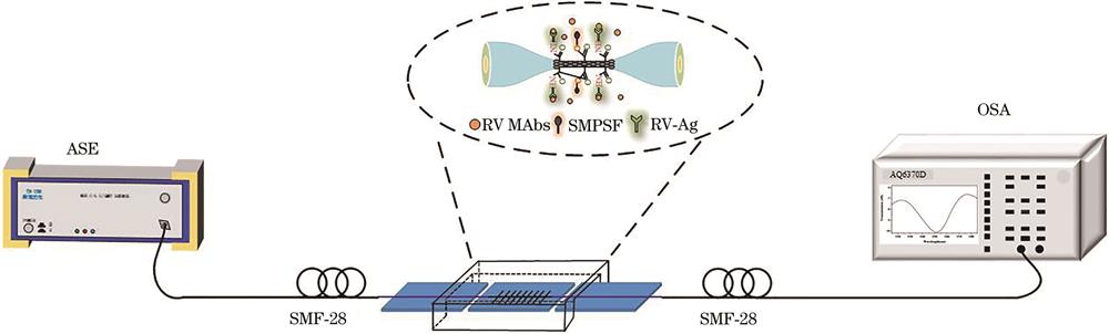
    Fig. 1. Schematic diagram of experimental system
    Microscopic image and spectrum of microfiber. (a) Diameter and length of microfiber;(b)interference spectrum of microfiber in water
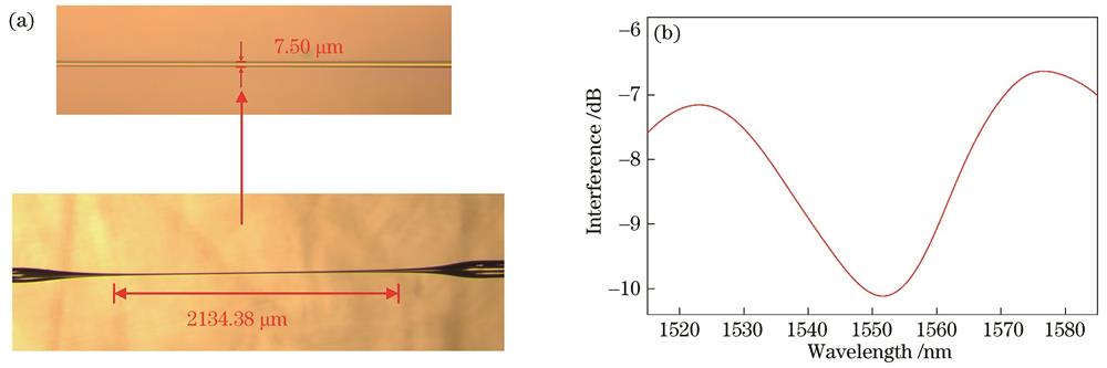
    Fig. 2. Microscopic image and spectrum of microfiber. (a) Diameter and length of microfiber;(b)interference spectrum of microfiber in water
    RI sensitivity calibration of microfiber. (a) Transmission spectra in different RI solutions;(b)RI sensitivity of microfiber
    Fig. 3. RI sensitivity calibration of microfiber. (a) Transmission spectra in different RI solutions;(b)RI sensitivity of microfiber
    Biofunctionalization process of microfiber
    Fig. 4. Biofunctionalization process of microfiber
    FESEM photos of GO-coated microfiber. (a) 3KX; (b) 30KX
    Fig. 5. FESEM photos of GO-coated microfiber. (a) 3KX; (b) 30KX
    Energy spectrum of microfiber surface modified with GO
    Fig. 6. Energy spectrum of microfiber surface modified with GO
    Spectra detection results in surface modification and functionalization of immunosensor. (a) Spectral evolution of microfiber; (b) corresponding wavelength red shift
    Fig. 7. Spectra detection results in surface modification and functionalization of immunosensor. (a) Spectral evolution of microfiber; (b) corresponding wavelength red shift
    RV antibody detection experiment of immunosensor. (a) Change curves of spectrum with RV antigen concentration; (b) variation curve of resonance wavelength shift with time
    Fig. 8. RV antibody detection experiment of immunosensor. (a) Change curves of spectrum with RV antigen concentration; (b) variation curve of resonance wavelength shift with time
    Relationship between wavelength red shift of microfiber immunosensor and concentration of RV antibody. (a) Langmuir curve fitting diagram; (b) detection sensitivity of microfiber immunosensor
    Fig. 9. Relationship between wavelength red shift of microfiber immunosensor and concentration of RV antibody. (a) Langmuir curve fitting diagram; (b) detection sensitivity of microfiber immunosensor
    Experimental results of clinical immunodetection of microfiber immunosensors. (a) Evolution curve of spectrum; (b) changes of resonance wavelength shift
    Fig. 10. Experimental results of clinical immunodetection of microfiber immunosensors. (a) Evolution curve of spectrum; (b) changes of resonance wavelength shift
    ChemicalMass fractionAtomic percentage
    Amount100100
    C51.4167.85
    O11.0810.98
    Si37.5121.17
    Table 1. Elemental mass fraction of microfiber surface modified with GO
    Qi Yang, Binbin Luo, Zhipeng Gu, Shengxi Wu, Shenghui Shi, Mingfu Zhao. Graphene Oxide Microfiber-Based Immunosensor for Rabies Virus[J]. Laser & Optoelectronics Progress, 2023, 60(7): 0728004
    Download Citation