• High Power Laser and Particle Beams
  • Vol. 32, Issue 11, 112001 (2020)
Jie Xu1、2, Baozhong Mu1、2、*, Liang Chen1、2, Wenjie Li1、2, Xinye Xu1、2, Xin Wang1、2, Zhanshan Wang1、2, Xing Zhang3, and Yongkun Ding4
Author Affiliations
  • 1Key Laboratory of Advanced Micro-structured Materials, Ministry of Education, Tongji University, Shanghai 200092, China
  • 2School of Physics Science and Engineering, Tongji University, Shanghai 200092, China
  • 3Laser Fusion Research Center, CAEP, Mianyang 621900, China
  • 4Institute of Applied Physics and Computational Mathematics, Beijing 100088, China
  • show less
    DOI: 10.11884/HPLPB202032.200133 Cite this Article
    Jie Xu, Baozhong Mu, Liang Chen, Wenjie Li, Xinye Xu, Xin Wang, Zhanshan Wang, Xing Zhang, Yongkun Ding. Progress of grazing incidence X-ray micro-imaging diagnosis technology[J]. High Power Laser and Particle Beams, 2020, 32(11): 112001 Copy Citation Text show less
    Ideal and actual implosion fuel compression
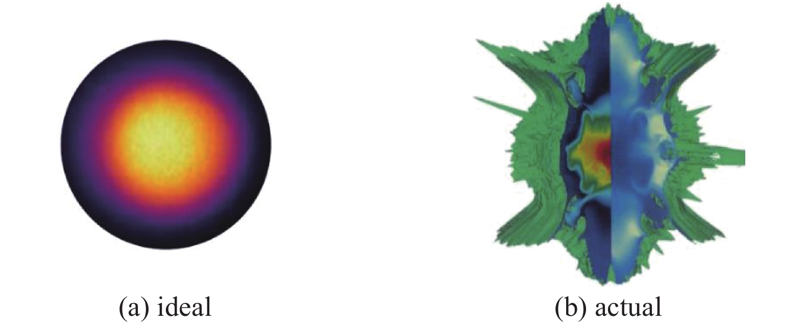
    Fig. 1. Ideal and actual implosion fuel compression
    Optical structure of KB microscope
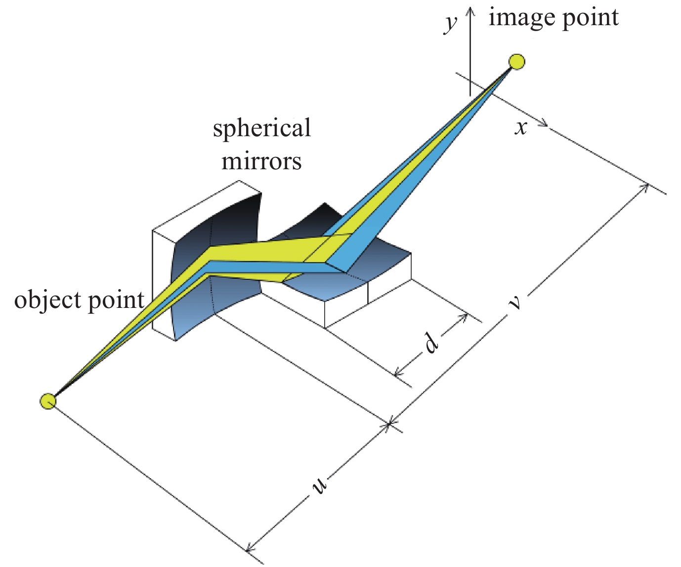
    Fig. 2. Optical structure of KB microscope
    Structural design drawing of four-channel KB microscope deployed in NIF
    Fig. 3. Structural design drawing of four-channel KB microscope deployed in NIF
    Experimental results of imaging calibration of Ni grid with four-channel KB microscope
    Fig. 4. Experimental results of imaging calibration of Ni grid with four-channel KB microscope
    (a)The special-shaped mirror used in the 16-channel KB microscope;(b)Example framed images obtained with KBFRAMED of a backlit Cu grid;(c)KBFRAMED images of hot-spot X-ray emission from a cryogenic target implosion.
    Fig. 5. (a)The special-shaped mirror used in the 16-channel KB microscope;(b)Example framed images obtained with KBFRAMED of a backlit Cu grid;(c)KBFRAMED images of hot-spot X-ray emission from a cryogenic target implosion.
    Picture of Wolter microscope objective applied to Z-pinch device
    Fig. 6. Picture of Wolter microscope objective applied to Z-pinch device
    Optical path diagram of Wolter micro-imaging system developed by NIF
    Fig. 7. Optical path diagram of Wolter micro-imaging system developed by NIF
    Multi-channel toroidal mirror X-ray microscope GXI-1 and grid backlight imaging results
    Fig. 8. Multi-channel toroidal mirror X-ray microscope GXI-1 and grid backlight imaging results
    Schematic diagram of three different types of films
    Fig. 9. Schematic diagram of three different types of films
    Reflectance curves of Ir single-layer film,W/B4C periodic multilayer film and non-periodic multilayer film
    Fig. 10. Reflectance curves of Ir single-layer film,W/B4C periodic multilayer film and non-periodic multilayer film
    Difference of X-ray imaging with single layer,period multilayer and non-period multilayer films
    Fig. 11. Difference of X-ray imaging with single layer,period multilayer and non-period multilayer films
    Schematic diagram of double-period multilayer film used for system assembly
    Fig. 12. Schematic diagram of double-period multilayer film used for system assembly
    (a)Schematic of the optical binocular system(OBS)and(b) its connection with the KB module
    Fig. 13. (a)Schematic of the optical binocular system(OBS)and(b) its connection with the KB module
    (a)SEM calibration results of four-quadrant grid;(b)Backlight imaging experiment results of four-quadrant grid;(c)Resolution calibration results
    Fig. 14. (a)SEM calibration results of four-quadrant grid;(b)Backlight imaging experiment results of four-quadrant grid;(c)Resolution calibration results
    Diagnostic experiments of X-ray KB microscope at Shenguang laser facility
    Fig. 15. Diagnostic experiments of X-ray KB microscope at Shenguang laser facility
    Optical structure for the time-gated four-channel KB microscope
    Fig. 16. Optical structure for the time-gated four-channel KB microscope
    X-ray microscope intensity calibration method based on “scanning pinhole+Si-PIN spectrum detector”
    Fig. 17. X-ray microscope intensity calibration method based on “scanning pinhole+Si-PIN spectrum detector”
    (a)Schematic of four-channel KB microscope;(b)4.75 keV four-channel KB imaging results of four-quadrant Cu grids at Shenguang II laser facility;(c)Diagnostic experiment of double turbulent amplitudes
    Fig. 18. (a)Schematic of four-channel KB microscope;(b)4.75 keV four-channel KB imaging results of four-quadrant Cu grids at Shenguang II laser facility;(c)Diagnostic experiment of double turbulent amplitudes
    Center field of view resolution of four-channel KB microscope
    Fig. 19. Center field of view resolution of four-channel KB microscope
    Results of hot-spot measurement with four-channel KB microscope
    Fig. 20. Results of hot-spot measurement with four-channel KB microscope
    Optical structure of eight-channel KB microscope and grid backlight imaging results
    Fig. 21. Optical structure of eight-channel KB microscope and grid backlight imaging results
    Experimental configuration for collaborative X-ray imaging diagnostics at Shenguang III laser facility
    Fig. 22. Experimental configuration for collaborative X-ray imaging diagnostics at Shenguang III laser facility
    Dual-channel microscope system sketch.
    Fig. 23. Dual-channel microscope system sketch.
    Static image of gold mesh target at 2.5 keV and 4.3 keV in implosion experiments
    Fig. 24. Static image of gold mesh target at 2.5 keV and 4.3 keV in implosion experiments
    Optical path diagram of STTS configuration aspherical KBA microscope
    Fig. 25. Optical path diagram of STTS configuration aspherical KBA microscope
    Simulation of optical performance of ultra-high resolution KB microscope
    Fig. 26. Simulation of optical performance of ultra-high resolution KB microscope
    grazing angle/(˚)field of view/µmresolution/µmreflectivity/%image field uniformityenergy resolution
    ir single layer0.425160660highnon
    W/B4C periodic multilayer 1.133200450low~30
    W/B4C non-periodic multilayer 1.133350510high<10
    Table 1.

    Comparison of KB microscope performance with different films

    不同膜系的KB显微镜性能比较

    Jie Xu, Baozhong Mu, Liang Chen, Wenjie Li, Xinye Xu, Xin Wang, Zhanshan Wang, Xing Zhang, Yongkun Ding. Progress of grazing incidence X-ray micro-imaging diagnosis technology[J]. High Power Laser and Particle Beams, 2020, 32(11): 112001
    Download Citation