
Juan He, Jie Cao, Wei Qian, Kun Huo, Chunqiao An, Fengze Dai. Processing Method of Graphite Surface Submicrostructure Based on Picosecond Laser[J]. Laser & Optoelectronics Progress, 2024, 61(5): 0514005
- Laser & Optoelectronics Progress
- Vol. 61, Issue 5, 0514005 (2024)
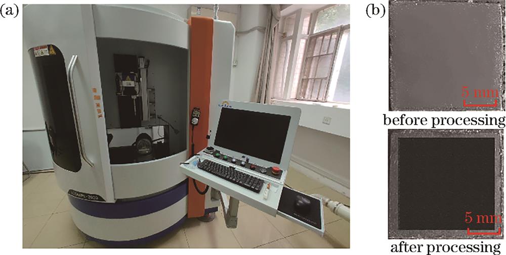
Fig. 1. Processing and characterization. (a) Image of picosecond laser processing system; (b) comparison of graphite before and after processing
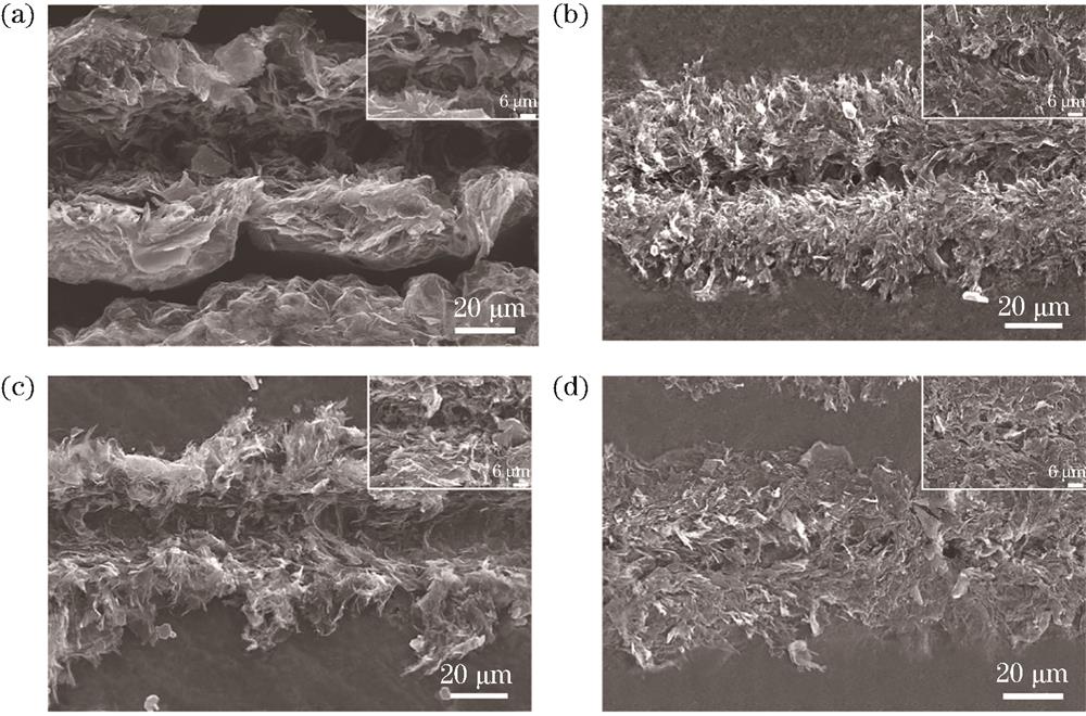
Fig. 2. Morphological characteristics of graphite surface under different scanning speeds. (a) 500 mm/s; (b) 1000 mm/s; (c) 1500 mm/s; (d) 2000 mm/s
Fig. 3. Morphological characteristics of graphite surface under different processing powers. (a) Change of depth and width of graphite surface texture under different powers; (b) surface groove topography of the sample surface when the processing power is 2.5 W; (c) corresponding partial enlarged view (2.5 W); (d) surface groove topography of the sample surface when the processing power is 3.5 W; (c) corresponding partial enlarged view (3.5 W)
Fig. 4. Morphological characteristics, width comparison and depth comparison of graphite surface under different number of scanning times. (a) Morphological of graphite surface under 2 scanning times; (b) morphological of graphite surface under 3 scanning times; (c) morphological of graphite surface under 8 scanning times; (d) morphological of graphite surface under 50 scanning times; (e) width comparison of graphite surface texture; (f) depth comparison of graphite surface texture
Fig. 5. Diffuse reflection properties of graphite surface under different laser processing parameters. (a) Different scanning speeds; (b) different processing powers; (c) different number of scanning times; (d) different scanning spacings
Fig. 6. Hydrophilic/hydrophobic characterization of graphite surface before and after processing. (a) Static contact angles at different scanning times; (b) Raman spectra of grooves on graphite surface under different scanning times; (c) ID/IG under different scanning times
Fig. 7. XPS spectrum of graphite C1s at different number of scanning times. (a) Original graphite surface; (b)‒(h) number of scanning time is 2, 3, 4, 5, 6, 8,10, respectively; (i) full XPS spectrum; (j) relationship between the content of three functional groups and the number of scanning times

Set citation alerts for the article
Please enter your email address
© Copyright 2018-2021 | Chinese Laser Press. All Rights Reserved 沪ICP备15018463号-20