• Advanced Photonics
  • Vol. 2, Issue 5, 055001 (2020)
Han Lin1、*, Scott Fraser1, Minghui Hong2, Manish Chhowalla3, Dan Li4、5, and Baohua Jia1、*
Author Affiliations
  • 1Swinburne University of Technology, Centre for Microphotonics, Faculty of Science, Engineering, and Technology, Hawthorn, Australia
  • 2National University of Singapore, Department of Electrical and Computer Engineering, Singapore
  • 3Rutgers University, Department of Materials Science and Engineering, Piscataway, New Jersey, United States
  • 4University of Melbourne, Department of Chemical Engineering, Melbourne, Australia
  • 5Monash University, Monash Centre for Atomically Thin Materials, Department of Materials Science and Engineering, Clayton, Australia
  • show less
    DOI: 10.1117/1.AP.2.5.055001 Cite this Article Set citation alerts
    Han Lin, Scott Fraser, Minghui Hong, Manish Chhowalla, Dan Li, Baohua Jia. Near-perfect microlenses based on graphene microbubbles[J]. Advanced Photonics, 2020, 2(5): 055001 Copy Citation Text show less
    On-demand generation and in situ characterization of the GO microbubbles. (a) Photoreduction setup with a femtosecond laser beam. The GO microbubbles are generated at the desired positions in an integrated platform. (b) Schematic of a GO microbubble. (c) The interference at the gas–bubble and gas–substrate interfaces. (d) Theoretical schematic of the Newton’s rings that can be observed using a CCD camera. (e) Experimental microscopic image of the observed Newton’s rings.
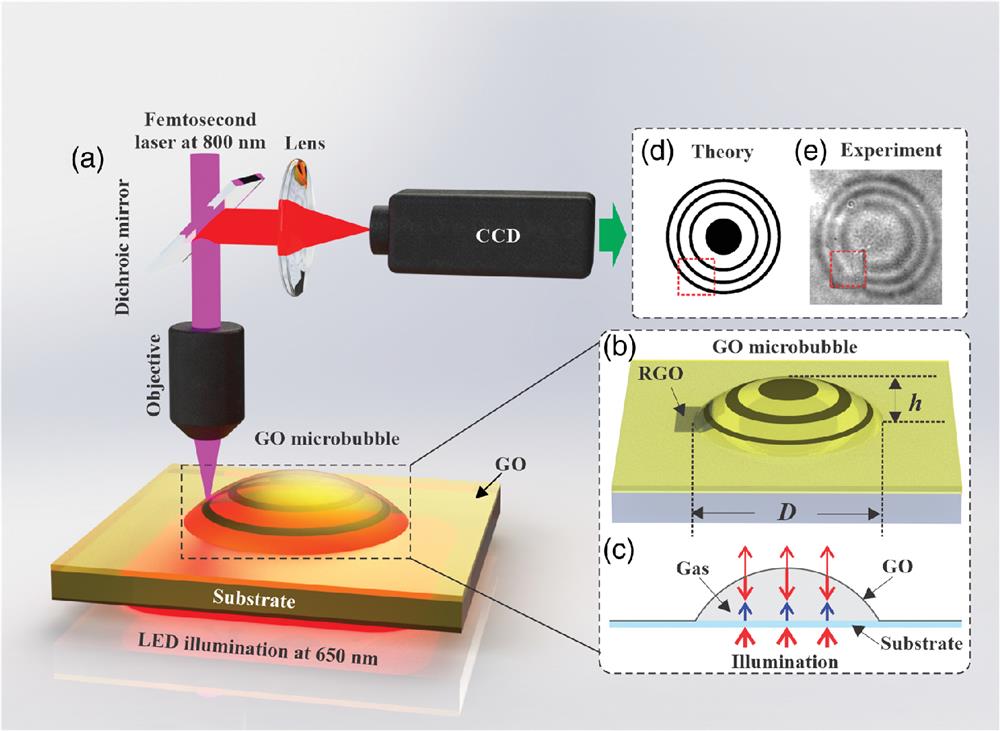
    Fig. 1. On-demand generation and in situ characterization of the GO microbubbles. (a) Photoreduction setup with a femtosecond laser beam. The GO microbubbles are generated at the desired positions in an integrated platform. (b) Schematic of a GO microbubble. (c) The interference at the gas–bubble and gas–substrate interfaces. (d) Theoretical schematic of the Newton’s rings that can be observed using a CCD camera. (e) Experimental microscopic image of the observed Newton’s rings.
    In situ optical microscopic images showing the process of the microbubble generation and elimination. (a) GO sample before photoreduction and (b)–(d) generated microbubbles corresponding to different reduced areas (Video S1). (e)–(h) Elimination of microbubbles by ablating the reduced area and measured at different times (Video S2). The reduced area is highlighted by the pink color (Video S1, MP4, 1.12 MB [URL: https://doi.org/10.1117/1.AP.2.5.055001.1]; Video S2, MP4, 1.00 MB [URL: https://doi.org/10.1117/1.AP.2.5.055001.2]).
    Fig. 2. In situ optical microscopic images showing the process of the microbubble generation and elimination. (a) GO sample before photoreduction and (b)–(d) generated microbubbles corresponding to different reduced areas (Video S1). (e)–(h) Elimination of microbubbles by ablating the reduced area and measured at different times (Video S2). The reduced area is highlighted by the pink color (Video S1, MP4, 1.12 MB [URL: https://doi.org/10.1117/1.AP.2.5.055001.1]; Video S2, MP4, 1.00 MB [URL: https://doi.org/10.1117/1.AP.2.5.055001.2]).
    Bubble characterization. (a) An in situ optical microscopic image of the Newton’s rings of the microbubble. The overall area of the microbubble is highlighted by the white circle and the x direction is marked by the yellow dash line. (b) Intensity profiles of the interference pattern of the microbubbles with different reduced areas. (c) Reconstructed surface profiles of the microbubbles with different reduced areas. A5 to A25 correspond to a photoreduced area of 5 to 25 μm2. (d) Reconstructed 3D profile of the A15 bubble [shown in (a)].
    Fig. 3. Bubble characterization. (a) An in situ optical microscopic image of the Newton’s rings of the microbubble. The overall area of the microbubble is highlighted by the white circle and the x direction is marked by the yellow dash line. (b) Intensity profiles of the interference pattern of the microbubbles with different reduced areas. (c) Reconstructed surface profiles of the microbubbles with different reduced areas. A5 to A25 correspond to a photoreduced area of 5 to 25  μm2. (d) Reconstructed 3D profile of the A15 bubble [shown in (a)].
    Focusing photonic jet with a GO microbubble. (a) Schematic of optical setup for characterizing the GO microbubbles. Left: Experimentally reconstructed 3D image of the characterized GO microbubble. (b) Schematic drawing of the shape of the microbubble as part of a sphere. h, height of the bubble; r, radius of the bubble; and R, radius of the entire sphere. Intensity distribution of the focal spot under white light illumination in the (c) x–y plane and (d) x–z plane (Video S3); the intensity plots along the (e) y direction and (f) z direction (Videos S3 and S4). (g) The surface plot of the experimentally measured 3D focal spot. The contours from inside correspond to the intensities of 0.8, 0.6, and 0.5 of the normalized peak intensity (Video S3, MP4 1.06 MB [URL: https://doi.org/10.1117/1.AP.2.5.055001.3]; Video S4, MP4, 606 KB [URL: https://doi.org/10.1117/1.AP.2.5.055001.4]).
    Fig. 4. Focusing photonic jet with a GO microbubble. (a) Schematic of optical setup for characterizing the GO microbubbles. Left: Experimentally reconstructed 3D image of the characterized GO microbubble. (b) Schematic drawing of the shape of the microbubble as part of a sphere. h, height of the bubble; r, radius of the bubble; and R, radius of the entire sphere. Intensity distribution of the focal spot under white light illumination in the (c) xy plane and (d) xz plane (Video S3); the intensity plots along the (e) y direction and (f) z direction (Videos S3 and S4). (g) The surface plot of the experimentally measured 3D focal spot. The contours from inside correspond to the intensities of 0.8, 0.6, and 0.5 of the normalized peak intensity (Video S3, MP4 1.06 MB [URL: https://doi.org/10.1117/1.AP.2.5.055001.3]; Video S4, MP4, 606 KB [URL: https://doi.org/10.1117/1.AP.2.5.055001.4]).
    Han Lin, Scott Fraser, Minghui Hong, Manish Chhowalla, Dan Li, Baohua Jia. Near-perfect microlenses based on graphene microbubbles[J]. Advanced Photonics, 2020, 2(5): 055001
    Download Citation