
Daquan Yang, Aiqiang Wang, Jin-Hui Chen, Xiao-Chong Yu, Chuwen Lan, Yuefeng Ji, Yun-Feng Xiao, "Real-time monitoring of hydrogel phase transition in an ultrahigh Q microbubble resonator," Photonics Res. 8, 497 (2020)
- Photonics Research
- Vol. 8, Issue 4, 497 (2020)
Abstract
1. INTRODUCTION
Monitoring and controlling the phase transition dynamics of materials is very important for both fundamental studies and practical applications [
Herein, real-time monitoring of the hydrogel phase transition (i.e., hydrophilic transition and hydrophobic transition) in WGM microbubble resonator (MBR)-based sensors is first demonstrated by continuously monitoring both wavelength shift and linewidth broadening simultaneously. Experimentally, the thermosensitive hydrogel phase transition is optically controlled by increasing/decreasing the irradiation light power (
2. MBR FABRICATION AND CHARACTERIZATION
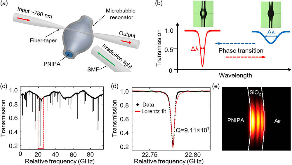
Figure 1.(a) Schematic of the MBR platform for real-time monitoring of the dynamic reactions of hydrogel phase transition. The thermosensitive phase transition of PNIPA is optically controlled by the irradiation light power (
As shown in Fig.
Sign up for Photonics Research TOC. Get the latest issue of Photonics Research delivered right to you!Sign up now
3. MBR FOR MONITORING HYDROGEL PHASE TRANSITION
![]()
Figure 2.Transmission evolution of the microbubble with the PNIPA hydrogel when the control power of the irradiation light first (a) increases from 0 to 3.00 mW, and then (b) decreases from 3.00 to 0 mW; (c) CCD images of a cycle of phase-transition process of the PNIPA hydrogel. The microbubble changes from transparent hydrophilic state to opaque hydrophobic state due to the increased scattering. Inset, the scale bar is 125 μm.
![]()
Figure 3.(a) WGM wavelength shifts and (b) linewidth broadenings as a function of control power of the irradiation light from 0 to 3.00 mW, when the MBRs are filled with air (blue line with triangular marker), DI water (black line with square marker), and PNIPA hydrogel (red line with circular marker). Compared with the result of microbubble cavities filled with air and DI water, note that a hydrophilic to hydrophobic transition process of PNIPA can be clarified as four stages: (i) pure hydrophilic state (0–1.44 mW); (ii) subtransition state (1.44–2.04 mW); (iii) transition state (2.04–2.52 mW); (iv) pure hydrophobic state (
![]()
Figure 4.(a) Real-time WGM resonance wavelength shift and (b) linewidth broadening during the PNIPA hydrogel phase transition (a hydrophilic to hydrophobic transition) monitored by an MBR. The control power of the irradiation light is switched on at
4. CONCLUSION
In summary, we experimentally characterize the thermosensitive PNIPA hydrogel phase transition via an ultrahigh Q MBR sensor. By controlling the output power of the irradiation light, the optical tuning of the PNIPA hydrogel phase transition has been successfully achieved. Furthermore, we reveal the refractive index and temperature changes during the different stages of the phase transition process by monitoring the wavelength shift and linewidth broadening in real time. Our work demonstrates that MBR-based biosensors are promising for further quantitatively investigating the energy change during a phase transition, thus providing insights into their dynamic reaction mechanisms.
Acknowledgment
Acknowledgment. The authors thank Qi-Tao Cao, Shui-Jing Tang, and Pei-Ji Zhang for helpful discussions.
References
[1] A. Onuki. Phase Transition Dynamics(2002).
[10] K. J. Vahala. Optical microcavities. Nature, 424, 839-846(2003).


Set citation alerts for the article
Please enter your email address

© Copyright 2018-2021 | Chinese Laser Press. All Rights Reserved 沪ICP备15018463号-20