• Laser & Optoelectronics Progress
  • Vol. 57, Issue 24, (2020)
Yuzhu Li1, Chuankang Li1, Xiang Hao1, Xu Liu1、3, and Cuifang Kuang1、2、3、*
Author Affiliations
  • 1State Key Laboratory of Modern Optical Instrumentation, College of Optical Science and Engineering, Zhejiang University, Hangzhou, Zhejiang 310027, China
  • 2Ningbo Institute, Zhejiang University, Ningbo, Zhejiang 315100, China
  • 3Collaborative Innovation Center of Extreme Optics, Shanxi University, Taiyuan, Shanxi 030006, China
  • show less
    DOI: 10.3788/LOP57.240002 Cite this Article Set citation alerts
    Yuzhu Li, Chuankang Li, Xiang Hao, Xu Liu, Cuifang Kuang. Review and Prospect for Single Molecule Localization Microscopy[J]. Laser & Optoelectronics Progress, 2020, 57(24): Copy Citation Text show less
    Research history of single molecule localization microscopy
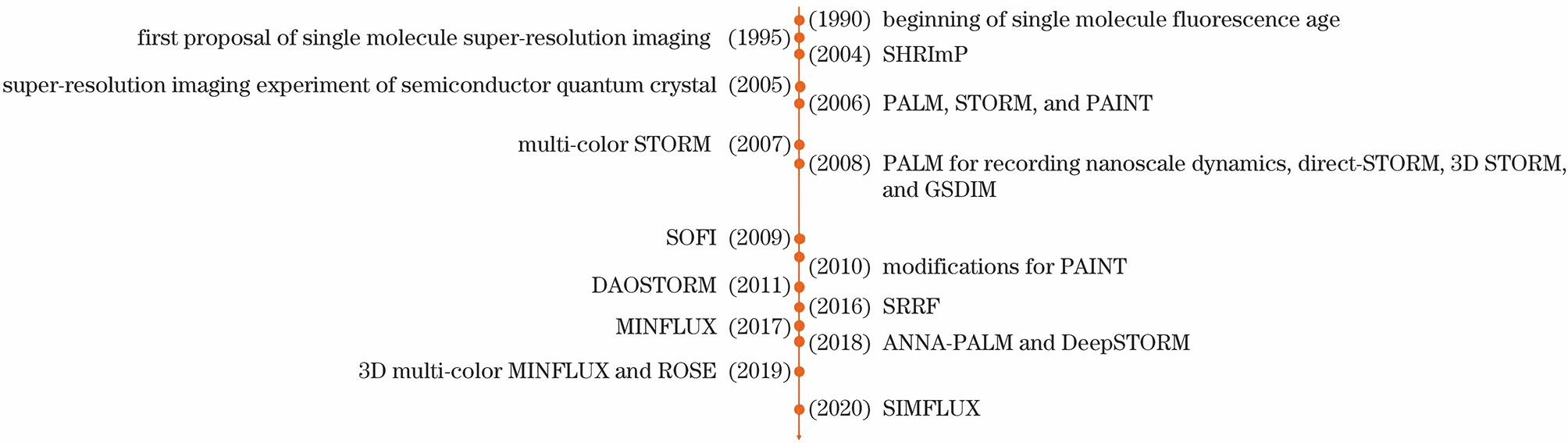
    Fig. 1. Research history of single molecule localization microscopy
    Principle of fluorescence emission
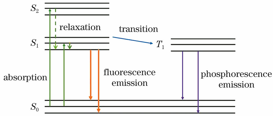
    Fig. 2. Principle of fluorescence emission
    Basic configuration of fluorescence microscopy
    Fig. 3. Basic configuration of fluorescence microscopy
    Schematic of three-dimensional STORM[20]. (a) Experimental setup; (b) relationship between axial lengths of elliptical PSF and z position of molecules
    Fig. 4. Schematic of three-dimensional STORM[20]. (a) Experimental setup; (b) relationship between axial lengths of elliptical PSF and z position of molecules
    Principle diagram of stochastic photobleaching super-resolution imaging [10]. (a) Attenuation of integrated intensity with time; (b) distinguishing two PSFs by photobleaching
    Fig. 5. Principle diagram of stochastic photobleaching super-resolution imaging [10]. (a) Attenuation of integrated intensity with time; (b) distinguishing two PSFs by photobleaching
    Setup and principle diagram of MINFLUX[29]. (a) Experimental setup of MINFLUX; (b) detection steps of MINFLUX
    Fig. 6. Setup and principle diagram of MINFLUX[29]. (a) Experimental setup of MINFLUX; (b) detection steps of MINFLUX
    Principle diagram of SIMFLUX[34]. (a) Experimental setup; (b) work flow chart
    Fig. 7. Principle diagram of SIMFLUX[34]. (a) Experimental setup; (b) work flow chart
    SMLMBest resolutionLocalizationprecisionAdvantageDisadvantage
    PALM [16]2-25nmσ/NSuper high precisionBeing time-consuming, large amount of calculation, being unsuitable for dynamic process imaging, severe photobleaching, and only exogenous proteins being observed
    STORM[17]About 20nmσ/NSuper high precisionBeing limited in in-vivo imaging, severe photobleaching, and being unable to quantify molecule numbers in a cell
    PAINT[18]About 25nmσ/NNo need to label sampleLarge amount of calculation, long post-processing time, and only obtaining surface information of sample, not internal information
    GSDIM[22]About 30nmσ/NNo need of special fluorophore, simple device, and shortening time of image acquisitionSevere photobleaching, complex processing, and being limited in real-time imaging of living cells
    SHRImP [10]About 10nmσ/NNo special fluorophore, and relatively simple processBeing unsuitable for cases where two or more neighboring molecules are bleached at the same time and being limited in 2D situation
    SRRF[28]50-106nmσ/NBeing able to be complemented in conventional microscopeBeing limited resolution
    SOFI[25]100nmσ/NNo phototoxicity and short processing timeBeing limited resolution
    MINFLUX[29]1-5nmAbout L/22NImproved photon utilization, improved localization precision, and being suitable for living cell imagingComplex processing and being limited in sparse solution and long acquisition time
    SIMFLUX[34]5-10nmσ/2.4NImproved localization precision and accuracyBeing limited in 2D situation
    ROSE[33]2-5nmAbout 50/NImproved localization precision and accuracyBeing time-consuming
    Table 1. Comparison among all SMLMs
    Yuzhu Li, Chuankang Li, Xiang Hao, Xu Liu, Cuifang Kuang. Review and Prospect for Single Molecule Localization Microscopy[J]. Laser & Optoelectronics Progress, 2020, 57(24):
    Download Citation