• Laser & Optoelectronics Progress
  • Vol. 58, Issue 24, 2400006 (2021)
Zhibing Xu, Wenxia Zhou, Dongdong Xu, Xiao Wang, Jianhua Yin, and Huijie Wang*
Author Affiliations
  • Department of Biomedical Engineering, College of Automation Engineering, Nanjing University of Aeronautics and Astronautics, Nanjing, Jiangsu 211106, China
  • show less
    DOI: 10.3788/LOP202158.2400006 Cite this Article Set citation alerts
    Zhibing Xu, Wenxia Zhou, Dongdong Xu, Xiao Wang, Jianhua Yin, Huijie Wang. Research Progress of Fluorescence Polarization Modulation Microscopy Imaging Technology[J]. Laser & Optoelectronics Progress, 2021, 58(24): 2400006 Copy Citation Text show less
    Absorption and radiative dipole moments of fluorescence dipole. (a) Dependence of fluorescence dipole absorption probability on angle between absorption dipole moment and electric field of exciting light; (b) emission intensity pattern of fluorescence dipole
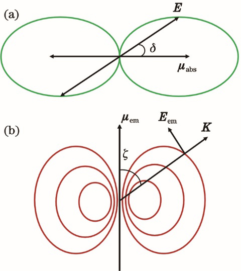
    Fig. 1. Absorption and radiative dipole moments of fluorescence dipole. (a) Dependence of fluorescence dipole absorption probability on angle between absorption dipole moment and electric field of exciting light; (b) emission intensity pattern of fluorescence dipole
    Schematic for measuring FA of molecules
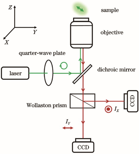
    Fig. 2. Schematic for measuring FA of molecules
    Schematic for measuring LD of molecules
    Fig. 3. Schematic for measuring LD of molecules
    Geometrical models with different dipole orientation distributions obtained by TP-FPM method [26]
    Fig. 4. Geometrical models with different dipole orientation distributions obtained by TP-FPM method [26]
    Schematic of experiment setup of SPoD
    Fig. 5. Schematic of experiment setup of SPoD
    Images of hippocampal neurons (scale bar: 1 μm) [33]. (a) WF images; (b) SPoD images
    Fig. 6. Images of hippocampal neurons (scale bar: 1 μm) [33]. (a) WF images; (b) SPoD images
    SDOM images of septin in live yeast cells (scale bar: 500 nm) [34]. (a) Orientation mapping images of septin with WF (upper-left) and SDOM (lower-right) under top view; (b) orientation mapping images under cross-sectional view
    Fig. 7. SDOM images of septin in live yeast cells (scale bar: 500 nm) [34]. (a) Orientation mapping images of septin with WF (upper-left) and SDOM (lower-right) under top view; (b) orientation mapping images under cross-sectional view
    Schematic of experiment setup of typical SIM
    Fig. 8. Schematic of experiment setup of typical SIM
    pSIM imaging results of actin in BAPE cells (scale bar: 5 μm) [35]. (a) Intensity images by WF and SIM; (b) pseudocolored images in dipole direction by PM and pSIM; (c) magnified view of boxed region in Fig.9(a)
    Fig. 9. pSIM imaging results of actin in BAPE cells (scale bar: 5 μm) [35]. (a) Intensity images by WF and SIM; (b) pseudocolored images in dipole direction by PM and pSIM; (c) magnified view of boxed region in Fig.9(a)
    Zhibing Xu, Wenxia Zhou, Dongdong Xu, Xiao Wang, Jianhua Yin, Huijie Wang. Research Progress of Fluorescence Polarization Modulation Microscopy Imaging Technology[J]. Laser & Optoelectronics Progress, 2021, 58(24): 2400006
    Download Citation