• Chinese Optics Letters
  • Vol. 16, Issue 7, 071701 (2018)
Myeongsu Seong1, Phuong Minh Mai2, Kijoon Lee3, and Jae Gwan Kim1、2、*
Author Affiliations
  • 1Department of Biomedical Science and Engineering, Institute of Integrated Technology, Gwangju Institute of Science and Technology, Gwangju 61005, Korea
  • 2School of Electrical Engineering and Computer Science, Gwangju Institute of Science and Technology, Gwangju 61005, Korea
  • 3School of Undergraduate Studies, Daegu Gyeongbuk Institute of Science and Technology, Daegu 42988, Korea
  • show less
    DOI: 10.3788/COL201816.071701 Cite this Article Set citation alerts
    Myeongsu Seong, Phuong Minh Mai, Kijoon Lee, Jae Gwan Kim. Simultaneous blood flow and oxygenation measurements using an off-the-shelf spectrometer[J]. Chinese Optics Letters, 2018, 16(7): 071701 Copy Citation Text show less
    Schematic of the proposed system. The system is configured with two lasers, a 1×2 fiber combiner/splitter, a fiber attenuator, and an off-the-shelf spectrometer coupled with detection fiber.
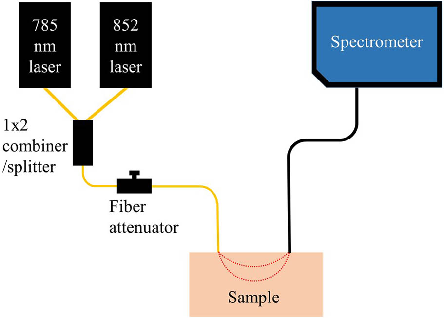
    Fig. 1. Schematic of the proposed system. The system is configured with two lasers, a 1×2fiber combiner/splitter, a fiber attenuator, and an off-the-shelf spectrometer coupled with detection fiber.
    Graphical explanation of the procedure to get the blood flow index (BFI) for each wavelength. One square means one pixel. Time varying intensity values for pixels of 785 and 852 nm were used to get the BFI for each wavelength in unit time.
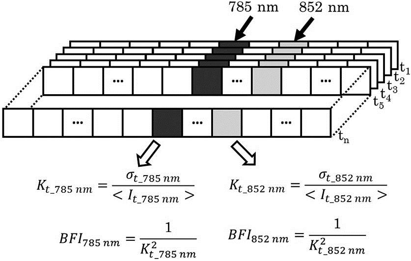
    Fig. 2. Graphical explanation of the procedure to get the blood flow index (BFI) for each wavelength. One square means one pixel. Time varying intensity values for pixels of 785 and 852 nm were used to get the BFI for each wavelength in unit time.
    Variation of normalized BFI of diffuse correlation spectroscopy, DCS (blue square), 785 nm signal (red dot), and 852 nm signal (green down triangle) of the spectrometer measured on the flow phantom while varying flow rate from 0 to 0.02 mL/s (with 0.01 mL/s of increment) and from 0.02 to 0.06 mL/s (with 0.02 mL/s of increment).
    Fig. 3. Variation of normalized BFI of diffuse correlation spectroscopy, DCS (blue square), 785 nm signal (red dot), and 852 nm signal (green down triangle) of the spectrometer measured on the flow phantom while varying flow rate from 0 to 0.02 mL/s (with 0.01 mL/s of increment) and from 0.02 to 0.06 mL/s (with 0.02 mL/s of increment).
    Variation of oxygenated (red square), deoxygenated (blue circle), and total hemoglobin (green up triangle) concentration to test the feasibility of oxygenation measurement of the system by the blood phantom test. 21% O2 and 79% N2 gases mimicking air (air), 100% N2 gas (N2), and 100% O2 (O2) were sequentially supplied for 10, 45, and 25 min, respectively. When N2 gas was supplied, the phantom was deoxygenated, and it was re-oxygenated when O2 gas was supplied.
    Fig. 4. Variation of oxygenated (red square), deoxygenated (blue circle), and total hemoglobin (green up triangle) concentration to test the feasibility of oxygenation measurement of the system by the blood phantom test. 21% O2 and 79% N2 gases mimicking air (air), 100% N2 gas (N2), and 100% O2 (O2) were sequentially supplied for 10, 45, and 25 min, respectively. When N2 gas was supplied, the phantom was deoxygenated, and it was re-oxygenated when O2 gas was supplied.
    Changes of oxygenated (red square for the suggested system and magenta up triangle for broadband NIRS), deoxygenated hemoglobin (blue circle for the suggested system and dark olive diamond for broadband NIRS) concentration (top) and percent variation of blood flow (bottom) for 785 nm (black up triangle with dotted line), 852 nm (red circle with short dashed line), and laser Doppler (blue solid square with solid line) during an arterial arm cuff occlusion (1 min of baseline, 3 min of occlusion with 220 mmHg, and another 3 min after cuff release).
    Fig. 5. Changes of oxygenated (red square for the suggested system and magenta up triangle for broadband NIRS), deoxygenated hemoglobin (blue circle for the suggested system and dark olive diamond for broadband NIRS) concentration (top) and percent variation of blood flow (bottom) for 785 nm (black up triangle with dotted line), 852 nm (red circle with short dashed line), and laser Doppler (blue solid square with solid line) during an arterial arm cuff occlusion (1 min of baseline, 3 min of occlusion with 220 mmHg, and another 3 min after cuff release).
    Myeongsu Seong, Phuong Minh Mai, Kijoon Lee, Jae Gwan Kim. Simultaneous blood flow and oxygenation measurements using an off-the-shelf spectrometer[J]. Chinese Optics Letters, 2018, 16(7): 071701
    Download Citation