• Acta Optica Sinica
  • Vol. 41, Issue 7, 0711001 (2021)
Yuye Wang1、2, Bozhou Jiang1、2, Degang Xu1、2、*, Guoqiang Wang1、2, Yifan Wang1、2, and Jianquan Yao1、2
Author Affiliations
  • 1School of Precision Instruments and Optoelectronics Engineering, Tianjin University, Tianjin 300072, China
  • 2Key Laboratory of Optoelectronics Information Technology (Ministry of Education), Tianjin University, Tianjin 300072, China
  • show less
    DOI: 10.3788/AOS202141.0711001 Cite this Article Set citation alerts
    Yuye Wang, Bozhou Jiang, Degang Xu, Guoqiang Wang, Yifan Wang, Jianquan Yao. Continuous Terahertz Wave Biological Tissue Imaging Technology Based on Focal Plane Array[J]. Acta Optica Sinica, 2021, 41(7): 0711001 Copy Citation Text show less
    Experimental setup for reflective terahertz focal plane imaging
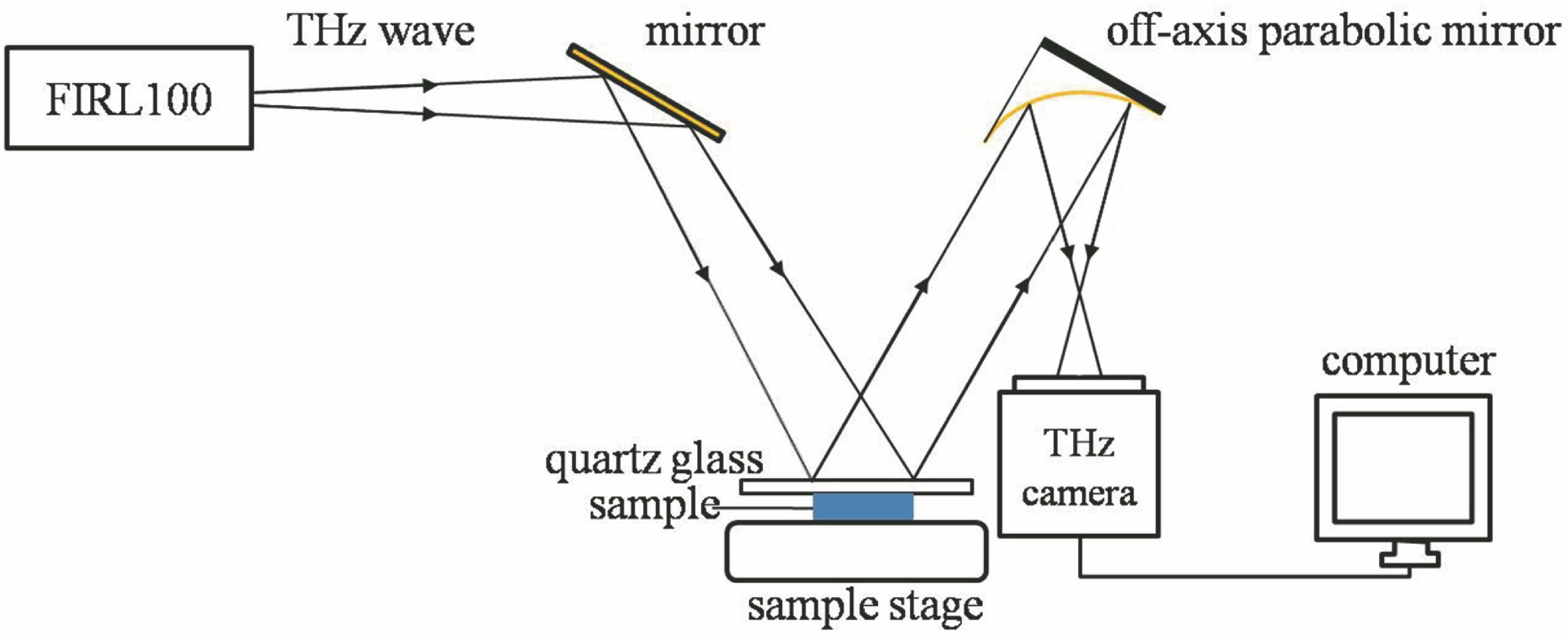
    Fig. 1. Experimental setup for reflective terahertz focal plane imaging
    Prediction of spatial resolution performance of terahertz focal plane imaging system. (a) Imaging spot; (b) metal sheet visible light image; (c) metal sheet terahertz image; (d) grayscale curve of vertical profiles, as indicated by the dotted line in Fig. (c)
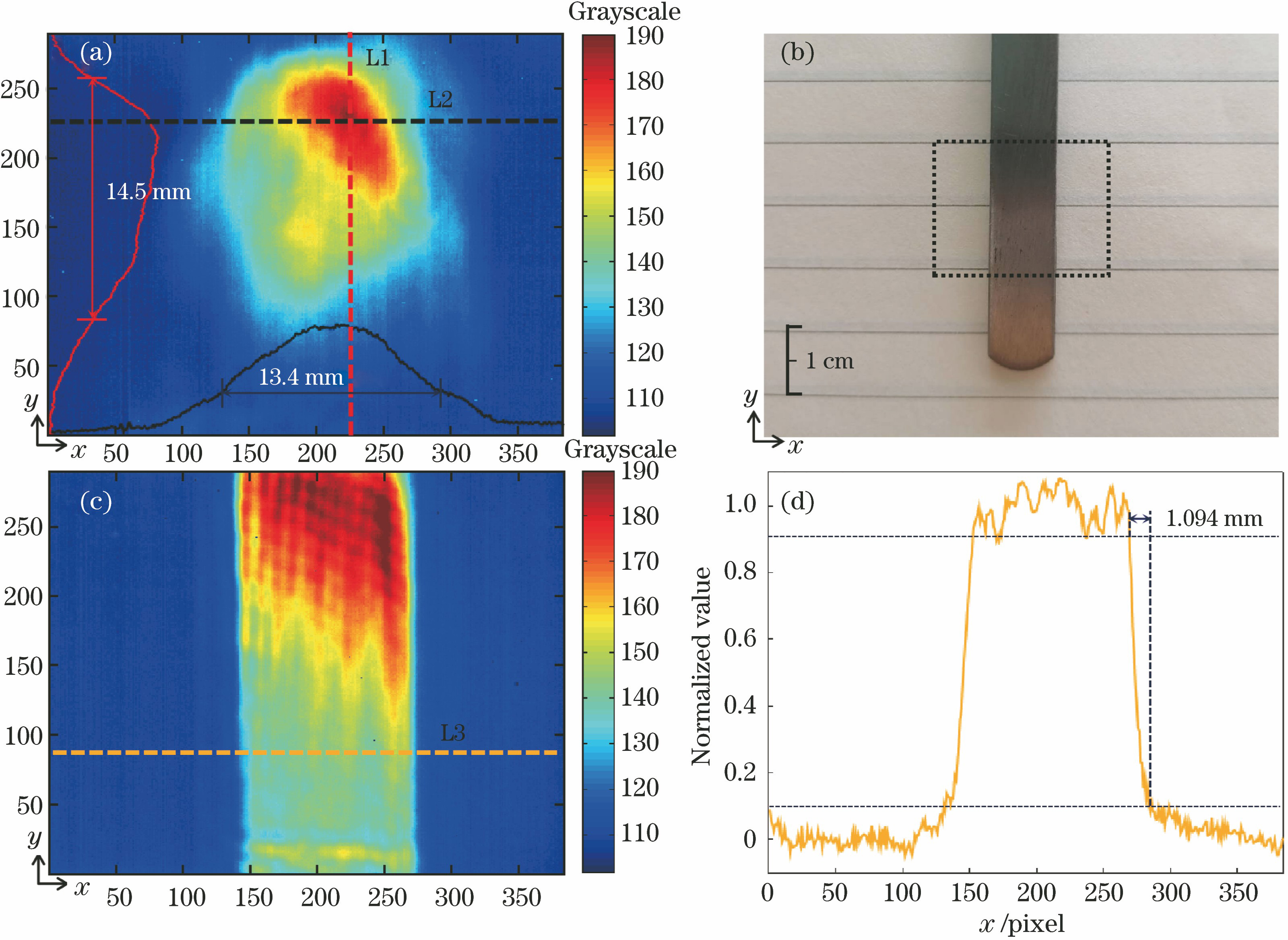
    Fig. 2. Prediction of spatial resolution performance of terahertz focal plane imaging system. (a) Imaging spot; (b) metal sheet visible light image; (c) metal sheet terahertz image; (d) grayscale curve of vertical profiles, as indicated by the dotted line in Fig. (c)
    Prediction of image contrast performance of terahertz focal plane imaging system. (a) Coin visible light image; (b) coin terahertz image before image processing; (c) terahertz image after image processing; (d) grayscale curves of vertical and horizontal profiles, as shown by solid and dotted lines in Fig. (b) and (c), respectively
    Fig. 3. Prediction of image contrast performance of terahertz focal plane imaging system. (a) Coin visible light image; (b) coin terahertz image before image processing; (c) terahertz image after image processing; (d) grayscale curves of vertical and horizontal profiles, as shown by solid and dotted lines in Fig. (b) and (c), respectively
    Terahertz imaging results of fresh pork tissue. (a)(c) Visible light images; (b)(d) terahertz images
    Fig. 4. Terahertz imaging results of fresh pork tissue. (a)(c) Visible light images; (b)(d) terahertz images
    Terahertz imaging results of fresh rat brain tissue. (a) Visible light image; (b) terahertz image
    Fig. 5. Terahertz imaging results of fresh rat brain tissue. (a) Visible light image; (b) terahertz image
    Terahertz imaging results of fresh human glioma tissue. (a) Visible light image; (b) H&E staining image; (c) terahertz image
    Fig. 6. Terahertz imaging results of fresh human glioma tissue. (a) Visible light image; (b) H&E staining image; (c) terahertz image
    Yuye Wang, Bozhou Jiang, Degang Xu, Guoqiang Wang, Yifan Wang, Jianquan Yao. Continuous Terahertz Wave Biological Tissue Imaging Technology Based on Focal Plane Array[J]. Acta Optica Sinica, 2021, 41(7): 0711001
    Download Citation