All-inorganic perovskite micro/nanolasers are emerging as a class of miniaturized coherent photonic sources for many potential applications, such as optical communication, computing, and imaging, owing to their ultracompact sizes, highly localized coherent output, and broadband wavelength tunability. However, to achieve single-mode laser emission in the microscale perovskite cavity is still challenging. Herein, we report unprecedented single-mode laser operations at room temperature in self-assembly microcavities over an ultrawide pumping wavelength range of 400–2300 nm, covering one- to five-photon absorption processes. The superior frequency down- and upconversion single-mode lasing manifests high multiphoton absorption efficiency and excellent optical gain from the electron–hole plasma state in the perovskite microcavities. Through direct compositional modulation, the wavelength of a single-mode microlaser can be continuously tuned from blue-violet to green (427–543 nm). The laser emission remains stable and robust after long-term high-intensity excitation for over 12 h (up to excitation cycles) in the ambient atmosphere. Moreover, the pump-wavelength dependence of the threshold, as well as the detailed lasing dynamics such as the gain-switching and electron–hole plasma mechanisms, are systematically investigated to shed insight into the more fundamental issues of the lasing processes in perovskite microcavities.
1. INTRODUCTION
Miniaturization and integration of photonic components hold great promise for advancing the field of optoelectronics [1–3]. There has been a long-standing and continuous pursuit of highly stable, wavelength-tunable semiconductor lasers on the micro/nanoscale due to the rapid progress of optical integrated systems. In particular, semiconductor lasers oscillating at a single frequency, such as single-mode lasers, have attracted great interest due to their potential for commercial applications in on-chip optical interconnects [4], quantum information processing [5,6], sensing [7,8], super-resolution imaging [9,10], and ultradense data storage [11]. Over the past decades, a wide range of micro/nanowire lasers has been reported, consisting of a multitude of compositions including various II-VI and III-V compound semiconductors [12]. Unfortunately, synthesis of these nanowires commonly requires expensive high-temperature or low-pressure conditions. Moreover, although most of these materials are stable in ambient atmosphere, only a few of them have demonstrated broad wavelength tunability, and the so-called “green gap” problem has not been solved in these material systems. Recently, studies on miniaturized lasers from perovskite semiconductors have experienced explosive growth and have led to great advances in lasing performance due to their high optical gain and tunable luminescence over the whole visible wavelength range [13–31]. From a historical perspective, Xing et al. reported low-threshold room-temperature (RT) amplified spontaneous emission in perovskite films in 2014 [32]. At almost the same time, lasing from perovskite-based vertical cavity surface emitting architecture was observed by Deschler et al. [16]. Shortly afterwards, Zhang et al. achieved near-infrared whispering-gallery-mode (WGM) nanolasers based on perovskite nanoplates [13], while Zhu et al. observed high-quality-factor Fabry–Perot (FP) lasing in perovskite nanowires with ultralow threshold of [14]. By conformally coating perovskite film on a silica microsphere, Sutherland and co-workers also demonstrated WGM lasers [15]. Besides, amplified spontaneous emission and lasing from colloidal nanocrystals of cesium lead halide perovskites were realized during nearly the same period by Yakunin et al. [33]. Since then, the lead halide perovskite micro/nanostructures served as gain materials with tunable lasing wavelengths that have been dramatically developed. Very recently, Qin et al. reported the first stable quasi-2D perovskite lasers under continuous-wave optical pumping in air at RT [34], which is expected to pave the way to the realization of future current-injection perovskite lasers. Huang et al. demonstrated the ultrafast control of perovskite-based vortex microlasers with ultralow energy consumption and simultaneously ultrahigh speed [35], providing a route to develop high-speed classical and quantum communication systems. Compared to organic and organic–inorganic perovskite components, all-inorganic cesium lead halide perovskites (, , Br, I, or their mixture) present better physical and chemical stability with respect to temperature, moisture, and light exposure, representing promising prospects of perovskites for the development of stable and high-performance devices [36,37]. Indeed, perovskite lasers have been achieved in many micro/nanostructures with different geometries, such as micro/nanowires [14,17–20,24,38,39], nanoplatelets [13,21,22,40], microspheres [15,23], microcubes [25,41,42], and hemispheres [27], for both single- and multiphoton excitations. However, most of these microcavity lasers are subject to random fluctuations and instabilities, showing typical multimode lasing. In principle, multiple modes in a resonant cavity are competitive with each other, and the one with the highest gain will dominate, but inhomogeneous gain saturation caused by spatial hole burning or crystal/cavity inhomogeneity can sustain multiple lasing modes [14,43]. Despite the extensive efforts that have been devoted to this enterprise, the realization of single-mode lasing in a microcavity with narrow spectral linewidth and broadband tunability remains a substantial challenge.
To date, the most developed preparation approaches for -based micro/nanostructures can be divided into two strategies: one is the liquid-phase method [14,18–20,38], and the other is the chemical vapor deposition (CVD) method [17,21–27,42]. Generally, the CVD route requires high temperature and an inert atmosphere, which need high energy consumption, a long preparation period, and effective waste management, and thus inevitably increase the cost and could limit the output in mass production. Recently, on the basis of the supersaturated recrystallization mechanism, Zheng et al. reported a low-temperature liquid-phase synthesis method to synthesize inner-defect-free inorganic perovskite micro/nanoflake single crystals [44]. The very thin supersaturated solution layer serves to promote heterogeneous nucleation of the material on the substrate due to the lower activation energy for nucleation induced by the production of ordered adlayers of anions on the surface [45]. Recrystallization takes place upon liquid transfer, and the nanocrystals grow rapidly due to the presence of the highly supersaturated state of the ions. Accordingly, the facile and high-compatibility liquid-phase self-assembly growth method based on the supersaturated recrystallization mechanism is very promising in achieving low-cost and environment-friendly micro/nanostructures [19,44].
In this work, we report unprecedented single-mode lasing with broad wavelength tunability in self-assembly microcavities, including microplates, microrods, and microcubes, over an ultrawide pumping wavelength range of 400–2300 nm at RT by using a modified low-temperature liquid-phase growth method. The as-grown microcrystals are single crystalline with well-formed facets and act as high-quality laser cavities. The superior frequency down- and upconversion single-mode lasing via one- to a giant five-photon absorption process demonstrates high multiphoton absorption (MPA) efficiency and excellent optical gain from the electron–hole plasma (EHP) state in the perovskite microcavities. By simple compositional modulation, high-quality single-mode lasing can be extended from blue-violet to green (427–543 nm) with low threshold and high stability. Additionally, the pump-wavelength-dependent lasing thresholds and the time-resolved kinetic emissions of the laser pulses are explored to reveal the physical mechanisms underlying the lasing behaviors.
Sign up for Photonics Research TOC. Get the latest issue of Photonics Research delivered right to you!Sign up now
2. EXPERIMENTAL SECTION
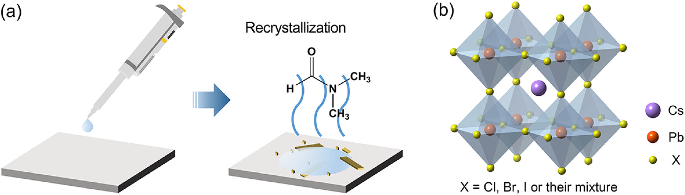
A. Synthesis Process of Single Microcrystals
The compounds single microcrystals were synthesized using a modified low-temperature liquid-phase growth method. Typically, the -dimethylformamide (DMF)– solution was prepared by mixing CsX ( or Br, 99.99%, Aladdin) and ( or Br, 99.99%, Aladdin) powders with a suitable stoichiometric ratio in DMF solvent, while the DMF– solution was obtained by mixing CsX ( or I, 99.99%, Aladdin), ( or I, 99.99%, Aladdin), and additive β-cyclodextrin (β-CD, 99%, TCI) powders in DMF, and then we kept stirring for 4 h at a temperature of 80°C. Upon cooling to RT, a drop of the DMF– solution (30 μL) was dispensed onto a Si, sapphire, or quartz substrate, which was preheated to a relatively low temperature (e.g., 40°C), and we kept the temperature unchanged to evaporate the solvent at a controllable rate. After the DMF solvent completely evaporated, high-quality single microcrystals with microplate, microrod, or microcube geometries were achieved on the substrate. In the contrary, microcrystals were synthesized by directly dropping the DMF– solution with β-CD (30 μL) onto the substrate, which was previously heated to a fairly high evaporation temperature of 330°C to ensure the perovskite crystals staying in the black cubic phase.
B. Characterization
Structural and morphological characterizations of the as-grown microcrystals were all conducted at RT (20°C). The crystal phase of the investigated samples was investigated using an X-ray diffraction (XRD) spectrometer, (Cu α, D8-Avance, BrukerAXS, Germany). The morphology micrographs and the elemental analysis of the microcrystals were carried out using a high-resolution scanning electron microscope (SEM) with an energy-dispersive X-ray spectroscopy (EDS) system (XL30, Philips, the Netherlands). The optical absorption spectroscopy was measured using a UV-Vis-NIR spectrophotometer (Cary 5000, Varian, America).
C. Optical Measurements
All the photoluminescence (PL), lasing, and time-resolved PL experiments were performed with the home-built confocal μ-PL system at RT by a Ti:sapphire femtosecond (fs) pulse laser operating at 800 nm (35 fs, 1 kHz, Verdi G8, Coherent, America). The impulsive 400 nm pulses were generated by frequency doubling the 800 nm fs pulses via a β-barium-borate (BBO) crystal, while the impulsive 1300, 1800, and 2300 nm pulses were generated from an optical parametric amplifier (OPA) system pumped by the 800 nm fs pulses. The pump laser beam was introduced into the confocal system and focused to a spot through a objective lens. The time-resolved kinetic investigation on pulse laser outputs was carried out by a streak-camera system (C10910, Hamamatsu, Japan) with a temporal resolution of about 10 ps. The luminescence signals were dispersed by a triple-grating spectrometer (Shamrock SR-303, Andor) and were recorded by a cooled charge-coupled device (CCD) camera.
3. RESULTS AND DISCUSSION
A. Direct Synthesis and Characterizations of Single Microcrystals
The schematic diagram of facile liquid-phase synthetic strategy for high-quality single microcrystals is presented in Fig. 1. Briefly, a drop of solution (μ) containing dissolved solvent (CsX and compounds with a target stoichiometric ratio) and DMF solvent is dispensed onto single-crystal Si, sapphire, amorphous quartz, or other substrates, which were preheated to a given temperature (e.g., 40°C) by a heater in ambient atmosphere. After the solution spreads out naturally, the DMF solvent begins to evaporate due to the higher substrate temperature. As the DMF continues to evaporate, the solution gradually experiences a highly supersaturated state. Once the supersaturation reaches the critical value for nucleation, the nuclei start to form on the substrate, and the concentration at the solid–liquid interface is reduced to the saturation concentration of the solution [45]. A concentration gradient is then established in the solution, which serves as the driving force for the ion or molecule diffusion that results in the continuous growth of the microcrystals until the DMF solvent completely evaporates. The crystal geometry and size, as well as chemical composition, can be controlled by adjusting the evaporation temperature, reactant concentration, substrate material, and stoichiometric ratio. The general chemical form of the perovskite structure is illustrated in Fig. 1(b).
Figure 1.Preparation of single microcrystals. (a) Illustration of the synthesis of microcrystals by the liquid-phase method. First, drop a certain amount of DMF– solution onto the substrate, which was preheated to a given temperature , and keep the temperature unchanged in ambient air. Then, evaporate the DMF solvent under the given temperature (). The solution gradually reaches supersaturation, and the nuclei start to form on the substrate. After the solvent is completely evaporated, the perovskite single microcrystals are obtained on the substrate surface. (b) Schematic chemical form of perovskite structure.
Figures 2(a)–2(c) show the top-view SEM images of the self-assembled single microcrystals based on the liquid-phase recrystallization on single-crystal Si, sapphire, and amorphous quartz substrates, respectively, with the reactant concentration of 40 mmol/L at a evaporation temperature of 40°C. It is seen that the end facets of the crystals are smooth, which is critical for efficient emission confinement by a naturally forming high-quality optical cavity. Besides, it is found that the perovskite microcrystals on Si substrate tend to form rectangular microplates that are pressed to the substrate surface. In contrast, the perovskites show microrod geometry on sapphire, while tilted microcubes appear on amorphous quartz under the same synthesis condition. By simply varying the halide composition, single microcrystals with the abovementioned three geometries can be easily obtained. The chemical composition and uniform spatial distribution of corresponding elements over the whole crystals, e.g., or , are confirmed by EDS mapping as shown in Figs. 2(e) and 2(f). The EDS analysis indicates that the atomic ratios of Cs/Pb/Cl and Cs/Pb/Br are respectively 1.06:1.02:2.92 and 0.96:0.96:3.08 [Fig. 2(d)], which are in good agreement with the ideal 1:1:3 stoichiometry of a crystal.
Figure 2.Geometry of the single microcrystals on different substrates. SEM micrograph of on (a) single-crystal Si, (b) sapphire, and (c) amorphous quartz, with the reactant concentration of 40 mmol/L at an evaporation temperature of 40°C. Scale bars: 50 μm. (d) EDS spectra of the and microplates. Inset: atomic ratios of Cs/Pb/Cl and Cs/Pb/Br (). (e), (f) EDS elemental mapping of the corresponding and microplates, respectively.
B. Broad Wavelength Tunable Emission from Microcrystals
Through direct compositional modulations, the band-gap energies and emission spectra of the microcrystals can be tuned over the entire visible spectral region from 420 nm (blue-violet) to 710 nm (red) [Fig. 3(a)]. The corresponding linear optical absorption spectra of pure , , and crystals are concomitantly presented. The PL experiments were conducted under low excitation intensity at RT by fs impulsive optical pumping at 400 nm, namely, a frequency downconversion (one-photon absorption, 1PA) PL process, as illustrated in Fig. 3(b). Notably, the crystal increase, and then the valence and conduction (δ) phase with a wide band gap () at RT, while a preferred black cubic (α) perovskite phase with a narrow band gap () requires high temperature atmospheres () [46,47]. Upon cooling, the cubic α- undergoes immediate transformation to the orthorhombic phase when exposed to ambient conditions [48]. Such intrinsic lack of stability is due to autodegradation and susceptibility to hydrolysis from atmospheric water and oxygen [49,50]. To achieve cubic α- () crystals with high phase stability, the supramolecular β- is introduced into the DMF– precursor solution prior to thermal evaporation at 330°C. After cooling to RT, the crystals with β- remain “frozen” in the black photoactive phase due to the drastically enhanced framework of the synthesized perovskites through the supramolecular interaction [51,52]. Such beneficial interaction tremendously improves the crystalline properties, humidity stability, and illumination stability of the perovskite materials. The molecular structure and approximate hydrophobic-cavity dimensions, as well as the schematic of the water-resistant property of the perovskite with β-, are respectively displayed in Figs. 3(c) and 3(d). Remarkably, such a new class of promoter for perovskite formation demonstrates the great potential of β- for a wide range of applications in perovskite-based optoelectronic devices, such as light-emitting diodes, lasers, and detectors. Figure 3(e) shows the XRD pattern evolution of the microcrystals with varying stoichiometric ratio of halide ions (e.g., Cl/Br and Br/I ratios). By comparing the measured XRD patterns with the standard XRD patterns of cubic and orthorhombic , it is found that the possesses the orthorhombic crystal phase, showing observable peak splitting, while the counterparts exhibit the cubic crystal phase. The identifiable blueshifts of the diffraction peaks from to are caused by the increasing lattice constant [20]. When the halide elemental weight increases from Cl to I, the energies of the halide orbital increase, and then the valence and conduction bands move closer [21], leading to the decrease of optical band gap from (420 nm, ) to (710 nm, ). The band-gap variations of the microcrystals are plotted in Fig. 4(f) as a function of chemical composition. By linearly fitting the experimental data, the dependence of band gap on composition () for the crystals can be quantitatively obtained as follows:
Figure 3.Tunable band-gap energies and emission spectra of the microcrystals. (a) Tunable emission wavelength and absorption of the (, Cl/Br, Br, Br/I, and I) mixed halide systems. (b) Schematic of the frequency downconversion (1PA) PL process. (c) Molecular structure and approximate cavity dimensions of β-. (d) Schematic diagram to show the mechanism for the water-resistant property of the () with β-. (e) XRD patterns of the as-grown mixed halide perovskites. (f) Band gaps of the with composition . The filled circles are the experimental data, and the solid lines are the linearly fitted curves.
Figure 4.Frequency downconversion single-mode lasing from microcavities based on 1PA. Single-mode lasing of the (a) microplate, (b) microrod, and (c) microcube. Insets: corresponding fluorescence microscopy images of the microcrystals. (d) Integrated emission intensity and FWHM as a function of pump fluence showing the lasing threshold at μ and gain saturation at μ. (e) Gaussian fitting of the lasing mode near the threshold, giving the FWHM of the lasing peak () and the factor . (f) Polarization characteristics of the laser emission at a pump fluence of . The DOP is estimated to be 73%, corresponding to an orthogonal polarization suppression ratio of 16 dB. (g) Multicolor single-mode laser emissions and corresponding fluorescence microscopy images of the microcrystals. (h) Integrated emission intensity of a microplate under 400 nm fs laser excitation at a constant pump density of for over 12 h while exposed to ambient atmosphere.
C. Robust Single-Mode Lasing from Microcavities Based on 1PA
The as-grown microcrystals are directly used for lasing experiments without transferring them to other substrate by a micromanipulator. Owing to the excellent crystal quality and intrinsic high material gain, single-mode lasing from microplates, microrods, and microcubes is successfully demonstrated under impulsive pumping at 400 nm. The pump laser beam is introduced into the confocal system and focused to a spot with the beam waist adjusted to be larger than the crystal facets using a objective lens. The end facets of these crystals naturally form a high-quality FP cavity or WGM cavity for optical amplification and oscillations. Under high pump fluence, along with the carrier density in the cavity achieving population inversion, amplified stimulated emission can be established. The lasing spectra and corresponding fluorescence microscopy images (see the insets) are shown in Figs. 4(a)–4(c) for the microplate, microrod, and microcube, respectively. The bright emission localized at the ends is consistent with a strong waveguiding effect and directly indicates that these microcrystals themselves form high-quality optical cavities. Figure 4(d) plots the representative integrated emission intensities (on log–log scales) and the full width at half-maximum (FWHM) as a function of pump fluence (). The light input–light output (L−L) behaviors exhibit the typical “-shaped” nonlinear characteristic of the transition from original spontaneous emission (SPE) to stimulated emission with the increase of excitation intensity. The laser threshold is derived at about μ, which is comparable to those reported in previous works [25,53]. Meanwhile, an abrupt narrowing of the spectral linewidth is clearly seen due to spontaneous coherence buildup in the microcavity for the lasing onset. As the excitation density increases above μ, the lasing intensity increases slowly due to gain saturation. The difference between the heights of the emission intensities before and after lasing roughly corresponds to an SPE coupling factor of , which physically represents the coupling efficiency of the SPE to the lasing mode [54]. Note that this value of factor is comparable to those reported in well-known vertical-cavity surface-emitting lasers [55,56], indicating the efficient enhancement of the SPE into a lasing mode by the microcavity effect in the crystals. By well fitting the data with a Gaussian function, the lasing peak linewidth can be identified as 0.27 nm [Fig. 4(e)]. The cavity quality factor is then calculated to be according to the relationship of [14], where is the peak center wavelength and is the peak width. Figure 4(f) depicts the polarization characteristics of the laser pulses. The integrated emission intensities are plotted as a function of detection polarization angle while keeping the pump fluence at and fitted with a function. The degree of polarization (DOP), given by [57] where and are respectively the maximum and minimum relative light intensities, is estimated to be 73%, showing a highly polarized excitation response with an orthogonal polarization suppression ratio of 16 dB [58,59]. Additionally, by mixing different amounts of cesium bromide and chloride in the precursor solution, we have realized high-quality single-mode laser emission from microcrystals over the spectral range from blue-violet to green (427–543 nm) as shown in Fig. 4(g). Note that the lasing operation is hardly achieved for the mixed halide system due to the irregularity in crystal morphology at a high synthesis temperature of 330°C. Besides, the additive β- may possibly enhance the light scattering and thus destroys the phase coherence of photons in the crystal. Nevertheless, the crystals with β- present substantially improved water stability and illumination stability, which are unattainable by conventional methods, and should be very valuable in a wide variety of new application areas, especially for high-efficiency light-emitting devices and solar cells [60,61]. To probe the stability of lasing outputs, the microplate was constantly pumped by the 400 nm fs laser source with 35 fs duration and 1 kHz repetition rate in the ambient atmosphere (20°C, 45% relative humidity) at for over 12 h (up to excitation cycles). As shown in Fig. 4(h), the laser emission remains stable and robust after long-term high-intensity excitation, demonstrating that the microlasers have great potential for integration onto on-chip optoelectronic circuitry.
D. Superior Frequency Upconversion Single-Mode Lasing from Microcavities Based on MPA
MPA processes enable many technologically important applications such as biomedical imaging [62,63], photodynamic therapy [64], and optical limiting [65]. During the past few years, MPA upconversion luminescence and lasing for perovskite materials have received extensive attention, and the nanocrystals and bulk phase materials have already been experimentally confirmed to be efficient multiphoton materials with large absorption cross sections [66] and resonantly enhanced multiple exciton generation [67]. However, up to now, although two- and three-photon absorption (2PA and 3PA) upconverted lasing has been recently achieved in all-inorganic perovskite micro/nanocavities [39–42], higher-order nonlinear absorption (e.g., 4PA or 5PA) laser action has been rarely reported despite the tremendous efforts. Here we first demonstrate the amazing excellent frequency-upconverted single-mode lasing from the microcrystals through 2PA to giant 5PA processes, covering an ultrawide pumping wavelength region of 800–2300 nm. Figure 5(a) shows the variation of emission spectra for the microplate under progressively higher pump fluences. It is clearly seen that one single sharp peak abruptly appeared above the SPE background and grew drastically with increasing pump density, and no other resonant peaks were found in this process. The distinct evolvement from SPE to stimulated emission indicates the achievement of an upconversion single-mode laser. The 2PA excitation process via virtual energy levels is schematically illustrated in the inset of Fig. 5(a). Figures 5(b) and 5(c) present the corresponding threshold behaviors of emission intensity and FWHM evolution, as well as the Gaussian fitting of the lasing linewidth, respectively. From the “s-shaped” curve and the fitting result, the 2PA lasing threshold can be derived as μ, and the lasing linewidth is about 0.28 nm. The cavity factor is then calculated to be , which is comparable to that of 1PA scheme. Figures 5(d)–5(f) show the 3PA, 4PA, and 5PA upconversion single-mode laser emissions for the microplates that are pumped by 1300, 1800, and 2300 nm, respectively, together with the corresponding MPA and fluorescence mechanisms (see the insets). Specifically, higher-order nonlinear absorption (4PA or 5PA) is especially desirable for next-generation multiphoton imaging due to its high spatial resolution, large penetration depth, and little photodamage and photobleaching [66,68]. Figures 5(g)–5(j) display the fluorescence microscopy images of a microrod via 5PA with increasing pump fluence. As is shown, at excitation densities below the lasing threshold, SPE dominates, and the microrod is uniformly emissive. Upon surpassing the threshold, however, stimulated emission takes over, and the coherent emission from the two end facets is clearly observed, indicating a strong optical confinement effect in the microrod cavity. On the basis of the abovementioned experiments, the pump-wavelength dependence of the lasing threshold for the microrod via MPA is exhibited in Fig. 5(k). The statistical analysis reveals that the lasing threshold of the microrod is superlinearly increased with increasing pumping wavelength. The approximate values of the thresholds are , μ, and , , for the respective 400, 800, 1300, 1800, and 2300 nm excitations. Such superlinear increase of threshold can be ascribed to the relatively lower conversion efficiency due to higher-order nonlinear multiphoton effects [66,67]. Remarkably, despite this efficiency degeneration, the superior upconversion single-mode lasing has been unambiguously realized, for the first time, in all-inorganic microstructures via 2PA to giant 5PA processes, indicating excellent optical gain and efficient feedback within the microcavities. Further analysis of the carrier behaviors reveals that the formation of EHP in the cavity eventually contributes to the stimulated emission from the microcrystals for the 1PA and MPA processes [39,69,70], which is discussed in more detail in Section 3.E.
Figure 5.Nonlinear frequency upconversion single-mode lasing from microcavities based on MPA. (a) Variation of the emission spectra for the microplate pumped by 800 nm (2PA). (b) Integrated emission intensity and FWHM versus pump fluence, and (c) Gaussian fitting of the lasing mode at based on 2PA. (d)–(f) Single-mode lasing from microplate pumped by 1300 nm (3PA), 1800 nm (4PA), and 2300 nm (5PA), respectively. Insets: corresponding MPA and fluorescence mechanisms for the respective pump wavelengths. (g)–(j) Fluorescence microscopy images of the microrod via 5PA with increasing pump fluence, showing the transition from original SPE to lasing. () Pump-wavelength-dependent lasing threshold of the microrod.
E. Lasing Dynamics of Single-Mode Laser Pulses from Microcavities
To gain additional insight into the lasing dynamics, time-resolved kinetic studies of pulse laser emission were performed using a streak-camera system. Figure 6(a) shows the representative streak-camera images of the detected PL from the microplate when the excitation fluences are below () and above () the lasing threshold. It is clearly seen that, below the threshold, SPE with a broad emission band and longer decay time dominates the emission process, while a stimulated emission with a much narrower emission band and an ultrashort decay time dominates the process upon surpassing the threshold. Figure 6(b) plots the decay curves at the excitation densities of 0.5Pth, 0.9Pth, and , from which the lifetimes can be obtained through the exponential decay fitting. As is shown, the SPE lifetime is as long as at ; with an increase of the pump density, a very fast decay with an ultrashort lifetime of 17 ps is observed above the threshold (), further manifesting the lasing occurrence in the microcavity. Moreover, the pump-density-dependent kinetic emission processes of the laser pulses above the lasing threshold are investigated to reveal the time-integrated spectral response of the perovskite microcavity. The streak-camera images and corresponding spectrally integrated waveforms of the pulses under progressively higher pump densities are shown in Fig. 6(c). As the pump fluence increases from 1.1Pth to , the delay time of the output pulse (or called laser onset time) is gradually reduced from 47.9 to 37.5 ps. At high excitations, the pump pulse is strong enough to enable carrier inversion in the microlasers, whose initial distribution of photoexcited carriers depends on the pump power when the pump photon energy is fixed [71]. This nonequilibrium distribution relaxes by various interaction processes such as carrier–phonon interaction. The fast () energy redistribution with the hot electron (hole) gas causes almost a Fermi distribution with high electron (hole) temperatures much greater than the ambient temperature. The hot electrons and holes then equilibrate by interaction with the lattice, mainly by carrier–phonon interactions [72]. Thus, the carrier relaxes to the respective band extremum by interactions with phonons, and the electrons and holes recombine by stimulated emission of photons with energies around the band-gap energy, which is therefore delayed relative to the pump pulse by a few picoseconds. Such onset time delay strongly depends on the pump power, that is, the onset delay of the stimulated emission decreases with increasing carrier density and saturates at a lower minimum when the carrier density is high enough that the spectral gain density becomes independent of any temporal carrier redistribution/relaxation close to the band gap. Conclusively, it is indeed an efficient way to tune the onset delay of microlasers by directly modulating the pump power and the respective carrier density. This could also encourage more conceptual thinking for technologically relevant microlaser applications.
Figure 6.Lasing dynamics of the microcavity lasers. (a) Streak-camera images of the microplate at the pump densities of (top panel) and (bottom panel). (b) Typical PL decay curve obtained at three different excitation densities of , , and . (c) Streak-camera images and the corresponding spectral integrated waveforms of output pulses of gain-switched microcavity lasers for various pump fluences. (d) Wavelength dependence of the delay time and pulse width of the laser outputs extracted from the streak-camera image (inset) with a pump density of . (e) Spectral evolution of the lasing pulses from the microplate, showing the obvious peak blueshift and linewidth broadening with increasing pump fluences from to . (f) Peak position and FWHM as a function of pump fluence extracted from (e).
Figure 6(d) presents the wavelength dependence of pulse width and delay time of the output pulses at a pump density of . The delay time and pulse width at each wavelength are directly extracted from the streak-camera images (shown in the inset) for quantitative analysis. It is disclosed that, over the wavelength range from 541.1 to 541.5 nm, the delay time increases almost linearly with increasing wavelength (linear down chirp) with a slope (i.e., wavelength chirp) of 42 ps/nm. In the meanwhile, the pulse width for wavelengths below 541.3 nm remains almost constant at , while it gradually increases to with increasing wavelength for wavelengths longer than 541.3 nm. Actually, the reduction of delay time (laser onset time) at elevated excitation levels, the dependence of pulse width on wavelength, as well as the exponential rise and decay of the pulses are the typical characteristics of gain-switched pulses in semiconductor lasers [73,74]. Thanks to their outstanding optical gain, perovskites should be very fascinating for advancing the field of low-cost, compact, and short-pulse semiconductor lasers based on the gain-switching technique for a variety of applications [75]. However, to the best of our knowledge, investigation on gain-switching characteristics in a perovskite laser is still lacking. Thus, great efforts should be devoted to both theoretical and experimental studies on this issue because of their significant academic and application value. Figures 6(e) and 6(f) show the variation of the normalized lasing spectra and corresponding peak position and FWHM with the increase of pump intensities, respectively. We can clearly identify the peak blueshifts and linewidth broadening with elevating pump levels, which can be unraveled by the chirping effect that results from changes in the carrier density and thus in the refractive index of the matrix [74,76].
Based on the above results, the EHP mechanism is believed to be responsible for the stimulated emission in our microcavities [19,39,69,70] because the formation of an EHP as the density of electron–hole pairs exceeds the Mott density () is expected to lead to a blueshift in the cavity modes due to a decrease in the refractive index that arises from exciton absorption saturation [77]. According to the previous systematic studies, the Mott density for perovskites is reported from to [19,21,70,78]. The carrier density () at the threshold of the microplate in our system is estimated to be by the relationship of [79] where is the PL quantum yield, the average excitation fluence, the reduced Planck constant, the angular frequency of excitation photon, and the effective active volume of the . This value of is almost 2 orders of magnitude higher than the reported Mott density, resulting in the stimulated emission from the EHP. Note that the lasing peak above the threshold appears only on the low-energy side of the PL spectrum, which further indicates the transition from the excitonic state to the EHP state that leads to the observed lasing, as reported for many other semiconductor nanostructures [80–83]. It is noteworthy that, for the multiphoton excitation conditions, the lasing thresholds are much higher than in the case of 1PA, as shown in Fig. 5(k), and thus the corresponding carrier density at the threshold far exceeds the Mott density for the MPA scenarios [84]. Consequently, we can reasonably conclude that the formation of EHP is the origin of the contribution from stimulated emission to optical gain in our microcavities in the high-density regimes for both single- and multiphoton excitations.
4. CONCLUSION
In summary, we have realized unprecedented single-mode laser operations at RT in self-assembly single microcrystals (including microplate, microrod, and microcube geometries) over an ultrawide pumping wavelength region of 400–2300 nm, covering 1PA to 5PA processes. The as-grown microcrystals are single crystalline with well-formed facets and act as high-quality FP or WGM optical cavities. The higher-order nonlinear absorption (4PA or 5PA) single-mode laser action manifests high MPA efficiency, efficient feedback, and excellent optical gain in the perovskite microcavities. The superlinear increase in lasing threshold for the microstructures with increasing pumping wavelengths is mainly attributed to the relatively lower conversion efficiency induced by higher-order nonlinear multiphoton effects. Through direct compositional modulation, the single-mode lasing emission can be continuously tuned from blue-violet to green (427–543 nm) with low threshold and high stability. The time-resolved kinetic studies of pulse laser emission provide additional insight into the lasing dynamics, including gain-switching and chirp characteristics, and the EHP recombination is considered the most convincing mechanism for the stimulated emission in the microcavities. These results suggest that self-assembly microcavities could be more promising and fascinating candidates for use in versatile optoelectronic integrated devices.