• Journal of Infrared and Millimeter Waves
  • Vol. 39, Issue 5, 553 (2020)
Li-Min WU1、2, Bin LIAO3, De-Gang XU1、2, Yu-Ye WANG1、2、3、*, Mei-Lan GE1、2, Chao-Nan ZHANG4, Jia-Hua LI4, Zhong-Cheng SUN1、2, Tu-Nan CHEN3, Hua FENG3, and Jian-Quan YAO1、2
Author Affiliations
  • 1Institute of Laser and Optoelectronics, School of Precision Instruments and Optoelectronics Engineering, Tianjin University, Tianjin300072, China
  • 2Key Laboratory of Optoelectronics Information Technology (Ministry of Education), Tianjin University, Tianjin300072, China
  • 3Institute of Neurosurgery, Southwest Hospital, Army Medical University, Chongqing;400038, China
  • 4School of Life Sciences, Tianjin Engineering Center of Micro-Nano Biomaterials, Tianjin University, Tianjin300072, China
  • show less
    DOI: 10.11972/j.issn.1001-9014.2020.05.004 Cite this Article
    Li-Min WU, Bin LIAO, De-Gang XU, Yu-Ye WANG, Mei-Lan GE, Chao-Nan ZHANG, Jia-Hua LI, Zhong-Cheng SUN, Tu-Nan CHEN, Hua FENG, Jian-Quan YAO. Study of in-vivo breast cancer in a subcutaneous xenograft mouse model using terahertz imaging[J]. Journal of Infrared and Millimeter Waves, 2020, 39(5): 553 Copy Citation Text show less
    Schematic diagram of continuous THz wave reflection imaging system
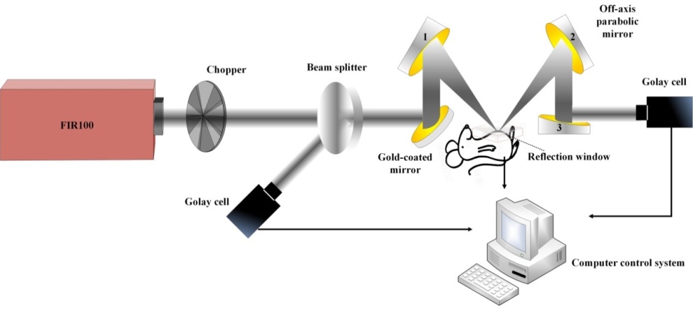
    Fig. 1. Schematic diagram of continuous THz wave reflection imaging system
    Measurement of the focal spot size by the knife-edge method
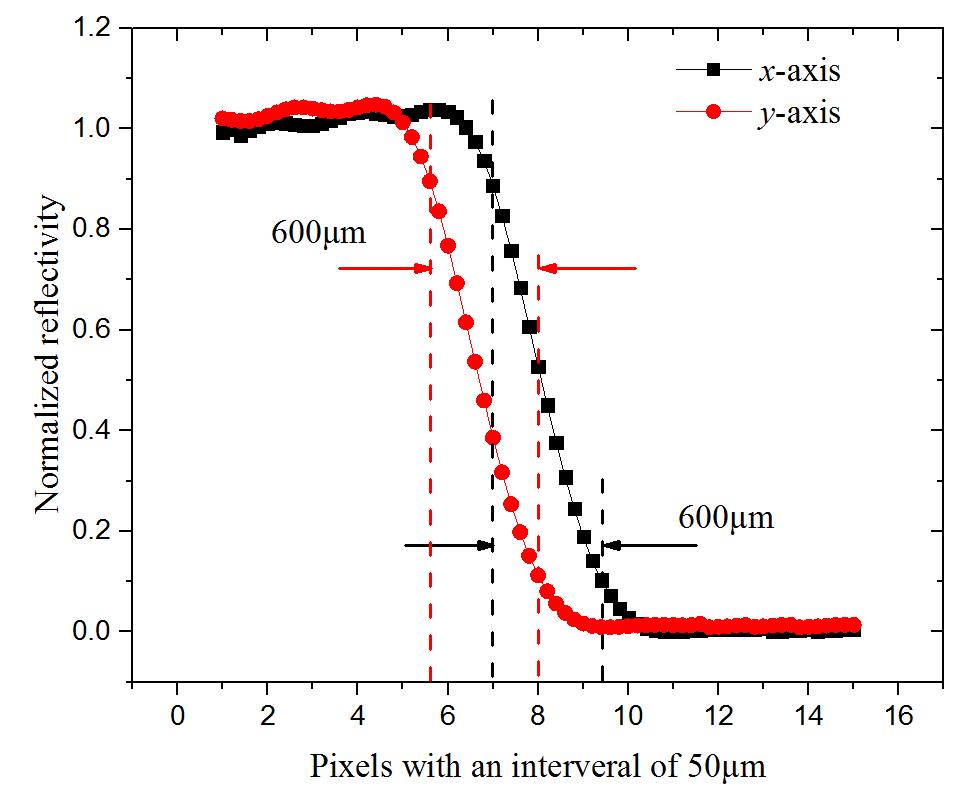
    Fig. 2. Measurement of the focal spot size by the knife-edge method
    The subcutaneous breast cancer of mouse model
    Fig. 3. The subcutaneous breast cancer of mouse model
    The sample is (a) not and (b) in close contact with the reflection window
    Fig. 4. The sample is (a) not and (b) in close contact with the reflection window
    Breast cancer tissue of mouse (a) the visual images of sample in contact with reflection window and (b) reflection images of THz wave. (a) The white dotted area is the tumor in contact with reflection window and (b) The black dotted area is the normal tissue in contact with reflection window.
    Fig. 5. Breast cancer tissue of mouse (a) the visual images of sample in contact with reflection window and (b) reflection images of THz wave. (a) The white dotted area is the tumor in contact with reflection window and (b) The black dotted area is the normal tissue in contact with reflection window.
    (a), (b) and (c) show the normalized relative reflectivity of the horizontal purple dotted lines of Fig.3 (b) NO.1, NO.2 and NO.3, respectively.
    Fig. 6. (a), (b) and (c) show the normalized relative reflectivity of the horizontal purple dotted lines of Fig.3 (b) NO.1, NO.2 and NO.3, respectively.
    The freshly excised breast cancer tissue of mice
    Fig. 7. The freshly excised breast cancer tissue of mice
    Staining images of freshly excised breast cancer tissue (NO.1) at different depths from skin surface
    Fig. 8. Staining images of freshly excised breast cancer tissue (NO.1) at different depths from skin surface
    d=2602.25d=4608.25d=86053.25
    d=3202.75d=5208.80d=92070.50
    Table 1. 距离皮肤表面不同深度组织病理染色图中的肿瘤面积
    Li-Min WU, Bin LIAO, De-Gang XU, Yu-Ye WANG, Mei-Lan GE, Chao-Nan ZHANG, Jia-Hua LI, Zhong-Cheng SUN, Tu-Nan CHEN, Hua FENG, Jian-Quan YAO. Study of in-vivo breast cancer in a subcutaneous xenograft mouse model using terahertz imaging[J]. Journal of Infrared and Millimeter Waves, 2020, 39(5): 553
    Download Citation