
Han Wang, Haofeng Zang, Yonghua Lu, Pei Wang. Mode Properties and Fluorescence Emission Mediation of Metal-Dielectric-Metal Nanoantenna Array[J]. Acta Optica Sinica, 2020, 40(4): 0426001
- Acta Optica Sinica
- Vol. 40, Issue 4, 0426001 (2020)
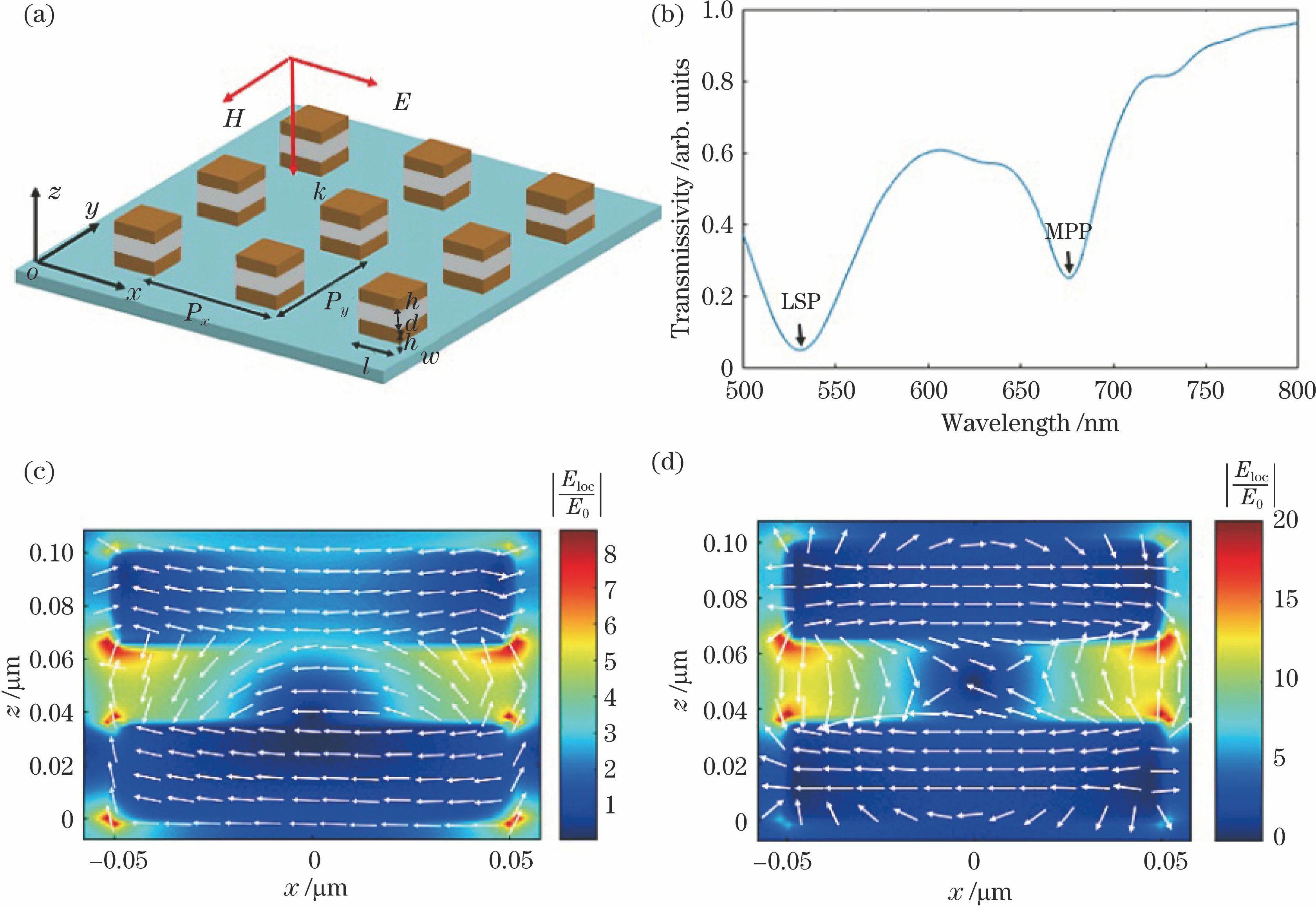
Fig. 1. Simulation model and simulation results. (a) Schematic of MDM nanoantenna array structure; (b) simulated transmission spectrum of white light when polarization direction of incident light is along x axis; (c) electric field intensity and electric vector distribution map on xoz plane at resonant position of 532 nm; (d) electric field intensity and electric vector distribution map on xoz plane at resonant position of 671 nm
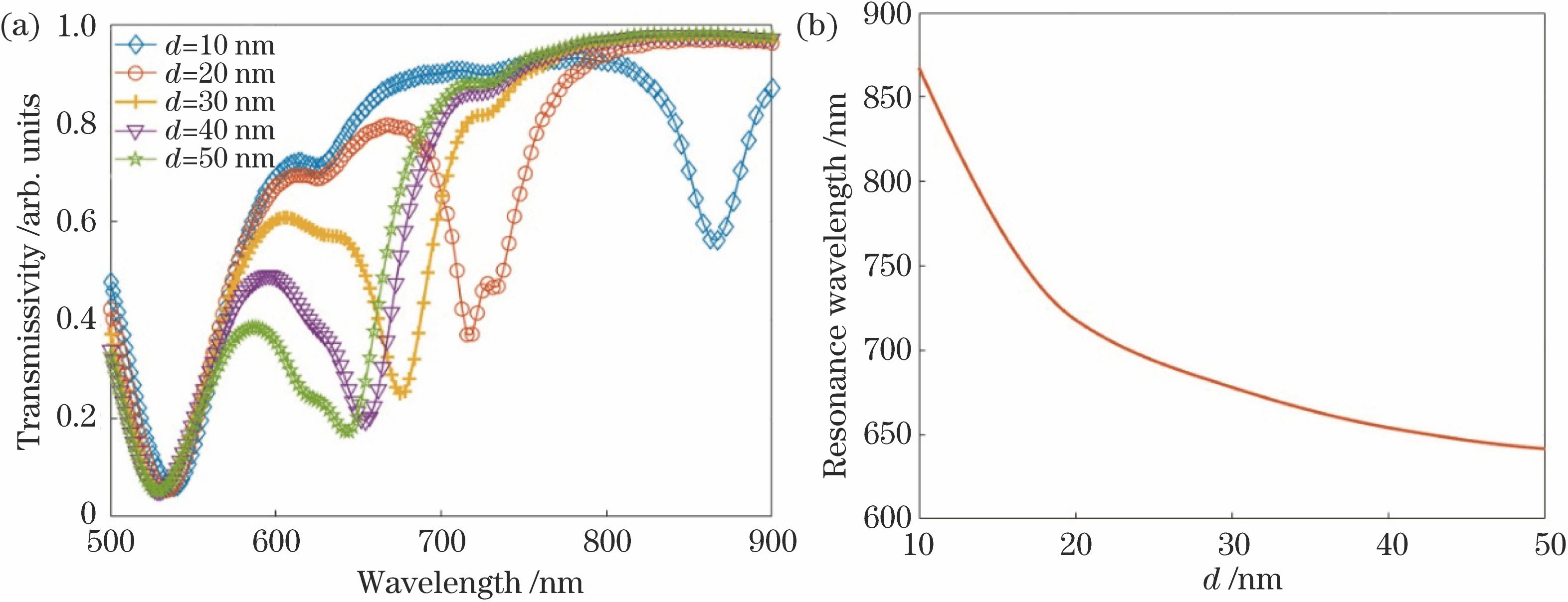
Fig. 2. Influence of structure parameters on resonance position. (a) Simulated transmission spectrum of the proposed structure; (b) change curve of resonance position of MPP mode with respect to d
Fig. 3. Simulated transmission spectra of proposed structure
Fig. 4. Influence of different length l and polarization angle on resonance position. (a) Simulated transmission spectrum of proposed structure; (b) variation of resonance position of LSP and MPP modes with l; (c) simulated transmission spectra of proposed structure, when l is 90 nm, w is 120 nm and step size is 15°
Fig. 5. MDM fishnet metasurface. (a) Schematic of MDM fishnet metasurface; (b) simulated transmission spectra when polarization direction of incident light is along x axis
Fig. 6. Total electric field and electric field in x, y, and z direction on xoy plane in dielectric layer when excitation light wavelength is 532 nm. (a)-(d) θ =0°; (e)-(h) θ=45°; (i)-(l) θ=90°
Fig. 7. Positions of dipole and corresponding properties. (a)(c)(e) Schematic when dipole is located at different positions; (b)(d)(f) radiative and non-radiative decay rate enhancement factor, Purcell factor and quantum efficiency of luminescent molecules of dipole
Fig. 8. Quantum efficiency of MDM fishnet metasurface and MDM nanoantenna structure when peak of fluorescent emission is at 650 nm
Fig. 9. Distribution of electric field intensity in each direction at x and y direction. (a)-(c) Results are obtained at 635 nm when dipole is excited by the x polarization laser; (d)-(f) results are obtained at 770 nm when dipole is excited by the y polarization laser

Set citation alerts for the article
Please enter your email address
© Copyright 2018-2021 | Chinese Laser Press. All Rights Reserved 沪ICP备15018463号-20