
Ke Wu, Ling Yang. Ultrasound Left Ventricular Segmentation Method Based on Multi-Phase Level Set[J]. Laser & Optoelectronics Progress, 2019, 56(16): 161014
- Laser & Optoelectronics Progress
- Vol. 56, Issue 16, 161014 (2019)
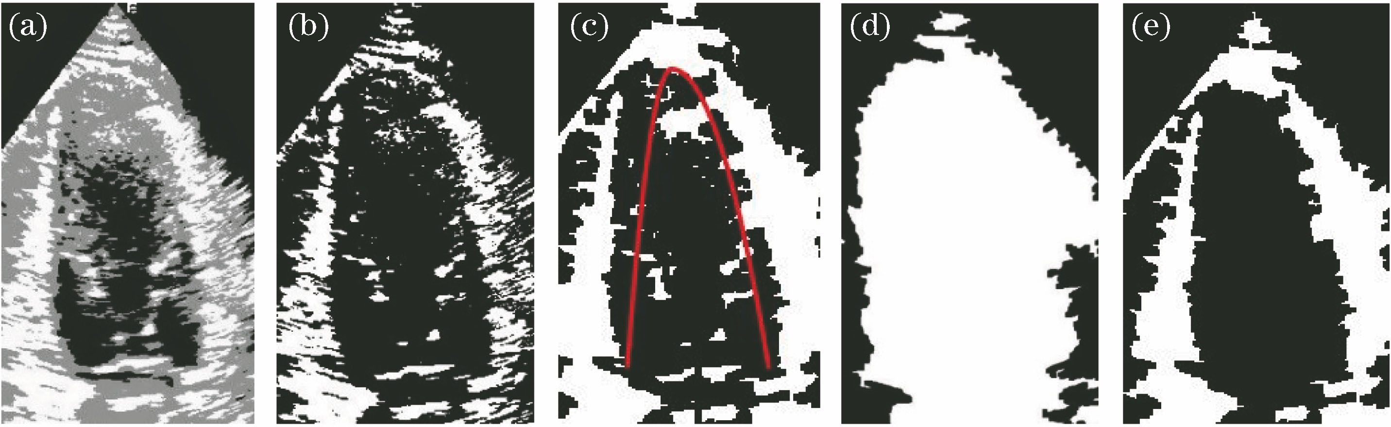
Fig. 1. Result of binary image processing. (a) Segmentation result of three-phase Level Set; (b) take out results of white part of Fig. 1(a); (c) parabolic model; (d) left ventricular full filling image; (c) result of binary image processing
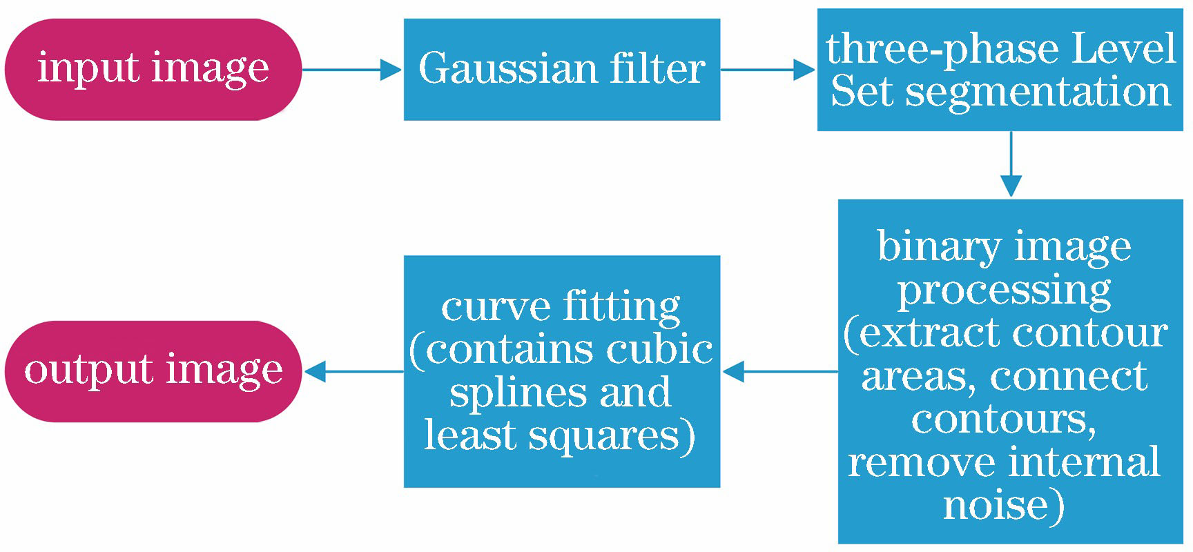
Fig. 2. Flow chart of algorithm
Fig. 3. Comparison of segmentation results of different algorithms with doctor's manual segmentation results. (a) Segmentation result of traditional Level Set method for low noise image; (b) segmentation result of proposed method for low noise image; (c) doctor's manual segmentation result for low noise image; (d) segmentation result of traditional Level Set method for high noise image; (e) segmentation result of proposed method for high noise image; (f) doctor's manual segmentation result for high nois
Fig. 4. Results of left ventricular segmentation. (a) Original image; (b) three-phase Level Set segmentation; (c) binarization; (d) binary image subsequent processing; (e) segmentation result
Fig. 5. Linear fitting results for different numbers of sample points. (a) 5; (b) 10; (c) 15; (d) 20; (e) 30
| |||||||||||||||
Table 1. Results of different evaluation parameter

Set citation alerts for the article
Please enter your email address
© Copyright 2018-2021 | Chinese Laser Press. All Rights Reserved 沪ICP备15018463号-20