• Laser & Optoelectronics Progress
  • Vol. 59, Issue 6, 0617029 (2022)
Wanxin Xiao1、2, Huafeng Li1、2, Yafei Zhang1、2、*, Minghong Xie1, and Fan Li1、2
Author Affiliations
  • 1Faculty of Information Engineering and Automation, Kunming University of Science and Technology, Kunming , Yunnan 650500, China
  • 2Yunnan Key Laboratory of Artificial Intelligence, Kunming , Yunnan 650500, China
  • show less
    DOI: 10.3788/LOP202259.0617029 Cite this Article Set citation alerts
    Wanxin Xiao, Huafeng Li, Yafei Zhang, Minghong Xie, Fan Li. Medical Image Fusion Based on Multi-Scale Feature Learning and Edge Enhancement[J]. Laser & Optoelectronics Progress, 2022, 59(6): 0617029 Copy Citation Text show less
    Basic framework of multi-scale feature learning and edge enhancement fusion network
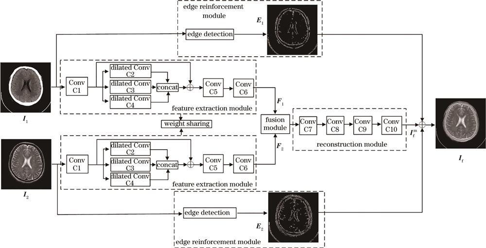
    Fig. 1. Basic framework of multi-scale feature learning and edge enhancement fusion network
    Fusion results obtained by the CNN and NSCT-PAPCNN methods. (a) Source CT image; (b) source MR-T2 image; (c) CNN; (d) NSCT-PRPCNN
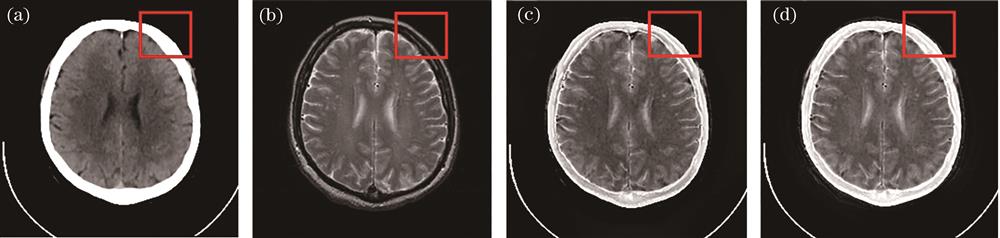
    Fig. 2. Fusion results obtained by the CNN and NSCT-PAPCNN methods. (a) Source CT image; (b) source MR-T2 image; (c) CNN; (d) NSCT-PRPCNN
    Source image samples in testing dataset
    Fig. 3. Source image samples in testing dataset
    Visual comparison of the different methods. (a) Source CT image; (b) source MR-T2 image; (c) FW-Net; (d) GF; (e) CNN; (f) NSCT; (g) NSCT-PRPCNN; (h) proposed method
    Fig. 4. Visual comparison of the different methods. (a) Source CT image; (b) source MR-T2 image; (c) FW-Net; (d) GF; (e) CNN; (f) NSCT; (g) NSCT-PRPCNN; (h) proposed method
    Fusion results without and with edge reinforcement branches. (a) Without edge reinforcement branch; (b) with edge reinforcement branch
    Fig. 5. Fusion results without and with edge reinforcement branches. (a) Without edge reinforcement branch; (b) with edge reinforcement branch
    Fusion results obtained by the proposed method in MR-T1 and MR-T2 image fusion task. (a) Source MR-T1 image; (b) source MR-T2 image; (c) proposed method
    Fig. 6. Fusion results obtained by the proposed method in MR-T1 and MR-T2 image fusion task. (a) Source MR-T1 image; (b) source MR-T2 image; (c) proposed method
    ModuleConvolutional layerSizeNumber of input channelsNumber of output channelsActivation layer
    Feature extraction module

    C1

    C2

    C3

    C4

    C5

    C6

    3

    1

    3

    5

    1

    3

    1

    64

    64

    64

    192

    64

    64

    64

    64

    64

    64

    64

    ReLU

    ReLU

    ReLU

    ReLU

    ReLU

    ReLU

    Reconstruction module

    C7

    C8

    C9

    C10

    3

    3

    3

    3

    64

    64

    32

    16

    64

    32

    16

    1

    ReLU

    ReLU

    ReLU

    ReLU

    Table 1. Structure of MFEnet

    Algorithm MFEnet training and testing algorithms

    Training

    Input:Training set source image Ii

    Output:Reconstructed image Io

    1)Randomly select m source images from the training set I1,,Im

    2)Input m source images into the feature extraction module to generate source image features F

    3)Input F into the reconstruction module to generate a reconstructed image Io

    4)Use Adam optimizer to update the parameters of the feature extraction module and reconstruction module:

    θ1WHIo-IiF2+αIo-IiF2

    5)If the number of iterations is equal to epoch,the training ends,otherwise repeat steps 1)-4)

    Testing

    Input:Testing set source images I1 and I2

    Output:Fused image If

    1)Input I1 and I2 into the feature extraction module to get the source image features F1 and F2

    2)Input I1 and I2 into the edge enhancement module to get the source image edge maps E1 and E2

    3)Input F1 and F2 into the fusion module and the reconstruction module to obtain the intermediate fusion image Ifm

    4)Combine IfmE1,and E2 to get the final fusion image If

    Table 2. Algorithms of MFEnet training and testing
    MethodFMIpixelSCDSSIMSLQYTime /s

    FW-Net

    GF

    CNN

    NSCT

    NSCT-PRPCNN

    Proposed method

    0.8182

    0.8595

    0.8637

    0.873

    0.8448

    0.8741

    0.7776

    0.8910

    1.1123

    0.969

    1.2690

    1.2879

    0.5108

    0.6612

    0.5717

    0.6160

    0.6350

    0.7412

    0.0072

    0.0167

    0.0134

    0.0155

    0.0122

    0.0069

    0.6600

    0.8407

    0.7165

    0.7311

    0.7575

    0.8422

    0.0199

    0.0912

    9.601

    3.43

    9.601

    0.0189

    Table 3. Average value of quality evaluation index of 20 fused images
    Fusion ruleFMIpixelSCDSSIMSLQY

    Addition35

    Average36

    Weighted average37

    Max value34

    0.8728

    0.8689

    0.8708

    0.8741

    1.1951

    1.1703

    1.2456

    1.2879

    0.7422

    0.7270

    0.7337

    0.7412

    0.0071

    0.0068

    0.0073

    0.0069

    0.8116

    0.8095

    0.8119

    0.8422

    Table 4. Average value of the quality evaluation index of fusion results with four different fusion rules
    Wanxin Xiao, Huafeng Li, Yafei Zhang, Minghong Xie, Fan Li. Medical Image Fusion Based on Multi-Scale Feature Learning and Edge Enhancement[J]. Laser & Optoelectronics Progress, 2022, 59(6): 0617029
    Download Citation