
Fei Wang, Hao Wang, Yaoming Bian, Guohai Situ. Applications of Deep Learning in Computational Imaging[J]. Acta Optica Sinica, 2020, 40(1): 0111002
- Acta Optica Sinica
- Vol. 40, Issue 1, 0111002 (2020)
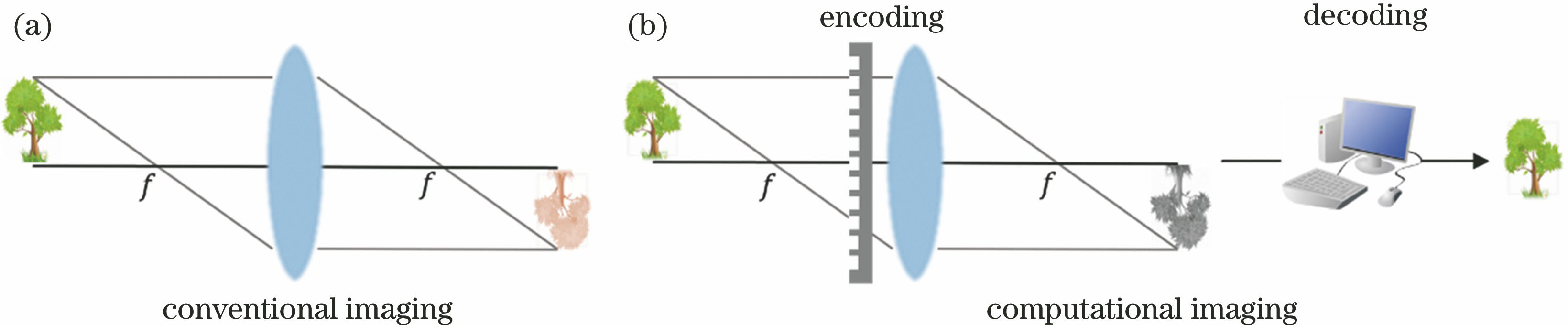
Fig. 1. Schematic diagram. (a) Conventional imaging model; (b) computational imaging model
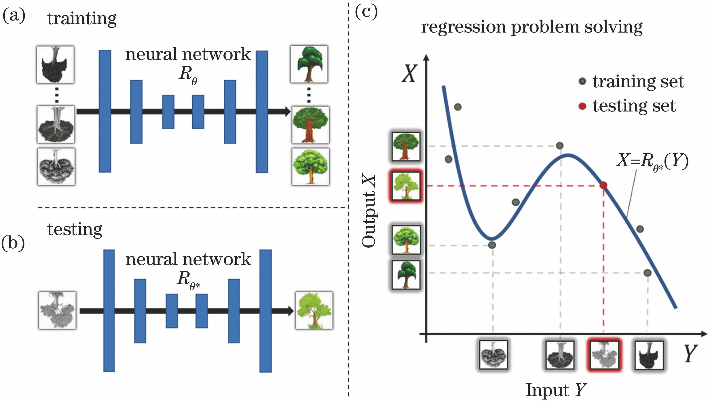
Fig. 2. Schematic of regression problem solved using neural network. (a) Training; (b) testing; (c) fitting process
Fig. 3. Diagram of data acquisition methods in computational imaging. (a) Images reconstructed by traditionally complex and costly methods; (b) presetting real objects with SLM; (c) data obtained by numerical simulation
Fig. 4. Schematic of fully connected neural network. (a) Single hidden layer; (b) multi hidden layers
Fig. 5. Simplified diagram of convolutional neural network. (a) Convolution: different convolution kernels corresponding to different feature maps; (b) pooling: replace data within the scope of operation with its maximum or mean; (c) deconvolution: interpolate data with zeros and then implement convolution; (d) convolution
Fig. 6. Main problems in neural network training. (a) Local minimum; (b) overfitting
Fig. 7. Flow chart of neural network training
Fig. 8. Applications of deep learning in scattering imaging. (a) Imaging through strong scattering media[28,74]; (b) imaging through different ground glasses[27]
Fig. 9. Applications of deep learning in digital holography. (a) Removing twin image[32]; (b) end-to-end phase reconstruction[34]; (c) end-to-end complex amplitude reconstruction[66]
Fig. 10. Applications of deep learning in computational ghost imaging. (a) Improving signal-noise-ratio[29]; (b) reconstructing objects by using intensity sequences[31]
|
Table 1. Typical applications of deep learning in computational imaging

Set citation alerts for the article
Please enter your email address
© Copyright 2018-2021 | Chinese Laser Press. All Rights Reserved 沪ICP备15018463号-20