• Laser & Optoelectronics Progress
  • Vol. 59, Issue 6, 0617024 (2022)
Yijia Geng1、3、5, Lili Cong1, Xiumian Cao1、4, Xin Guan2, Zepeng Huo1, Gang Chen1, Yu Liu3, Weiqing Xu1、5、***, Chongyang Liang2、**, and Shuping Xu1、5、*
Author Affiliations
  • 1State Key Laboratory of Supramolecular Structure and Materials, College of Chemistry, Jilin University, Changchun , Jilin 130012, China
  • 2School of Pharmaceutical Sciences, Jilin University, Changchun , Jilin 130021, China
  • 3State Key Laboratory of Applied Optics, Changchun Institute of Optics, Fine Mechanics and Physics, Chinese Academy of Sciences, Changchun , Jilin 130033, China
  • 4College of Physics, Jilin University, Changchun , Jilin 130012, China
  • 5Institute of Theoretical Chemistry, College of Chemistry, Jilin University, Changchun , Jilin 130012, China
  • show less
    DOI: 10.3788/LOP202259.0617024 Cite this Article Set citation alerts
    Yijia Geng, Lili Cong, Xiumian Cao, Xin Guan, Zepeng Huo, Gang Chen, Yu Liu, Weiqing Xu, Chongyang Liang, Shuping Xu. Preliminary Exploration of Plasmon-Enhanced Four-Wave Mixing Imaging and Its Possible Application in Antibody-Drug Metabolism in the Body[J]. Laser & Optoelectronics Progress, 2022, 59(6): 0617024 Copy Citation Text show less
    Multi-mode nonlinear optical imaging system. (a) Schematic diagram of the structure design of the multi-mode nonlinear optical imaging system; (b) picture of Olympus FVMPE-RS microscope
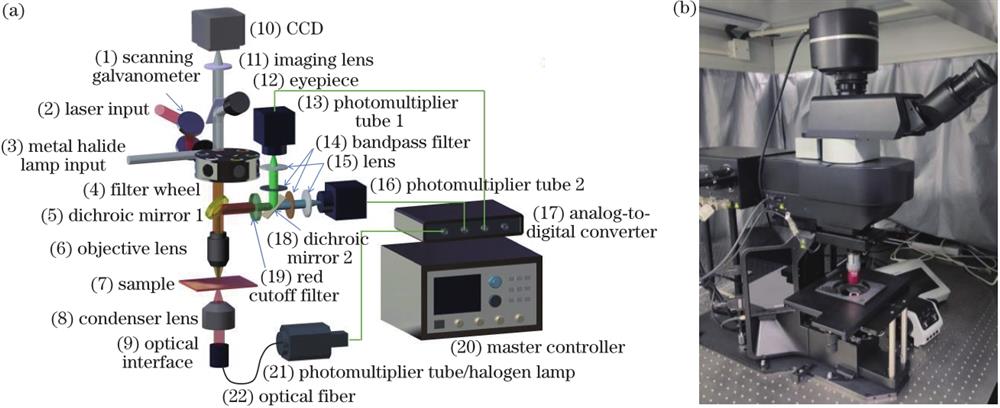
    Fig. 1. Multi-mode nonlinear optical imaging system. (a) Schematic diagram of the structure design of the multi-mode nonlinear optical imaging system; (b) picture of Olympus FVMPE-RS microscope
    Test for two-photon imaging function. (a) Two-photon imaging of anthracene crystals; (b) two-photon fluorescence spectra of anthracene crystals
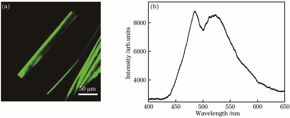
    Fig. 2. Test for two-photon imaging function. (a) Two-photon imaging of anthracene crystals; (b) two-photon fluorescence spectra of anthracene crystals
    Test for SRS imaging function. (a) Bright field imaging of PMMA; (b) SRS imaging effect; (c) Raman spectra of PMMA
    Fig. 3. Test for SRS imaging function. (a) Bright field imaging of PMMA; (b) SRS imaging effect; (c) Raman spectra of PMMA
    Optimization of SRS resonance conditions. (a) SRS images of PMMA varying with the delay time of two wavelength lasers; (b) SRS images of PMMA associated with the pump laser wavelength
    Fig. 4. Optimization of SRS resonance conditions. (a) SRS images of PMMA varying with the delay time of two wavelength lasers; (b) SRS images of PMMA associated with the pump laser wavelength
    Schematic diagram of plasmon enhanced FWM. (a) Diagram showing the coupling of surface plasmon resonance for energy levels for FWM; (b) local electromagnetic field simulations of Ag and Au nanoparticles in FWM; (c) FWM image of metal nanoparticles recorded by the self-built multi-photon microscope
    Fig. 5. Schematic diagram of plasmon enhanced FWM. (a) Diagram showing the coupling of surface plasmon resonance for energy levels for FWM; (b) local electromagnetic field simulations of Ag and Au nanoparticles in FWM; (c) FWM image of metal nanoparticles recorded by the self-built multi-photon microscope
    TEM image of gold nanoparticles
    Fig. 6. TEM image of gold nanoparticles
    Thin-section images of mouse liver and kidney tissue
    Fig. 7. Thin-section images of mouse liver and kidney tissue
    Yijia Geng, Lili Cong, Xiumian Cao, Xin Guan, Zepeng Huo, Gang Chen, Yu Liu, Weiqing Xu, Chongyang Liang, Shuping Xu. Preliminary Exploration of Plasmon-Enhanced Four-Wave Mixing Imaging and Its Possible Application in Antibody-Drug Metabolism in the Body[J]. Laser & Optoelectronics Progress, 2022, 59(6): 0617024
    Download Citation